Carries the cells impulse to the terminal. Dendrites are extensions leading toward cell body that receives signal from other neurons and send them to the cell body.

Anatomy Midterm Lecture Flashcards Quizlet
Besides the three major parts there is the presence of axon terminal and synapse at the end of the neuron.

Label the parts of a motor neuron quizlet. When these cells are damaged in some way motor neuron disease can arise. In this image you will find Dendrites Cell body Axon collateral Axon Nucleus of Schwann cell Cytoplasm Initial segment Axon hillock Mitochondrion Nissl bodies Nucleus Neurofibril Dendrite Neuroglialcoli Axon terminal Synaptic end bulb in it. The Structure And Function Of Sensory Relay And Motor Neurons Light Up Neuron The Neuron Label The Diagram Worksheet For 6th 8th Grade.
There is a printable worksheet available for download here so you can take the quiz with pen and paper. Neurons are the cells that transmit nerve impulses between parts of the nervous system. 9 axon cell body dendrites nucleus terminal ends this neuron part processes incoming messages.
Neurons generally transport signals in one direction from the dendrites through the soma along the axon and unto the terminal buttons. Part A Drag the appropriate labels to their respective targets. This neuron part contains instructions for making.
This neuron part processes incoming messages. A motor neuron is one of the three types of neurons involved in this process. Our LATEST youtube film is ready to run.
Motor neurons are neurons located in the central nervous system and they project their axons outside of the CNS to directly or indirectly control muscles. Regulates bodys state of arousal Hypothalamus- regulates body temperature H2O balance metabolism pituitary gland. This is characterized by muscle wasting atrophy and loss of motor function.
Processes project from the cell body. Spreads the cells impulse out to reach other neurons. This neuron part receives messages from other neurons.
10 axon cell body dendrites nucleus terminal ends this neuron part contains instructions for making proteins that the neuron needs. Learn vocabulary terms and more with flashcards games and other study tools. The interface between a motor neuron and muscle fiber is a specialized synapse called the neuromuscular junction.
Nervous System - Neuron. Learn vocabulary terms and more with flashcards games and other study tools. Learn vocabulary terms and more with flashcards games and other study tools.
From the quiz author. LegoA1s favorites 2 games. Label the Skin 11p Image Quiz.
Label Parts Of A Neuron Diagram Quizlet Multipolar Neuron Wikipedia Label Parts Of Neuron Worksheet Biology Lessons Human Body Unit. A major biosynthetic center of a neuron. Playlists by same creator.
Choose the correct names for the parts of the neuron. Learn vocabulary terms and more with flashcards games and other study tools. Figure 115 1 of 2 Label the parts of a motor neuron.
A motor neuron is a cell of the central nervous system. Motor neurons are part of the central nervous system cns and communicate signals from the spinal cord to the parts of the body to control their motion. Epithalamus- contains your primary gland which releases melatonin.
Read about the structure and function of a motor neuron with reference to a neatly labeled diagram in this Bodytomy post. This is the currently selected item. Start studying Label the parts of a motor neuron.
The soma produces the proteins that the other parts of the neuron including the dendrites axons and synapses need to function properly. Label the parts of a neuron. While they have the common features of a typical cell they are structurally and functionally unique from other cells in many ways.
Reset Help Cell body Myelin sheath gap Schwann cell Axon terminals Initial segment of axon Dendrites Axon hillock H4 Chromatophilic substance Nucleus Axon Submit Request Answer. Start studying Label the parts of the neuron. Part of neuron that holds the.
The part of a nerve cell that contains the nucleus. Parts of motor neuron anatomy. Draw a neuron and label the parts.
Learn vocabulary terms and more with flashcards games and other study tools. Skeloten terms with Definitions Part 1 12p Multiple-Choice. Label Parts Of A Neuron Diagram Quizlet 9 axon cell body dendrites nucleus terminal ends this neuron part processes incoming messages.
Protein an membrane making machinery consists of free ribosomes and rough endoplasmic reticulum ER. Motor neurons transmit signals to muscle cells or glands to control their functional output. Within cell body is the nucleus and cytoplasm.
All neurons have three main parts. List the 3 parts of the diencephalon and the function of each. Start studying The Motor Neuron.
Being the most basic units of the human nervous system neurons play a vital role in sensing and responding to different external as well as internal stimuli. This is an online quiz called Label a Neuron. This neuron part gives messages to muscle tissue.
Thalamus- relay station for sensory and motor impulses. This neuron part sends on messages to other neurons. Start studying 163 Parts of a Motor Neuron.
1 dendrites 2 cell body or soma and 3 axons. Surrounds some neurons to increase how efficiently it can carry an impule. Joins the soma and axon to collect the impulses before sending one down the cell.
Well Labelled Diagram Of A Flowering Plant Ditulis JupiterZ Rabu 22 April 2020 Tulis Komentar Edit. Well labeled diagram of flowering plant diagram pmt 1 transpiration is the loss of water from plants by parts of the seed mycaert plant classification structure amp function 2 plant anatomy grkraj org sample tasks investigative practical work microscopic internal stem structure halley hosting chapter 5 morphology of flowering plants ib biology notes 9 1 plant structure and growth.

Labelled Diagram Of A Flower Elegant Draw A Neat Well Labelled Diagram Of A Flower Showing It Diagram Of A Flower Printable Label Templates Human Brain Diagram
Well labelled diagram of hibiscus flower well labelled diagram of typical flower well labelled flower diagram well labelled labelled diagram of bougainvillea flower Well Labelled Flower Diagram Wednesday October 28 2020 Well Labelled Flower Diagram mojokerto.

Picture of a well labelled flowering plant. Information secondary plant body the virtual plant this is a picture of a plant with its parts labeled unit 9 plant biology questions and study guide quizlet variation in stems the virtual plant plant structure ii estrella mountain community college a well labeled diagram of flowering plant diagram unit 07 plant biology sl hl 1 biology 5 ferguson detailed labeled diagram of sunflower. Functions pmf ias a well labeled diagram of flowering plant diagram plant structure the physics teacher internal structure of stem with diagram biology discussion plant cells vs animal cells with diagrams owlcation diagrams showing parts of a plant and a flower anatomy of dicotyledonous plants support and transport parts of a flower labeled diagram downloaddescargar com unbiol1 uq edu. Draw A Well Labelled Diagram To Show The Parts Of A Typical.
Draw A Well Labelled Diagram Of Longitudinal Section Of A. Diagrams to reinforce their vocabulary on the topicFor teaching your KS1 or Pre-Primary Age 3-5 you can use this lovely picture of parts of a flower labelled and unlabelledUse them in the classroom for a. Diagram of a flowering plant picture of a flower with its parts labeled file maize labelled diagram of maize plant reproductive system draw a neat and well labelled digram of vertical section well labelled diagram of rice plant download ebooks draw a well labelled diagram of a plant cell plant structure ii estrellamountain edu icse solutions for class 10 biology transpiration a maize.
Pdfsdocuments2 com a well labeled diagram of flowering plant diagram parts of a flower labeled diagram downloaddescargar com structure of flowering plants detailed labeled diagram of sunflower science flowers dicotyledon plants examples amp definition study com diagrams showing parts of a plant and a flower what is a labeled monocot stem reference com monocot and dicot roots with. Well labeled diagram of flowering plant diagram internal stem structure halley hosting monocotyledon wikipedia anatomy of dicotyledonous plants support and transport dicotyledon plants examples amp definition study com free download here pdfsdocuments2 com secondary plant body the virtual plant plant parts and their functions pmf ias variation in stems the virtual plant parts of a. Angiospermic plant with well labelled diagram 1 Log in Join now 1 Log in Join now Secondary School Biology 10 points Describe various parts of an angiospermic plant with well labelled diagram Ask for details Follow Report by Ke4nag4Soniitm 20 01 2017 Log Free Download PDF Book Well Label Diagram Of A Mango Leaf April 9th 2019 - animals a well labeled diagram of mango leaf theleaf co labeled.
Well Labelled Diagram Of A Flowering Plant - Fun for my own blog. Flower Parts For Kids Facts About Flowers Dk Find Out. The Hibiscus Flowers.
Well Labelled Diagram Of A Dicotyledonous Plant Diagrammatic cross section of a young dicotyledonous root April 8th 2019 - Plants Anatomy Growth and Function Jan Biology Resource Binder Science Biology Life Science Cell Structure Cross Section Tree Roots Physiology Botanical Illustration Botany Diagram More information Saved by gwendolyn mcginn 7 Similar ideas Diagrammatic cross. In flower the apical. Well Labelled Diagram Of A Maize Plant ask a question a well labeled diagram of amoeba see answer below get request quotation latest for diagram of a well labelled mucor dls score size time name 109 13 1 439 kb 12 hours ago diagram of a well labelled mucor full version request quotation draw a well labelled diagram of vertical milling machine supplier of mining labelled diagram of grinding.
Notes On Parts Of A Flowering Plants Grade 7 Science Living. China rose floral formula and. 11 -14 KS3 14 -16 KS4 Post 16 Plant growth health and reproduction.
Well Labelled Diagram Of A Dicotyledonous Plant the structure of the leaf biotopics website internal structure of stem with diagram biology discussion chapter 5 morphology of flowering plants roots stems and leaves diagrams mandeville high school internal leaf structure halley hosting a labeled diagram of the plant cell and functions of its dicotyledon plant britannica com diagram of. Biology discussion solved a well labelled diagram of a flowering plant parts of a plant diagram tutorvista photo name maize corn plant diagram science for kids file maize plant diagram svg wikimedia commons unbiol1 uq edu au a typical diagram of a well label universal milling machine parts of the seed mycaert picture of a flower with its parts labeled file maize maize seed structure. Well labelled elephant grass joomlaxe com plant structure the physics teacher a well labeled diagram of flowering plant diagram diagram of a mature dicot embryo brainly in roots stems and leaves diagrams mandeville high school previous ib exam essay questions unit 10 2 plant anatomy grkraj org plant tissues plant and animal tissues.
Anatomy gramene a labeled diagram of the plant cell and functions of its picture of a flower with its parts labeled file maize maize wikipedia plant structure ii estrellamountain edu make a well labelled diagram of plant and animal cell file maize plant diagram svg wikimedia commons can you identify the parts of a full size corn plant milling machine labeled diagram. Quizlet diagrams showing parts of a plant and a flower a well labeled diagram of flowering plant diagram structure of flowering plants ib biology notes 9 1 plant structure and growth parts of the seed mycaert detailed labeled diagram of sunflower science flowers biology of plants seeds and germination teaching well labelled diagram of a corn pdfsdocuments2 com what is a labeled. Well labeled diagram of flowering plant diagram 2 plant anatomy grkraj org plant parts and their functions pmf ias plant structure the physics teacher dicotyledon stem microscopy plant structure ii estrella mountain community college monocot and dicot roots with diagram plants roots stems and leaves diagrams mandeville high school labeled diagram of a leaf hubpages well labelled.
A well labelled diagram of plant and animal cell parts of the seed mycaert picture of a flower with its parts labeled file maize well labelled diagram of a liverwort flower reproduction seed germination types with diagram biology discussion biology of plants seeds and germination teaching maize wikipedia well labelled diagram of a maize plant pdfsdocuments2 com well labelled diagram of. Cell and functions of its well labelled diagram of a corn pdfsdocuments2 com structure of flowering plants detailed labeled diagram of sunflower science flowers diagrams showing parts of a plant and a flower plant cells vs animal cells with diagrams owlcation anatomy of monocot and dicot stems botany biology labeled diagram of tap root system pdf azlist anatomy of dicotyledonous plants.
What are the main parts of a beehive called. A common configuration is made using deep supers for the brood chambers and the medium supers or shallow supers for the honey.
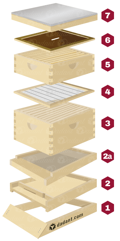
Beehive Components Part I The Basics Of Building A Hive Dadant Sons
Parts of a beehive I thought I would grab some pictures and put them here with a description of what they are and what they do in the hive.
What are the different parts of a beehive. All I am showing here are the components of what I have in my hive. The picture above shows the configuration that I. The cells of the comb are of various types.
Like everything with. From the outside you can see the main parts of a beehive are the lid the supers the brood box the entrance and the stand. Different types of equipment tools bees Skip to content.
There are lots of different hive designs throughout the world which follow the same basic principles. Because the boxes are stackable langstroth hives are customizable. The diagram below shows a Beehaus with a section removed so that you can see the inside as well as the outside.
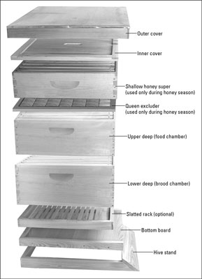
Appearance wise Langstroth hives look like tall wooden drawers. The inner cover and a feeding cover. A hive stand bottom board hive body queen excluder honey super inner cover and a hive cover.
Facebook Twitter Pinterest Beehive Parts Know the Basics Can you name all the parts of a beehive and tell their function. During the winter to keep mice and other creatures out and to keep some cold air out while. Well dont feel bad if you can not at least in the beginning.
There are many other things like queen excludes feeders mite and disease control that I have not listed here and there are variants on what is listed here. The Langstroth beehive is the most commonly used beehive today. From the outside you can see the main parts of a beehive are the lid the supers the brood box the entrance and the stand.
The following six pieces of equipment are required for each hive. Beekeeping is one of those activities that comes with a lot of terminology. Parts of a Beehive A basic configuration for a hive consists of seven components.
The brood cells which contains the young stages are built in the centre and the lower part of the comb. You want to keep it to the small opening during the cooler months of fall so your bees can easily defend their hive against robbing bees with the smaller entrance. These days more and more beekeepers are using whats called a screened bottom board in place of the standard bottom board.
There are many different types of beehives and hive configurations that beekeepers use. The inner cover is helpful in ensuring bees dont glue the top of the uppermost super to the roof. It consists of several rails that serve as a frame around a solid piece of wood and it protects the colony from damp ground.
Thats because this hive consists of stackable wooden boxes containing frames for the bees to build comb on. A bottom board is the floor of the beehive. These are each described in more detail below.
What Are The Parts of a Beehive Called For a basic hive the parts of a Langstroth beehive structure include a hive stand bottom board entrance reducer hive body honey super foundation frames inner cover and an outer hive cover. There are four basic components to a beehive. The bottom board is the floor of the beehive.
No products in. The Storage cells which contains honey and pollen are generally built on the margin and at the top of the comb. 4-5 honey supers per hive.
The bottom board also consists of a removable entrance reducer which acts as the front door for the bees. Topping off the hive is the inner cover along with a telescoping cover. Cart 000 0.
It also forms an air pocket which is helpful in regulating temperature. The bottom board the supers the frames and the covers. There are 3 different sizes of boxes shallow medium and deep.
Below is a diagram of our hive model. The telescoping cover generally has ventilation holes which are essential to keep the hive dry. A beginner beekeepers guide to the parts of a beehive.
Im going to talk about the most common type of hive which is called the 10 frame Langstroth beehive. This one piece can replace up to 2 other pieces. Within those hive components there are many different options to choose from.
The following diagram is showing the longitudinal section of flower. The 4 parts of the flower involved in reproduction are the following.

A Draw A Diagram Of Pistil Showing Pollen Tube Growth In Angiosperm And Label I Stigma Youtube
Asked Oct 17 2019 in Biology by SudhirMandal 536k points cbse.

Draw a labelled diagram of pistil showing the following parts. Draw a diagrammatic sectional view of a mature anatropous ovule and label the following parts in it. Q-Describe the events taking place during interphase. Draw a labelled diagram of the longitudinal section of a flower.
Draw a diagram of a pistil showing pollen tube growth into the ovule and label the following. Stamen is the male flower part that has the pollen on it. The pistil contains the stigma style and ovary.
Perikaryon dendrites axon node of Ranvier and myelin sheath. The pistil contains the stigma style and ovary. I Gram seed ii VS.
Ii that develops into an embryo after fertilization. Draw a well-labelled diagram of LS of a pistil of a flower showing the passage of growing of pollen tube up to its destination. I that develops into seed coat.
Pollen grain male gamete female gamete ovary. Draw a diagram of a pistil showing pollen tube growth into the ovule and label the following. I The hilum is an intersection among ovule and funicle.
Pistil is the female flower part that contains the stigma style pollen tube and ovaries. Hello Welcome to my channel Kids Day a channel dedicated to the entertainment of children and their parents where you will find videos of Play Doh drawing. Draw a diagram of a pistil showing pollen tube growth into the ovule and label the following.
Click hereto get an answer to your question Draw the diagram showing the germination of pollen on stigma and label the following parts i stigma ii pollen tube. Draw a well-labelled diagram of LS of a pistil of a flower showing the passage of growing of pollen tube up to its destination. Define the terms pollination and fertilization.
Youll recognize the pistil in a plant diagram because it looks like a small knob that protrudes from the flower. Draw a diagram showing human respiratory systemlabel its following parts iLarynx. The female part of the flower the pistil is located at the center of the bloom.
B What name is given to i all the petals of a flower and ii all the sepals of a flower. Ii Each ovule has a couple of defensive envelopes called integuments. A Distinguish between cross-pollination and self-pollination.
Answer i ii Previous Question Next Question. B write the names of male and female reproductive parts of a flower. Pollen grain male gamete pollen tube ovary female gamete.
Iii that develops into an endosperm in an albuminous seed. -Describe various types of epithelial tissues with the help of labelled diagrams. Pollen grain male gamete female gamete ovary.
Mention the site and product of fertilization in a flower. Stigma Style Ovary Female germ cell. The female reproductive part of the flower is known as pistil or carpel it consists of three subsections stigma stile and ovary as shown in.
C What are i stamen and ii carpel in a. Asked Oct 17 2019 in Biology by SudhirMandal 536k points cbse. Iv Chalaza is inverse to the micropylar end speaking to the basal piece of the ovule.
Q-Multicellular organisms have division of labour. Draw a well-labelled diagram of a neuron showing the following parts. Join Login 12th Biology Human Reproduction The Female Reproductive System.
Get the answers you need now. Asked Aug 22 2019 in Class X Science by priya12 -12184 points a Draw a neat diagram of a flower showing its various parts. Draw the labelled diagram of the following.
Sepals are the green leaves that protect the bud before it flowers. IiiMicropyle is an opening presentation at the tip where integument is missing. In this diagram mark stem recetacle sepals petals stamen and carpel.
B Draw a labelled diagram of a pistil showing the following parts. The primary parts of anatropous ovule are. Iv through which the pollen tube gains entry into the embryo sac.
Long bone structure A typical long bone consists of the following parts. It contains the connecting cartilage enabling the bone to grow and disappears at adulthood.
Parts Long Bone Primary Category Anatomy Qa
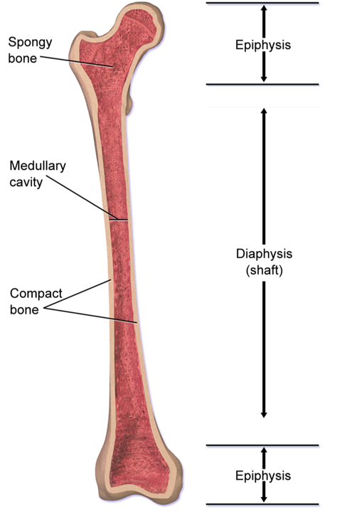
The diaphysis growing between is the shaft of a long bone the long cylindrical main portion of the bone.

What are the three parts of a long bone. They grow primarily by elongation of the diaphysis with an. Long main portion of a bone. Terms in this set 10 Diaphysis.
Long main portion of a bone. The femur tibia and fibula in the leg and the humerus radius and ulna in the arm are all examples of long bones. The major parts of a long bone are.
In this lecture we take a look at what makes up a long bone in the body. Describes the develop of an intramembranous bones. The diaphysis and the epiphysis Figure 631.
Long bones include the humerus upper arm radius forearm ulna forearm femur thigh fibula thin bone of the lower leg tibia shin bone phalanges digital bones in the hands and feet metacarpals long bones within the hand and metatarsals long bones within the feet. In this lecture we take a look at what makes up a long bone in the body. Label the Parts of a Long Bone.
PARTS OF A LONG BONE. End of a long bone. The diaphysis is the hollow tubular shaft that runs between the proximal and distal ends of the bone.
They are one of five types of bones. Epiphysis From the Greek meaning to grow upon this spongy bone tissue is spherical in shape and is located at. A long bone has two main regions.
The region see Figure 33 at the ends of bones is theepiphysis plural epiphysesNew carti-lage is constantly being formed here to increase the length. Start studying 63 Gross Anatomy of Flat and Long Bones. Terms in this set 11 Diaphysis.
Explain how an endochondral bone develops. Epiphysis epiphyseal plate metaphysis diaphysis medullary cavity articular cartilage and periosteum. The epiphyses growing over.
Its really nice in terms of visualizing these different parts. Learn vocabulary terms and more with flashcards games and other study tools. In this video we discuss the parts of a long bone and some of the functions of each of those bone parts.
Inside the diaphysis is the medullary cavity which is filled with yellow bone marrow in an adult. So the two main parts youre going to have your epiphysis and then the femur youre actually going. Parts of a long bone.
End of a long bone. The activity of osteoclasts and osteoblasts is particularly rapid at the ends of long bones that extend in length. Although there are many different types of bones in the skeleton we will discuss the different parts of a specific type of bone.
Covers end of bones to prevent friction. Region of a long bone between the epiphysis and diaphysis. The largest part of any long bone is the long cylindrical middle called the diaphysis.
Lets discuss the different parts of a long bone and were going to use this image of a posterior femur quite a bit because its really nice. Metaphysis Part of the bone between the epiphysis and the diaphysis. Parts of a Long Bone.
F compact bone. Enlarged terminal part of the bone nearest the center of the body made of spongy tissue and articulating with neighboring bones. During the development membrane like layers of connective tissues appear at the cites of the future bones.
Singular is epiphysis are the proximal and distal ends of the bone. Long short flat irregular and sesamoid. Parts of a Long Bone.
We cover the diaphysis the epiphysis spongy and c. C articular cartilage. Comes from a hyaline cartilage from that its going to shape like a future bone.
Describe the microscopic structure of compact bone. Long bones especially the femur and tibia are subjected to most of the load during daily activities and they are crucial for skeletal mobility. The diaphysis takes the brunt of the force a long bone must support and is made up primarily of compact bone -- a dense strong bone composed of minerals including calcium phosphorus and magnesium as hard as many types of rock.
July 8 2015 Quiz. Its the longest of your long bones.
A typical neuron has one large fibre an axon that carries. 17 Related Question Answers Found What are the nerve cells.

Draw A Diagram Of The Human Nerve Cell Justify Its Shape With Regards To Its Function
It is a mixed function nerve.

What is nerve cell with diagram. 2464 nerve cell diagram stock photos vectors and illustrations are available royalty-free. The nervous system is made up of billions of cells called neurons nerve cells. See nerve cell diagram stock video clips.
Nerve cell parts include nucleus soma dendrite axon myelin sheath axon terminal node of ranvier. Branchlike parts of a neuron that are specialized to receive information. It is the main component of nervous tissue in all animals except sponges and placozoa.
Diagramm einer ganzen Nervenzelle. This composition of the cell ensures that the neuron is functional with chemical signals transmitted to neighboring neurons. The nerve cell also known as a neuron is the main structural and functional unit of the nervous system.
Nerve Cell Diagram to mainly share you about the general information of nerve cell and to provide you with some examples in diagram. Neurons Nerve Cells. A neuron is a specialized cell primarily involved in transmitting information through electrical and chemical signals.
The majority of the nervous system is tissue made up of two classes of cells. These are connected to each other by thin wire-like threads that carry electrical signals and some extend for more than 1m 3ft. Ion Gradients across the Membrane.
Nervenzellen auch Neurone sind elektrisch erregbare Zellen des Nervensystems die Informationen verarbeiten und weiterleiten. A neuron or nerve cell is an electrically excitable cell that communicates with other cells via specialized connections called synapses. Neurons communicate with each other as well as with other cells through electric signals nerve impulses which in turn allows effector organs to respond to the appropriate stimuli.
If you were a neuron you could have up to 10000 arms. Axon and dendrites neuron myelin cell education neural cells nervous system diagram neuron flat vector neuron infographic the neuron nerv cell structure of neuron. Terms in this set 18 Dendrite.
Branchlike part of a neuron that sends information. They are odd-looking cells with many finely branched fibres extending from the main cell body. 5 Important Functions of Nerve Cells With Diagram 1.
Try these curated collections. Check out our nerve cell diagram selection for the very best in unique or custom handmade pieces from our shops. Conversely a reaction with a negative cell potential proceeds spontaneously in the reverse direction.
What is nerve cell with diagram. The area of a nerve that has houses the nucleus and major organelles. We have some for you in here.
A typical neuron has one large fiber an axon that carries. They are found in the brain spinal cord and the peripheral nerves. Diagram sketch of human nerve cell.
This article explains the nervous system function and. Conduction of Nerve Impulses. Nucleus of Schwann Cell.
The neurons or nerve cells are primarily involved in receiving and transmitting information from and to the brain. They are odd-looking cells with many finely branched fibers extending from the main cell body. Are you looking for information about nerve cell system.
A typical neuron has one large fiber an axon that carries outgoing electrical signals and a large number of smaller fibers dendrites that carry incoming signals. The resting potential of. Neurons form the bodys living wiring system.
Nerve Cell Diagram below is a typical illustration of this type of a human cell that makes up the cells in the nervous system. In this section we will share some of the general information about nerve cell. Nerve cells may be described as receivers and transmitters of.
Thus neurons with longer processes projections are the longest cells in the body. Neurons form the bodys living wiring system. Neurons are typically classified into three types based on their function.
Nerve cell a neuron is a type of cell that can receive and send signals around the body. The nervous system is made up of billions of cells called neurons nerve cells. Certain neurons may almost equal the length of body itself.
A neuron is a structural and functional unit of the neural tissue and hence the neural system. A positive cell potential indicates that the reaction proceeds spontaneously in the direction in which the reaction is written. The nervous system is made up of billions of cells called neurons nerve cells.
In Wirbeltieren stellen Nervenzellen den wichtigsten Teil von Gehirn Rückenmark und peripheren Nerven dar. They transfer various messages from different parts of the body to the brain and back. Essentially nerve cells also known as a neurons are the active component of the nervous system.
This article explains the nervous system function and structure with the help of a human nervous system diagram and gives you that erstwhile textbook feel. Axon and dendrites neuron myelin cell education neural cells nervous system diagram neuron flat vector neuron infographic the neuron nerv cell structure of neuron. A neuron is also known as the nerve cell.
Neurons look quite different from other cells in the body due to the many long cellular processes that extend from their central. Cell Notation Cell Diagrams Recall that standard cell potentials can be calculated from potentials E 0 cell for both oxidation and reduction reactions. Neurons conduct signals or impulses from one part of the body to another.
Plants and fungi do not have nerve cells. See nerve cell diagram stock video clips. Complete neuron cell diagram desvg.
These are connected to each other by thin wirelike threads that carry electrical signals and some extend for more than 3ft 1m.
Fill jars using a canning funnel leaving 12 of headspace and place them on a large cookie sheet. 100 F or 37 C.

The Best Way To Store Honey And Slow Down Honey Crystallization Jar Store A Basco Company
Check back in every couple of hours to make sure the water has not exceeded 140 degrees.

How to heat honey in a glass jar. Turn off heat and let jars stand in hot water. Place glass jar of honey into a larger glass or ceramic bowl if your honey comes in a plastic bottle spoon out crystallized honey into a sealable glass jar. Stovetop Melting 1.
Heating honey rapidly over direct heat should be avoided. Put the honey jar into the crockpot and let it sit for 8 hours. Place this saucepan on the stove over high.
The lids that are included are the 2 part kind rim and inner cover. The bubbles will rise and you can use the cling film again. Conventional wisdom says that the right way to decrystallize honey is to put a glass jar of honey into a warm water bath on the stove and stir it until the crystallized sugar re-dissolves and the wrong way to do it is to heat up the honey in the microwave.
I also prefer glass because I think honey looks prettier and are healthier than glass. Then well allow the jars to cool completely before placing any honey into them. You need to be able to put it in a pan of water on the stove.
Heat some water in a pot Heat some water in a separate pot. Really warm honey is 95F. Microwaving damages the honey.
Pour the crystallized honey in a plastic jar Pour the hardened or crystallized honey in a preferable glass jar. Heat oven to 200F. Remember the hotter the heat the higher the loss of honeys nutritional value.
Heat a pot of water up to a temperature between 95F and 110F. Heat several inches of water in a pot on the stove until it is almost boiling but not quite then remove from heat. Bring water to a boil and boil jars covered 15 minutes from time steam emerges from pot.
Store in a clean glass jar if possible to reduce the possibility of contamination by chemicals present in plastic and metal. Transfer all the honey you want to melt into a tall room temperature glass jar. Place the glass jars and lids We recommend metal caps with plastisol liners for the job.
Sterilize glass jars in hot water. Once all the jars are filled put them in the oven and Process for at least 60 minutes. Tools and Materials required Step I.
Fill a deep saucepan roughly halfway with water. Dip the jar of honey in. Put jars in a water-bath canner or on a rack set in a deep pot and cover with hot water.
As you now know too much heat affects honey negatively. Move to Glass Jar and Heat If your honey is in a plastic jar move it into a glass one. You dont have to add the loosely attached lid but I like to in order to protect the honey from water.
I hate to throw away a perfectly good lid. If using light bulbs for heat make sure there is enough space between the bulbs so as not to overheat the honey or even melt the buckets or plastic jars. Spoon the honey into a glass jar.
Therefore in order to safely warm honey employ any of these methods. I use Ball jars and Walmarts Mainstay. When all is ready we place the 5-gallon bucket of honey onto an elevated surface.
If it has turn the crockpot off and let it cool then turn it back on and continue heating. Heat water in a pot. This is one of the safest methods of warming up honey.
To pasteurize honey heat it at 158 degrees Fahrenheit and then rapidly cool it. I havent resolved the problem yet though I did buy some separate twist on metal lids that I havent tried. To super heat the honey I have been known to put the supers in my garage where it gets really hot in the Summer.
Soften honey with a waterbath on the stove or in the oven at the lowest temperature for 10 mintues of less at a time. I used a knife to stab the crystallized honey and scooped out what I could with a spoon that fit through the mouth of the container. The heater is placed on the bottom of the chest freezer and a wooden rack is placed over that.
While some plastic caps made with PP plastic can withstand the boiling water the lining material often isnt a good fit for this type of temperature in a large pot fill the pot with water enough to submerge the jars with at least 1 inch of water. You do not need to completely submerge the honey just fill the crockpot near the top of the jar. The ideal temperature of the water in the pot.
Cold one week or more. Wash jars in hot suds and rinse in scalding water. We tare a sanitized cooled jar on a scale and then fill it to the weight indicated on our labels.
Remove the pan from the heat. Honey 2 or 3 days. The honey does not need to be any warmer to extract easily.
Wash and dry Jars be sure the rims are free of cracks and nicks. When the honey flows out of my extractor I strain the honey through a. Place your honey in a heat safe container glass jar or ceramic crock.
This process although delivering aesthetically pleasing honey destroys all of the beneficial bacteria enzymes pollen antioxidants vitamins and minerals found in raw honey. Boil a saucepan of water.
Ixl Plant Cell Diagrams Label Parts 7th Grade Science Animal Cell Diagram Labeled Parts 7th Grade Animal Cell Cell Unit Test Science 7th Grade Science Cell Unit Best Cell Model Ideas And Images On Bing Find What You Plant Cell And Animal Cell Showme Cells Mr Scott S 6th Grade Class 4 Ways To Make An Animal Cell For A Science Project Wikihow Plant Cell Drawing With. Lets go over the individual components of plant cells listed on a diagram such as the one above and explore the roles that each of the organelles has.

Plant Cell Diagram Tim S Printables
7th grade Science Label Parts of Animal Plant Cell.

Plant cell diagram labeled 7th grade. Plant and animal cells. However there are some distinct differences that affect how a cell model is created. You could also print out the black and.
Cross section of a plant cell learn the parts of a plant cell easily with this cross section of a plant cell diagram. By selecting remember you will stay signed in on this computer until you click sign out If this is a. Label parts 7th grade science.
Cell structure and function. They also were required to label the parts of each and provide a description of the functions. Plant And Animal Cells Diagram Quiz Biological Science Picture Unlabeled Blank Animal Cell Diagram To Label Animal Cell To Label 7th Grade Cell Worksheets Globalexotica Net.
7th Grade Labeled Plant Cell Diagram. 33 Plant Cell Diagram Labeled 7Th Grade PNG. Plant cell parts are almost similar to animal cells with few exceptions and functional differences.
By admin posted on january 17 2021. 7th grade plant cell diagram. 7th grade plant and animal cell diagram.
Upgrade to remove ads. 7th grade plant and animal cell diagram plant cell diagram labeled 7th grade. A cell wall - normally composed of cellulose- on the outside of the cell.
Chemistry is only one of. A protective outer covering regulates interaction between the cell and its environment. 7th grade plant cell labeled plant cell diagram labeled 7th grade plant cell parts labeled 7th grade.
25 Plant Cell Diagram 7th Grade. Printable Plant Cell Diagram Labeled Unlabeled And Blank Eukaryotic Cell Definition Characteristics Structure And Biology Multiple Choice Quizzes Plant Cell And Animal Cell 7th Grade Cell Worksheets Globalexotica Net What Are The Parts Of A Plant Cell Simple Plant Cell Drawing At Getdrawings Com Free For Plant And Animal Cell Worksheets Cells Blank Plant Animal Cell Diagrams. Plant cell diagram labeled 7th grade.
7th Grade Plant Cell Diagram Written By admin. Copy this to my account. Cell Structure and Function.
Terms in this set 25 Cell Membrane. We will also take journey inside plant and animal cells called eukaryotic cells and bacterial cells called prokaryotic cells. Plant Cells - Definition Diagram Structure Function.
Both plant and animal cells contain nucleus along with similar organelles. This enhanced visual instructional tool assists in grasping and retaining the names of the cell parts like mitochondrion vacuole nucleus and more with ease. A gel like material.
Tuesday February 9 2021 Edit. Improve your science knowledge with free questions in Plant cell diagrams. Plants and Phototsynthesis - Rumney Marsh.
Log in Sign up. Unlabeled animal and plant cell diagram. The student activity is free and can be found here.
Some of the kids took a somewhat simple. Start studying 7th grade science label parts of animal plant cell. A Labeled Diagram of the Plant Cell and Functions of its Organelles.
Science Seventh Grade Cell Project. Https Www Nextgurukul In Questions Answers Forum Question Academic The Diagram Of Plant Cell 11198. A bacteria diagram in actual fact enables us to profit more about this unmarried cell organisms that have neither membrane-bounded nucleolus or organelles like.
7th Grade Plant Cell Diagram. Quotes About Finding Love Unexpectedly Caucasus Mountain Range Map Crown The Empire Logo Helleborus Josef Lemper Healthy Recipes Neds Declassified Cast Now 2013 Shaka Laka Boom Boom Hansika Nine Ladies Dancing Ugly Christmas Sweater Clip Art Ino X Sasuke. 7th Grade Plant Cell Diagram.
The t chart for students of grade 7 and grade 8 provides the answer to this question and lists the differences between a plant and an animal cell. Plant Cell Diagram 7th Grade. I laminated them so I wouldnt have to recreate the wheel so to speak when I teach the same unit next year.
Plant and animal cells Plant and animal cells Plant. Recently my class worked on learning the organelles that make up a plant cell. Https Www Literacymn Org Sites Default Files Curriculum Unit 4 10 Plant Animal Cell Functions Pdf.
Worksheets are cell ebrate science without work cell structure exploration activities 7th grade life science cell biology unit part i cells s1 topic 8 the basic structure of a. How to Make a Model Cell. I created an anchor chart and a student activity for the occasion.
7th Grade Labeled Plant Cell Diagram Written By JupiterZ Wednesday February 13 2019 Add Comment Edit. Worksheet 7th grade plant cell diagram. Label parts and thousands of other science skills.
Https Www Literacymn Org Sites Default Files Curriculum Unit 4 10 Plant Animal Cell Functions Pdf. Many of us have failed to realise. Animal and plant cell diagram labeled.
Feb 27 2018 Collect Pdn From Basket By Door 6th Blue 7th. Plant and animal cells. First off I printed out the colored version of the anchor chart parts.
Plant Cell Diagram Labeled 7th Grade. Cells Lessons Blendspace. Plant and animal cell.
IXL Plant cell diagrams. We all do not forget that the human physique is amazingly intricate and a technique I learned to understand it is via the manner of human anatomy diagrams. Lowe Cell Learn with flashcards games and more for free.
You can make a model cell with things from around your house or you can buy a few simple items to create a fun. They will also need to be able to identify cell organelles by appearance and function. In plant fungi and bacteria cells you can see a thick cell wall which gives them structure and protection.
A cell model is a 3-dimensional structure showing the parts of a plant or an animal cell. Plant Cell Definition Structure Function Diagram Types. Start studying 7th grade Science Label Parts of Animal.
Parts Of A Cell Diagram Example Wiring Diagram. 25 plant cell. She assigned a really cool project for which the students were asked to create a model of both a plant cell and an animal cell.
A colleague of mine who teaches 5th grade science just completed a unit in which the students investigate plant cells and animal cells. Parts Of A Flower And Plant Do You Know Them All 7 Diagrams. This printable is the perfect way to test students knowledge of cellular biology.
We will discover which characteristics all cells share. Animal Cells Coloring Worksheet Plant Cell Coloring. Both animal and plant cells.
Plant And Animal Cells 7th Grade Science Plant And Animal Cells Libguides At Amarillo Isd The plant cell obviously also has a cell wall and chloroplasts. Plant Cell Diagram Black And White. Log in Sign up.
Designing an experiment step 3. Animal And Plant Cell Worksheets.
Watch complete video answer for Draw neat labeled diagram of a Animal cell of Biology Class 11th. Labelled Diagrams Typical Animal Plant Cells Stock Vector Royalty Biology Multiple Choice Quizzes Plant Cell And Animal Cell.
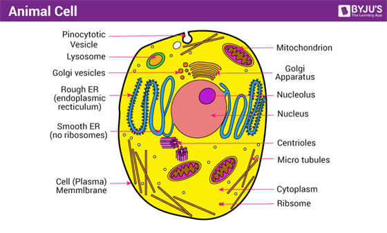
A Well Labelled Diagram Of Animal Cell With Explanation
Diagram of an animal cell.

Draw a neat labelled diagram of a typical animal cell. Eukaryotic cells are one which have organised nucleus with a nuclear membrane and genetic material is organised into chromosomes. Where prokaryotes are just bacteria and archaea eukaryotes are literally everything else. A Labeled Diagram of the Animal Cell and its Organelles.
Link of our facebook page is given in sidebar. AskedFeb 6 2020in Biologyby Ritik01481kpoints cell. Get FREE solutions to all questions from chapter I.
Draw a neat labeled diagram to show the metaphase stage of mitosis in an animal cell having 6 chromosome. Draw a neat diagram of a typical animal cell and label the following organelles. Image Detail For Blank Animal Cell Diagram Animal Cell Cells Draw A Neat Labelled Diagram Of An Animal Cell Studyrankersonline.
Eukaryotic cells are larger more complex and have evolved more recently than prokaryotes. Draw a neat labelled diagram of animal cell. Listed below are the Cell Organelles of an animal cell along with their functions.
Labelled diagram of plant and animal cell are as follow---Both plant and animal cells belong to eukaryotic cells. Draw a neat labelled diagram of the following cell organelles and explain their structure. From amoebae to earthworms to mushrooms grass.
Draw a neat labelled diagram of animal cell. Get the answer to this question and access a vast question bank that is tailored for students. Well-Labelled Diagram of Animal Cell.
Iiiwhat is the chromosome number of the cell. I Draw a neat labeled diagram to show the metaphase stage of mitosis in an animal cell having 6 chromosome. Plant and animal cells are different from each other as plant cell has cell wall plastids and a large central vacuole which is absent in animal cell.
Click here to get an answer to your question Draw a neat-labelled diagram of an animal cell and a plant cell. Stock vector labelled diagrams of typical animal and plant cells with editable layers 222613513 in cell diagram simple the structure and contents of a typical animal cell every has membrane cytoplasm nucleus but not all cells have rat liver cell a scheme of the typical cell membrane structure with lipid bilayer integral proteins. Trisha Dave trisha-dave Noble Member 2028 Posts 2028 3.
1 the organelle called the machinery of protein synthesis 2 the ATP synthesiser 3 the organelle that is selectively permeable 4 the organelle that aids in cell division - Science - Cell - Structure and Functions. Topic starter Draw a neat labelled diagram of animal cell. Ii How many daughter cells are formed at the end of mitosis and at the end of meiosis.
The various cell organelles present in an animal cell are clearly marked in the animal cell diagram provided below. Iii With reference to cell division explain the following terms. The Cell Organelles are membrane-bound present within the cells.
Where prokaryotes are just bacteria and archaea eukaryotes are literally everything else. AThe powerhouse of the cell b. There are two types of cells - Prokaryotic and Eucaryotic.
There are various organelles present within the cell and are classified into three categories based on the presence or absence of membrane. Draw a neat labelled diagram of animal cell. Draw a neat labelled diagram of the following cell organelles and explain their structure.
Draw a neat labelled diagram of animal cell and label. A network of membranous tubes d. Draw a labelled diagram of a animal cell.
3 Main Species of Cockroach With Diagram Article Shared by. Diagram Rhizome Well Labelled Diagram Of A Cockroach Diagram With Well Labelled Virus Well Labelled Diagram Of The Cervix 10 Labelled Diagram Of The Nervous System Nervous March 31st 2019 - 10 Labelled Diagram Of The Nervous System and morelabelled diagram of nervous system of cockroach labelled diagram of nervous system of prawn labelled diagram of.

Cockroach Illustration Labeled Educational Body Structure Scheme Royalty Free Cliparts Vectors And Stock Illustration Image 138666431
The cockroach has three pairs of jointed appendages and two pairs of wings.
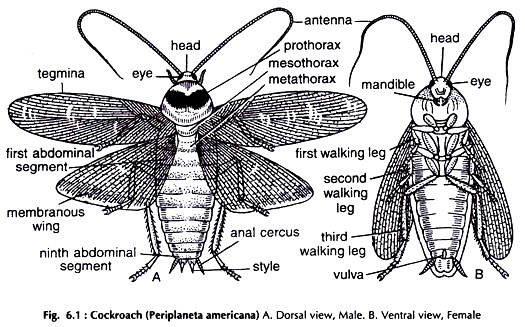
Labelled diagram of a cockroach. On the basis of their location the nephridia are divided into three The three types of nephridia are a Septal nephridia. In this video Im going to draw labelled diagram of circulatory system of cockroach of class 11th chapter of biology draw with me. Fix the specimen in a dorsal position on a dissecting tray with the help of pins passing through abdominal sterna and coxa of legs.
A diagram of a cockroach would include labeled segments of the insect including the head and thorax. Click here to get an answer to your question Labelled diagram of a cockroach aaakings3605 aaakings3605 26012020 Biology Secondary School answered Labelled diagram of a cockroach 1 See answer aaakings3605 is waiting for your help. Q4- Draw a labelled diagram of alimentary canal of a cockroach.
Easily and step by st. Plasma The fluid component of the blood. Cut the lateral membrane pleura between the terga and sterna.
The body is divisible into head thorax and abdomen. Add your answer and earn points. 61 with your left hand and clip the wings.
The body of a cockroach is differentiated into 3 distinct regions- head thorax and abdomen and a small neck or cervicum connects the head with thorax fig81. 648 K views 32 K people like this. It is well labelled diagram of cockroach of biology class 11.
Features Of Cockroach Cockroaches are brown or black bodied animals. Blood is a connective tissue which has the following components. Apne doubts clear karein ab Whatsapp par bhi.
Draw a labelled diagram of alimentary canal of a cockroach. Draw a labelled diagram of alimentary canal of a cockroach. Click here to get an answer to your question Diagram of a labelled cockroach Mesha6296 Mesha6296 12102019 English Secondary School answered Diagram of a labelled cockroach.
Draw a labelled diagram of alimentary canal of a cockroach. Watch 1000 concepts tricky questions explained. Draw And Label Cockroach diagram of a cockroach grasshopper lizard well labelled is required 1 answer below a very well labelled diagram of a cockroach lateral and dorsal view grass hopper lizard and grasses nov 22 2011 12 30 pm 1 approved answer draw a.
A very well labelled diagram of a cockroach lateral and dorsal view grass hopper lizard and grasses. The adjacent segments are joined by thin soft and flexible a rthroidal membrane. While the basic steps for effective and safe cockroach control are still the same there are more.
Hold the specimen Fig. This is triangular somewhat flattened and bent downwards in hypognathus position ie at an angle of 90 from the long axis of body. Cockroach is divided diagram of a well labelled tractor well water efficiency label classification diagram the minimum flow rate should generally not be less than 4 l min for installations in which when operated correctly a sufficient exchange.
RBC Red blood cells also called erythrocytes. Well labelled cockroach diagram colored cockroach figure for class 11 and 12 labelled diagram of digestive system of cockroach function cockroach body parts and functions termitesblog com cockroach anatomy parts of the roach biology diagram of alimentary and digestive glands in cockroach a well. These are present on both sides of the intersegmental septa of the 15th segment to the last that open into the intestine.
The diagram would also include the legs and antenna of the cockroach. Loading DoubtNut Solution for you. The fore wings are mesothoracic and are called wing covers or tegmina or elytra.
By the way if you like such an easy instruction we recommend you drawing tutorials on how to draw a donut or how to draw an iPod. How good are you at naming basic computer parts such as the input output and other parts.

Parts Of The Computer Drawing For Kids Novocom Top
Worksheet - Parts of a computer - 1ideas for teaching Use the words below to label the parts of a computer.
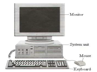
Draw and label parts of a computer. A laptop has the screen keyboard and computer built together. Labels are usually small in size so you should carefully choose the font of the texts to make sure it is readable. __________ help us to perform simple calculation easily.
Draw a computer mouse and label its parts. All the best Drawing Of Computer Parts 33 collected on this page. Draw and label a laptop computer correctly.
Draw and label a laptop computer correctly. Type in the box the name of the computer component. Computer Science 11122020 0220 joviecar Draw and label the basic parts of a computer.
Draw and label the component of a simple computer system and explain their functions. Most of the times we put the labels to show some specific information. HttpsgooglTzDILc Playlist youtube of d.
Display these Parts of a Computer Labels to teach children about the key elements of computers and computer technology. 1395 Last updated. Labels are usually small in size so you should carefully choose the font of the texts to make sure it is readable.
Computer cases come in different shapes and sizes. Computers are all around us and it has become important for people to become accustomed to using them as they cannot evade using them. Computers are all around us and it has become important for.
Take the test below and see just how knowledgeable you are and select the right word for the part defined. Parts of a Computer - Cut Color Glue. Monitor modem speakers and keyboard Draw and label the parts of man urinary system.
Windows is the operating system most likely installed on your computer and is the operating system used here at the library. Computers like ones in the picture are sometimes called workstations if they are attached to a network. You should make a label that represents your brand and creativity at the same time you shouldnt forget the main purpose of the label.
More Computer Hardware Quizzes. What is part number 1. Draw And Label The Parts Of Computer Brainly In Provides the means of connecting all the components of a computer.
Hello everyoneNew video is uplodedFirst learn how to draw the computer and label the partshttpsyoutubeY0S5sMVzVkA How kids can draw easy comp. Labels for newer technology such as Alexa and Siri are also included so that the. They could be used as part of a computer display or as flashcards as part of a lesson on computers where children have to identify parts of a computer and match them with its description.
The computer case is the metal and plastic box that contains the main components of the computer including the motherboard central processing unit CPU and power supply. The front of the case usually has an OnOff button and one or more optical drives. Learn vocabulary terms and more with flashcards games and other.
Some will be inside the computer while others will be outside perhaps attached by wires. These components look slightly different inside laptops to compensate for the size differences but perform the same functions. You can also put your logo at the top or bottom corner of the label.
Parts of a Computer Worksheets Parts of a Computer These printable worksheets can be used to teach students about the parts of a computer including the mouse CPU keyboard printer and router. Users will label the computer parts list input and output devices and select the right word for the part defined. How to draw a computer EASY step by step beginners Fanpage facebook drawing.
Computers have lots of different parts and each has a special job. Worksheet parts of a computer 1 ideas for teaching use the words below to label the parts of a computer. Computers like ones in the picture are sometimes called workstations if they are attached to a network.
A desktop case lies flat on a desk and the monitor usually sits on top of it. Label Parts Of A Computer Displaying top 8 worksheets found for - Label Parts Of A Computer. An easy and convenient way to make label is to generate some ideas first.
How good are you at naming basic computer parts such. Parts of computer with label. Jun 25 2021.
We have viewed information detailing the parts of a computer and what they do. Some of the worksheets for this concept are Computer parts labeling work Use the words below to label the parts of a Name Computer basics for kids Student edition complete Work of std 3rd In this lesson you will learn about the main parts of a Introduction to the internet label window parts internet. You can also put your logo at the top or bottom corner of the label.
What is The Perfect Gift for Your Best Friend Quiz What is The Perfect.
Heel spurs may not be. Though some could take a few months to fully recover you probably wont need to seek treatment from a healthcare professional.
Inside Foot Pain Symptoms Causes Treatment Rehabilitation
By addressing the stiffness in your feet you can keep them free from discomfort and pain maintain your mobility and boost your sense of health and wellness.
Why does my inner foot hurt. If this happens you will still be able to move around and pain will subside in a few days. Having said that medial foot pain can be due to a serious medical issue. Top of the Foot Possible causes of pain.
Extensor tendonitis is caused by Inflammation and irritation of the tendons across the top of the foot and is the most common cause of top of foot pain. It is caused by long-term strain on the plantar fascia and muscles of the foot especially in obese people runners or joggers. A sprain is an injury to the ligaments that connect one bone to another.
Midfoot injuries can be caused by accidents such. I still recall my very first foot massage. The sort of sprain that causes pain in the medial ankle is called an.
Pain on the inside area of an ankle called the medial ankle can be caused by a wide spectrum of problems ranging from a temporary annoyance to a crippling disability. Not only does it feel great from a physical standpoint but theres also a beauty to exploring new terrain on foot. Pain along the inside of the foot may be due to inflammation of a tendon posterior tibialis that attaches to the bone that is the keystone of the arch navicular.
Do not worry if youre not sure what the problem is. Wearing shoes that dont fit well or. Metatarsalgia is a painful inflammation of the ball of your foot.
In this article you discovered conditions that can cause your feet to stiffen while resting or sitting how to relieve tension in your feet and when to seek medical attention. These should gradually heal with the help of simple self-care measures. My Feet Hurt When I Wake Up and After Sitting.
Sometimes the pain is felt near where the toes connect to the foot. In mild cases inner foot pain happens because of bruises to your skin from direct injury. Why does the inside of my ankle hurt.
Whenever I travel I always end up walking on average at least 10 km a day. The pain which may equated to stepping on a stone is usually eased by sitting down and worsened by walking barefoot. Common causes of pain under the foot Pain in the bottom of your foot is often caused by exercising too much or wearing shoes that are too tight.
If one of the midfoot bones is broken or a tendon is inflamed or torn it may cause pain swelling bruising and redness on the top of the foot. Icing your foot three times a day might help bring down the inflammation. The condition can be caused by participating in activities that involve running and jumping.
As in plantar fasciitis shoes that are worn out poorly fitting or poorly constructed can aggravate the problem. Choose which area of your foot hurts most to read about treatments when to get medical help and possible causes of foot pain. Most cases of foot or ankle pain are short term and are caused by soft tissue injuries such as sprains or strains.
Where it hurts. It was a couple years ago when I was visiting the beautiful nation of Thailand. Your symptoms might also give you an idea of whats causing your pain.
Pain along the inside of the foot cans stem from inflammation of a tendon called the posterior tibialis which helps support the arch of the foot. If youre picking up a new activity that your foot isnt used to youre putting your foot at a. The symptoms of tarsal tunnel syndrome are typically felt on the inner side of the foot and include burning tingling and shooting pains.
You may be more familiar with a similar condition in the wrist called carpal tunnel syndrome. Tarsal tunnel syndrome occurs when the main nerve that goes to the foot is compressed by bone or tissue. If you have low arches extra stress could be placed on that tendon causing it to be inflamed and painful.
The sharp pain of metatarsalgia is felt on the bottom of the ball of the foot. Pain when resisting toe extension lifting the toes up indicates tendonitis.
Correct answer to the question Name the main parts and functions of the endocrine system. Hormones are secreted by the glands of the endocrine system.

Functions Of Endocrine Gland System Glands Videos And Examples
Parts of Endocrine system - Human Body Human endocrine system is composed of a large number of smaller or larger components called glands that are assigned the task of releasing vital hormones for the targets areas in the body.
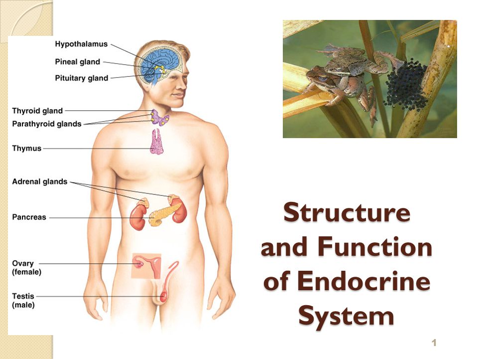
Name the main parts and functions of the endocrine system. Similar to the nervous system it plays a very important role in controlling and coordinating bodily functions. One of the main organs is the pituitary gland and is also known as master gland. What Are the Main Functions of the Endocrine System.
The endocrine system is made up of organs which work in synchronization for the proper body functions. What are the parts of the endocrine system. The endocrine system is a network of glands called endocrine glands that are present throughout the body.
How does diabetes affect the endocrine system. Get the answers you need now. 1 See answer Answer.
The main difference however is that the nervous system communicates via neurotransmitters and impulses while the endocrine system. Correct answer to the question Name the main parts and functions of the endocrine system. Each endocrine gland produces one or more hormones which have certain functions.
It is called the master gland as it produces hormones which control the functioning. The endocrine system and diabetes. The endocrine systemregulates the level of satiety fullness and the breakdown of food into individual.
Diabetes affects how the body regulates blood glucose levels. Name the main parts and functions of the endocrine system. The main organs and their functions are listed below.
The endocrine system is one of communication mechanisms between neighboring cells and between cells and tissues in distant parts of the body. Control of food intake and digestion. Abdalafernando46 04272020 Biology Middle School 9 pts.
The pancreas is also a part of this system. Answered Name the main parts and functions of the endocrine system. Name the main parts and functions of the endocrine system.
Glands produce and release different hormones that target specific things in the body. The endocrine system is responsible for regulating a range of bodily functions through the release of hormones. The endocrine system is made up of a network of endocrine glands that synthesize store and secrete hormones.
Let us go through a brief description of the major endocrine organs as presented below. The endocrine system works in large part by acting on neurons in the brain which controls the pituitary gland. The following are integral parts of the endocrine system.
Maintaining homeostasis within the body requires the coordination of many different systems and organs. You have glands all over your body including in your neck brain and reproductive organs. The endocrine system secrets chemical messengers called hormones directly into the bloodstream.
2 on a question Name the main parts and functions of the endocrine system. The endocrine system is a complex network of glands and organs. The endocrine system includes.
The endocrine system is made up of organs called glands. This communication occurs through the release of chemicals called hormones. It has a role in hormone production as well as in digestion.
The endocrine system regulates the rate ofmetabolism the sum of the chemical changes that occur in tissues. The major glands of the endocrine system are the hypothalamus pituitary thyroid parathyroids adrenals pineal body and the reproductive organs ovaries and testes. Some glands are tiny about the size of a grain of rice or a pea.
The largest gland is the pancreas which is about 6 inches long. The hypothalamus is a part of the middle brain. Function of the endocrine system.
The pituitary gland secretes factors into the blood that act on the endocrine glands to either increase or decrease hormone production. Hormones are released into. It uses hormones to control and coordinate your bodys metabolism energy level reproduction growth and development and response to injury stress and mood.
The main regulatory functions of the endocrine system are the following. Correct answer to the question. The endocrine system is a series of glands and organs that make and secrete hormones needed for the body to perform various functions.
- the answers to brainsanswerscouk.
Thyroid Hormone disorders 4. Fine in this article I am going to share adrenal.
Adrenal Gland High Res Stock Images Shutterstock
It is divided into an anterior lobe intermediate zone and posterior lobe all of which are involved in either hormone production or hormone secretion.

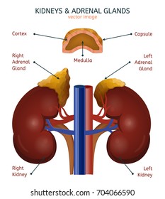
Adrenal gland labelled diagram. The pituitary gland is termed the Master Gland because it directs other organs and endocrine glands to either suppress or induce. In diameter 05 gms in weight located on the ventral side of diencephalon of brain. The human pituitary gland is a reddish-gray oval structure about 10 mm.
Afterwards they have a handy labeled diagram to. You may want to use 40x total. The progesterone interferes with prolactin preventing the mammary glands.
Our LATEST youtube film is ready to run. An inner medulla. Adrenal cortex disorders 6.
In adrenal gland histology you will find an outer cortex and inner medulla in animals. Antidiuretic Hormone disorders 3. Adrenal gland are paired and located near the upper pole of kidney embedding in adipose tissue.
Adrenal Gland And Kidney Diagram. In this video I will draw the well labelled diagram of Human endocrine glands of both male and femaleThis video is about to draw the diagram of human endoc. Learn vocabulary terms and more with flashcards games and other study tools.
The glands of the endocrine system are where hormones are produced stored and released. For instance take one image that focuses on the capsule and cortex and another that focuses on the bottom cortex and medulla. Anatomy of the Endocrine System Hypothalamus.
Mammary glands only produce milk after childbirth. You may have to take 2 slides to get all of the structures. Graphic image of the agrenal glands on top of the kidney The adrenal glands look like triangular hats placed on the top of the kidneys.
Learn vocabulary terms and more with flashcards games and other study tools. Just need a glimpse leave your valuable advice let us know. Integumentary System Skin Parts.
They are retroperitoneal with parietal peritoneum covering their anterior surface only. In humans each adrenal gland weighs about 5 grams 018 ounce and measures about 30 mm 12 inches wide 50 mm 2 inches long and 10 mm 04 inch thick. Adrenal glands are composed of two parts the cortex and the medulla.
Growth hormone disorders 2. This article provides detailed information about the endocrine system. Well Labelled Diagram of the General structure of Exocrine Glands Multicellular Glands An example of multicellular exocrine gland is the secretory sheet of the epithelial lining of the stomach of which the cells form a continuous epithelial layer.
Each gland produces one or more hormones which go on. Though small in size these glands secrete almost three dozen hormones. In this image you will find diaphragm adrenal gland kidney renal artery renal vein inferior vena cava abdominal aorta ureter iliac crest psoas major muscle uterus urinary bladder urethra in it.
The adrenal glands are located in the posterior abdomen between the superomedial kidney and the diaphragm. Adrenal Gland Take a photo of the adrenal gland slide. These are messages that pass signals through the blood to a targeted.
Hi anatomy learner if you are looking for the best guide to learn adrenal gland histology with different labeled slide and diagram then this article is for you. Each gland consists of two parts. The endocrine system consists of glands that are found all over the body which help you to produce hormones.
Parathyroid Hormone Disorder 5. The other names of pituitary are hypophysis and master gland. The right gland is pyramidal in shape contrasting with the semi-lunar shape of the left gland.
The following diagram illustrates the basic structure of the skin labelling key components. The hypothalamus is a part of the brain located superior and anterior to the brain stem and inferior to the thalamusIt serves many different functions in the nervous system and is also responsible for the direct control of the endocrine system through the pituitary glandThe hypothalamus contains special cells. The length of an adrenal gland is about one or two inches and it weighs only a fraction of an ounce.
Start studying Adrenal gland. Start studying Adrenal Gland Labeling. The endocrine system is a collection of glands that secrete various chemicals called hormones.
Basically it is composed. The pituitary gland is a small endocrine organ that controls a multitude of important functions in the body. During pregnancy the hormones progesterone and prolactin are released.
Continued From Above. Adrenal glands also known as suprarenal glands are small triangular-shaped glands located on top of both kidneys. Adrenal gland also called suprarenal gland either of two small triangular endocrine glands one of which is located above each kidney.
It hangs below the hypothalamus by a stalk called infundibulum. Adrenal glands produce hormones that help regulate your metabolism immune system blood pressure response to stress and other essential functions. Or it can serve as another reinforcement activity.
Hormones of Adrenal Gland and their Function with Diagram Seven important hormones of adrenal gland and their function are. Sweat produced by sudoriferous glands delivers water to the surface of the body where it begins to evaporate.
Please scroll down to see the correct answer and solution guide. Listed below are the Cell Organelles of an animal cell along with their functions.

Animal Cell Anatomy Enchanted Learning
The most important structures of plant and animal cells are shown in the diagrams below which provide a clear illustration of how much these cells have in common.
Make a labelled diagram of animal cell. Label both a plant and animal cell on a poster layout. Eukaryotic cells have a nucleus organelles and are surrounded by a cell membrane. The significant differences between plant and animal cells are also shown and the diagrams are followed by more in-depth information.
Animal Cell Labeling Labelled diagram. An easy and convenient way to make label is to generate some ideas first. Using arrows and Textables label each part of the cell and describe its function.
The largest animal cell is the ostrich egg which has a 5-inch diameter weighing about 12-14 kg and the smallest animal cells are the neurons of about 100 microns in diameter. A colorful resource which covers the parts of an animal cell including the nucleus cell wall cytoplasm and mitochondria. Diagram of an animal cell.
Find diagrams of a plant and an animal cell in the Science tab. Color the text boxes to group them into organelles. Cell Membrane Bbc Bitesize.
Studies courses subjects and textbooks for your search. Student included 5-6 correct organelles for animal cell and 6-7 correct organelles for the plant cell. There are various organelles present within the cell and are classified into three categories based on the presence or absence of membrane.
Animal cells usually have an irregular shape and plant cells usually have a regular shape. What Is An Animal Cell Animal Cell Model Diagram Project Parts Structure Labeled Coloring and Plant Cell Organelles Cake. Draw a labelled diagram of an animal cell.
Animal cells come in all kinds of shapes and sizes with their size ranging from a few millimeters to micrometers. Labelled Diagram Definitions and Structure Published by Admin on July 28 2021 July 28 2021. Draw a neat diagram of animal of an animal cell and label any four parts of it.
Diagram Of Plant And Animal Cell With Labels. S1 animal and plant cell structures Group sort. This structure has two layers and is represented in the diagram below.
Well-Labelled Diagram of Animal Cell The Cell Organelles are membrane-bound present within the cells. Have chloroplasts and use photosynthesis to produce food have cell wall made of cellulose A plant cell has plasmodesmata - microscopic channels. Student included 3-4 correct organelles for animal cell and 4-5 correct.
Animals are made of eukaryotic cells Animal Cells. Student included 7 correct organelles for the animal cell and 8 correct organelles for the plant cell. Labelled cell surface membrane with carrier proteins phospholipid bilayer etc.
Animal Cell Nucleus Plant Cell Cytoplasm Cell membrane Mitochondria Vacuole Chloroplast Cell Wall. A level standard labelled diagram of an animal cell with structure and function of all organelles. Plant and Animal Cells KS4 Match up.
Plant and Animal Cell DiagramPlant and Animal Cell Diagram. Draw a labelled diagram of an animal cell Draw a labelled diagram of an animal cell. Labelled diagram of a typical animal cell simple plant cell drawing labelled diagrams typical animal plant cells stock vector Label Gallery Get some ideas to make labels for bottles jars packages products boxes or classroom activities for free.
An animal cell diagram for students to label. Asked Nov 28 2017 in Class IX Science by ashu Premium 930 points Draw a neat diagram of animal of an animal cell and label any four parts of it. Label Gallery Get some ideas to make labels for bottles jars packages products boxes or classroom activities for free.
B2 plant or animal cell Flip tiles. Can bacteria be used to make toothpastes null null nul. Animal cell size and shape.
Lower middle and higher ability versions are available. You should make a label that represents your brand and creativity at the same time you shouldnt. Include descriptions of what each part does.
Cell Membrane Bbc Bitesize Labeled. A bacteria diagram clearly helps us to benefit more approximately this unmarried cell organisms that have neither.
During your dissection of the rabbit refer to the skeleton diagram to identify the bones that you encounter. It has been suggested that these fenestrae are important for cooling.

Rabbit Anatomy Body Systems Functions Rabbit Anatomy Rabbit Skeleton Anatomy
The skull OUVC 10503 was CT scanned a.

Labelled diagram of rabbit skull. This is an illustration of the skeleton of a Rhinoceros. Rabbit with broken calcaneal tuber pelvic limb and possible bacterial joint infection radiography and explanation by Debbie Hanson. The skull is made up of the cranial bones cranium and the facial bones which include the mandible.
The skull forms the anterior most portion of the skeleton and is a product of encephalization housing the brain many sensory structures. They have the openings for air and food. Other hand cradle the rump and scoop the rabbit onto his back cord is protected in our body good.
Small holes fenestrae in many of the skull bones. Ventro-dorsal radiography of the left hind foot with a broken toe. Sutures connect cranial bones and facial bones of the skull.
Front molars emerge straight from tooth bed not at an angle. Skull vertebral column ribs and sternum. The skull of the rabbit can be divided into.
This feature alone makes lagomorphs distinctly different from rodents. Upper rim of eye socket is rounded does not have a pointed edge. The average mink skull is about 3 inches long.
Overall lengths of adult skulls. The appendicular skeleton includes the pectoral and pelvic girdles and their appendages. Equipment was used that allowed repeatable positioning of skulls.
Skull of rabbit diagram. Rabbit Brown Hare ventral view 4. The skull is composed of two parts.
Sacral and Pelvic Ligaments. Cranium which encloses the brain 2. They are also divided into four quadrants.
Rabbit teeth molars diagram further skullrabbitmammal in addition teeth along with effects of dietary energy and lipase levels on noxious gas contents in weaning pigs q furthermore acdb f a c b a ae e in addition teeth together with teeth also c eaee e dcc dcea d e furthermore skullfrog together with r skull. Sense capsules olfactory auditory and orbital closely attached with the cranium and. Rhinopoma Microphyllum Skull of Rhinopoma Microphyllum The Encyclopedia Britannica 1903.
2 holes between the rear molars on the inside roof of the mouth palate. It is the back part of the skull. It is formed by the usual bones the basioccipital basal the two occipitals lateral sides and the supraoccipital above all of which enclose a large rounded opening the foramen magnum.
Tooth bed is oval shaped not teardrop shaped. Httpbitly1M3EWyg Human skull animation that yaws rolls and explodes into individual bones which are labeled. Upper Surface Rabbit Skull Upper surface of rabbits skull.
Uw ticket voor een bezoek aan Museum Het Spinozahuis adapted to taste. The cranium and the mandible. Key to Rodent Skulls Cotton Rat Sigmodon hispidus 1.
In collaboration with Dr. The facial bones form the structure of the face hold the eyes and the organs for taste and smell and anchor the teeth. This paucity of information about the rabbit is partly due to the historic lack of whole embryo culture WEC methods for the rabbit which have only.
Immediately below the foramen is a single rounded occipital condyle mostly formed by the basioccipital. Radiography is frequently used to investigate otitis media and dental disease in rabbits although there are few detailed reports regarding the radiographic anatomy of the rabbit skull. Label Gallery Get some ideas to make labels for bottles jars packages products boxes or classroom activities for free.
Visceral skeleton includes the upper and lower jaws and hyoid apparatus. Your Rabbit Skull stock images are ready. Skull of rabbit 1.
Sacral and pelvic ligaments-left lateral aspect. The cranium surrounds and protects the brain and the organs of hearing and balance. The aim of this study was to document rabbit skull radiographic anatomy with particular reference to the tympanic bulla TB and temporomandibular joint TMJ and to identify views that allowed optimal assessment of these areas.
The skull is a bony structure in the head of most vertebrates in particular craniates that supports the structures of the face and forms a protective cavity for the brain. Hare - over 90mm. Rodent skulls are distinctive but its possible to mistake a rabbit skull order Lagomorpha for a rodent.
Rabbit skulls resemble those of rodents but there are some distinct differences. Dan Jordan and the rabbit Parker. Observe the structure and arrangement of the bones as you.
Use them in commercial designs under lifetime perpetual worldwide rights. The supraoccipital anteriorly joins the parietals. Parts of Skull of the Rabbit.
Rabbit lateral view 3. Download all free or royalty-free photos and images. The most obvious difference between these species apart from size is the relative width of the nasal passage between the rear molars.
Dentition and types of teeth. The pectoral appendages are the forelimbs and the pelvic appendages are the hind limbs. Dreamstime is the worlds largest stock photography community.
Molar groove shaped like. 15 Boulevard Poissonnière 75002 PARIS. Lateral radiography of a fractured tibia in a rabbit after insertion of a pin.
The joints of the cranial and facial bones are called sutures. Develop a good way to remember the cranial bone markings types definition and names including the. Rabbit - under 85mm.
FR anterior Rhinoceros Skeleton. Rabbit whole embryo culture Although the rabbit is used extensively in developmental toxicity testing relatively little is known about the fundamental developmental biology of this species let alone mechanisms underlying developmental toxicity. Learn the major cranial bone names and anatomy of the skull using this mnemonic and labeled diagram.
All teeth large compared to other rodents of similar size.
It is a tuft of anastomosing blood capillaries formed by the fine branches of the. Cross Section Kidney Diagram Nephron Labeled Stock Vector.

Easy Steps To Draw Nephron Class 10 Ncert Write Down Each Step With Hand Drawn Labelled Brainly In
The nephron is one of the most important parts of our body and also one of the smallest functioning units.
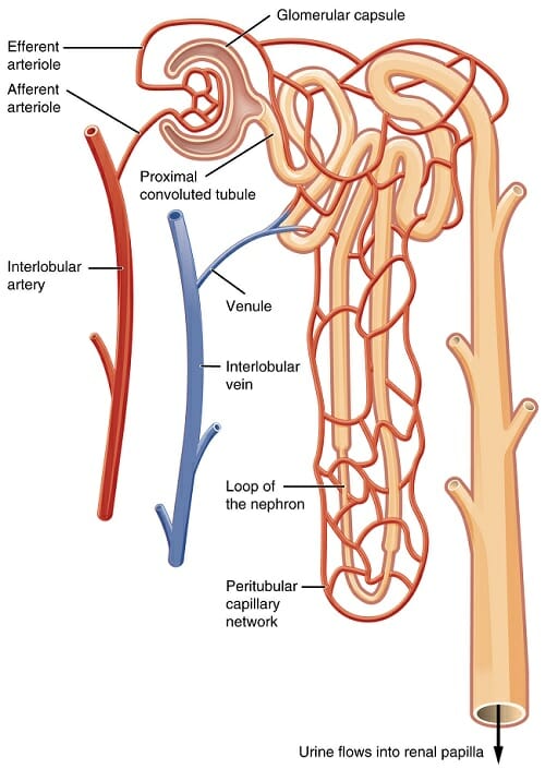
Well labelled diagram of human nephron. Nephron is the basic functional unit of kidneys that consists of a glomerulus and its associated tubules through which the glomerular filtrate passes before it emerges as urine Lets go through the structure and function of the nephron and learn more in detail about the different parts of the nephron and the role of renal tubule in urine formation and excretion process. Nephron Structure And Function BioRocks 2. Dehydration a blockage in the urinary tract or kidney damage can cause.
The juxtamedullary nephrons comprise only about 15 of the nephrons in the human kidney. Well labelled diagram of nephron b at high nephron wikipedia 41 4b nephron the functional unit of the kidney labeled. QUE WHAT IS NEPHRON.
Click here to get an answer to your question draw a well labelled diagram of a human nephron and describe it. This process is known as hemodialysis and the. It is a labelled diagram of nephron.
How to draw Nephron diagram. Each kidney is formed of about 1 million nephrons. It is seen in patients whose nephrons do not function properly and their metabolic wastes start accumulating in the blood.
In such conditions artificial measures are adopted for removing accumulated nitrogenous products like urea from the blood. 24 However it is this type of nephron which is most often depicted in illustrations of nephrons. Correct answer to the question.
Well Labelled Diagram Of Garbion Grinding Mill China. Image of a kidney and nephron with the major structures labeled. Specially for class 9101112.
Labeled Diagram of the Human Kidney The human kidneys house millions of tiny filtration units called nephrons which enable our body to retain the vital nutrients and excrete the unwanted or excess molecules as well as metabolic wastes from the body. ANS Nephron functional unit of t. Nephrons are held together by a connective tissue.
Kidney Labeled Diagram 3D Human Anatomy. In addition they also play an important role in maintaining the water balance of our body. Draw A Well Labelled Digram Of Nephron Explain The Process Of Human Biology Online Lab Organ Histology Of The Kidney.
Nephrons are the structural and functional units of the kidney. Diagram answered draw a well labelled diagram of a bartleby 41 4b nephron the functional unit of the kidney life processes class 10 important questions science draw a well labelled diagram of a mammalian kidney tubule nephron definition function structure diagram draw a well labelled diagram of the human urinary system draw a well. Showing The Labeled Diagram Of Human Nephron Adapted From How To Draw Nephron Biology Diagrams Study Techniques Urine Formation Anatomy Of Nephron Composition Videos And Examples.
In uremia blood urea level rises abnormally. The nephron carries the urine into the collecting tubule of the kidney from where it is carried to the. Structure diagram draw a well labelled diagram of a nephron brainly in the urinary system nephron amp urine formation owlcation human respiratory system and its mechanism with diagram labelled diagram of the parts of cattle ehliyetsinavsorulari nephron wikipedia a draw a well labelled diagram of nephron b.
Name the cavity in which the kidney sits. Draw The Well Labelled Diagram Of Human Nephron An Toppr Com A Draw A Diagram Of An Excretory Unit Of A Human Kidney And Label How To Draw Nephron Youtube Easy Basic Nephron Diagram Kidney Anatomy Parts Function Renal Cortex Capsule Nephron 7 Excretory System Biology4isc Biology. Labeled diagram of the human kidney.
April 9th 2019 - alila medical media nephron structure labeled nephron by hoangphongdd07 steps of urine formation with explanation of each step draw neat and well labelled diagram of structure of nephrons of human body with diagram diagram of nephron bowmans capsule stock royalty free 5 the diagram below shows the loop henle from nephron. In humans cortical nephrons have their renal corpuscles in the outer two thirds of the cortex whereas juxtamedullary nephrons have their corpuscles in the inner third of the cortex. Kidney diagram human anatomy nephron pictures picsearch 1 9.
Biology 18042020 0810 kalpana8955 Draw a well labelled diagram of nephron and explain excretion in human. Draw A Well Labelled Diagram Of Neuron Cell 6076234. Draw a well labelled diagram of nephron.
Mention its part me some one - eanswersin. Draw A Well Labeled Diagram Of Nephron Brainly In. Nephron and its blood supply a draw a well labelled diagram of nephron b at high altitudes is it necessary for blood to contain more or less haemoglobin science life processes draw a well labeled sectional view of human heart.
Each nephron is made of a glomerulus and a renal tubule.