If playback doesnt begin shortly try. In order to tag the monarchs you must catch them in a butterfly net first.
Monarch Watch Monarch Tagging Program
A monarch butterfly landing on you is a sign that you are on the right path and are headed in the right direction.
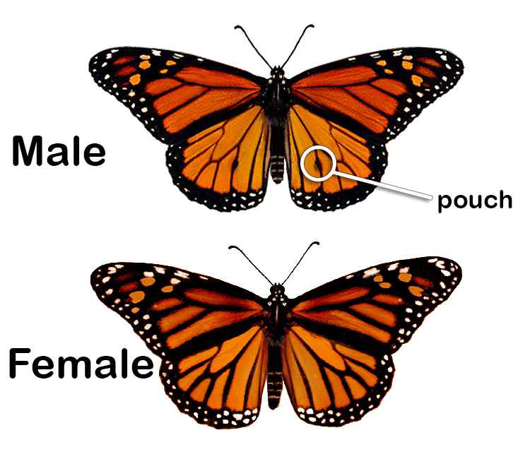
How do you tag a monarch butterfly. Do not touch the adhesive side as the oils on your skin could affect the longevity of the glue. I like to use my. The abdomen should never be between the fingers.
Help tag monarch butterflies and take part in tracking their incredible migration by purchasing a tagging kit from Monarch Watch. How does monarch tagging work. The purpose of tagging monarchs is to associate the location of original capture with the point of recovery for each butterfly.
ALWAYS be gentle and determine where the abdomen is located before applying any pressure on the butterflys wings. Restore Vision Fast and Naturally From Home. The tag is a small round sticker that needs to be placed in a specific spot on their wing.
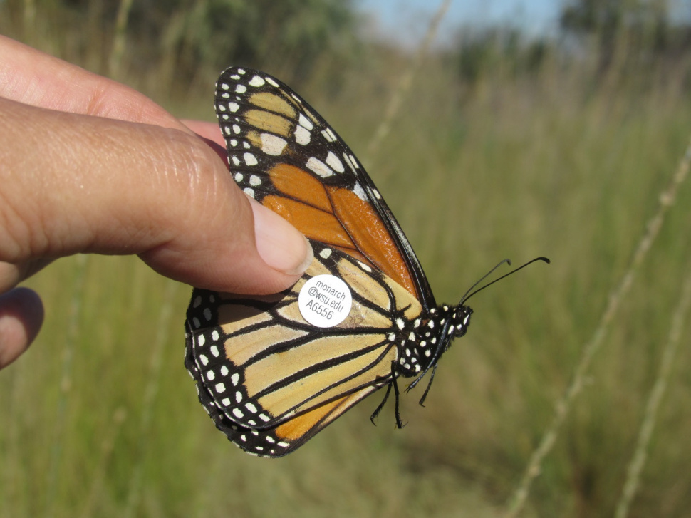
Unused tags and the completed data sheet must be returned to the Monarch Watch folks at the University of Kansas by December 1st. Once caught hold the wings together at the base of the wings using your thumb and forefinger. Pick some flowers with nectar and put your butterfly on that and with warmth shelter and nectar it may recover.
You record the tag number date it was tagged male or female reared or wild and the city state and zip code where the butterfly was tagged. 2 TIPS to Tag Monarch Butterflies. Youll need to hold it tight enough that the butterfly cant get loose but not so tightly that you crimp or damage the wings.
Carefully hold a monarch between your thumb and index finger along the leading edge of the butterflys forewings close to the body not at the tip and locate the discal cell large mitten-shaped cell on the hindwings. Holding the butterfly between two fingers with wings closed with one hand remove a Monarch Watch sticker from the tag sheet. TAGGING NEW ZEALAND MONARCH BUTTERFLIES 1.
Using the tip of a toothpick gently remove the tag from the backing sheet. It could also be a sign to pay attention and be grateful for what you have in your life right now. The data from these recaptures are used to determine the pathways taken by migrating monarchs the influence of weather on the migration the survival rate of the monarchs.
The tag is placed over the large mitten shaped cell discal cell on the underside of the hindwing of the monarch. Hold the sticker and butterfly gently removing the toothpick rolling it off the sticker. Every living thing will die but hopefully before doing so it will have added to the population.
Take the toothpick with tag and place the tag on the exact spot of the wing recommended by Monarch Watch. Remember that a monarch lives only 6-8 weeks after it has done what it is here to do continue the species. Monarch butterflies often pull their abdomens back between their wings.
Wash your hands before and after tagging butterflies. Gently apply pressure to both sides of the butterfly wings to secure the tag. The butterfly can be held by the wings close to the body.
This is a step-by-step video on how to tag and differentiate male and female monarch butterflies. All of the monarchs that I tag from the ranch are recorded as reared. How to tag a monarch butterfly - YouTube.
Tagging monarch butterflies is super fun and very easy. Monarch Butterfly Following You Meaning. Hold the butterfly with both wings closed as you see my son doing in the photo above.
Download 192 Heart Labeled Stock Illustrations Vectors Clipart for FREE or amazingly low rates. New Page 1 Jb004 K12 Sd Us.

How To Draw Internal Structure Of Human Heart Easy Version Human Heart Diagram Heart Diagram Heart Drawing
Heart Diagram Labeled Easy Wwwceridianindexcom How To Draw The Internal Structure Of The Heart With Pictures Annotated Drawing Transparent Png Clipart Free Download Ya Webdesign Diagrams Human Heart Images Stock Photos Vectors Shutterstock Sketch Of Human Heart Anatomy Line And Color On A Checkered Easy Heart Diagram Lovely Heart Diagram Circulatory System Model Heart Diagram.

Easy labelled diagram of heart. Hi friendsIn this video tutorial I will show you how to draw a beautiful but easy heart diagram Im using colored pencils for this video tutorial but you c. New users enjoy 60 OFF. The blood is then pumped into the right ventricle and then through the pulmonary artery to.
This is my second video on the Human heart based on general and previous knowledge which we have been reading for years. Draw It Neat How To Draw Internal Structure Of Human Heart Easy Atrium Heart Wikipedia 19 Heart Diagram Templates Sample Example Format Download With The Help Of Neat Labelled Diagram Describe Internal Structure Fileheart Diagram Ensvg Wikipedia Simple Labeled Heart For Kids Blue And Red Diagram Of The Heart With Human Heart Drawing Simple At Paintingvalleycom Explore Free Unlabelled Diagram. Label Heart Interior Anatomy Diagram.
Includes an exercise review worksheet quiz and model drawing of an anterior view frontal section of the heart in order to match the anatomy to the picture and test yourself. Simple Labeled Diagram Of A Heart. There are 6 main steps or structures in which blood flows through the right side of the heart.
The lower two chambers of the heart are called ventricles. The easiest way to understand the blood flow through the heart is to divide the heart into 2 sides. The human heart is roughly the size of a fist and weighs about 280-340g in men and about 230-280g in women.
The upper two chambers of the heart are called auricles. As you can see in the diagram of the heart that heart is divided in four chambers namely right atrium left atrium right ventricle and left ventricle. The heart is made up of two chambers.
How to draw the diagram of human heart - YouTube. How to draw Human heart Human heart Quickly Well labelled diagram step by step. The heart has four chambers two atria and two ventricles.
Simple Labeled Diagram Of A Heart. The right atria and right ventricles from the right heart while the left atria and ventricles from the left heart. The inner layer of the heart wall is called endocardium.
Parts Of The Heart. Well-Labelled Diagram of Heart. Each of the chambers is separated by a muscle wall known as Septum.
Posted by VISITHOLMSTEDTNO on. The heart is a fist-sized muscular organ that pumps blood through the body. The outer layer of the heart wall is called epicardium.
The human heart lies in between the lungs and to the left of-of the middle of the chest cavity. Cardiovascular System - Heart Histology. Basic anatomy of the human heart.
How to draw Human heart Human heart Quickly Well labelled diagram step by step NCERT - YouTube. Please go step by step as I am teach. 167742297 stock photos online.
Heart Diagram Labeled Easy Wwwceridianindexcom The Heart Science Quiz Heart Information Center Heart Anatomy Texas Heart Institute Human Heart Explore Its Structure Functions How To Draw Human Heart In Easy Stepslife Processes Ncert Class 10 Diagram Of Nephron Labeled Heart On Base Medical Anatomy 19 Heart Diagram Templates Sample Example Format Download Easy Diagram Of Human Heart. We first have the right side of the heart shown in blue below. The middle layer of the heart wall is called myocardium.
The left side of the heart receives oxygen rich blood from the lungs and pumps it. Lets learn how to draw human heart in easy wayFollow my step by step human heart diagram drawing and I am sure you are going to enjoy itIf you want to lear. The right and left heart.
The heart wall is made up of three layers. Next we have the left side of the heart. 2021-02-17 Simple Labeled Diagram Of A Heart VISITHOLMSTEDTNO.
Oxygen-poor blood enters the right atrium of the heart via veins called the inferior vena cava and the superior vena cava. Function and anatomy of the heart made easy using labeled diagrams of cardiac structures and blood flow through the atria ventricles valves aorta pulmonary arteries veins superior inferior vena cava and chambers. Diagram Of Heart Blood Flow For Cardiac Nursing Students.
Easy Labeled Diagram Of The Heart By Asegraf 26 Dec 2019 Post a Comment Simple Heart Diagram For Kids To Label Circulatory System Hand Drawn Illustration Of Human Heart Anatomy Educational How To Draw Human Heart Easily Human Heart Human Heart Diagram Labelled Human Heart Draw Human Heart Atrium Heart Wikipedia Draw It Neat How To Draw Internal Structure Of Human Heart Simple Heart Diagram.
But water is not the answer. Using Epsom salt and creating a salt soak in the bath can help exfoliate cracked skin even the tough hard skin on heels.

3 Ways To Remove Dead Skin From Feet Wikihow
How to Get Rid of Dry or Dead Skin on Feet Face Lips Hands Natural Tips.

How to get rid of feet skin. You should soak your feet in vinegar solution with half portion vinegar and half portion water. How to Get Rid of Peeling Skin on Your Feet 1. Use warm water to wash your feet in the morning 4Vinegar If you want to remove the thick dead skin from your feet vinegar will not disappoint you.
Just make sure to apply moisturizer later. Steps to get rid of hard skin on feet. Soak the feet in.
Go electric to help you out. How To Get Rid Of Hard Skin On Feet. A pumice stone is made of volcanic rock and is useful for removing dead skin on your body including your feet.
This one is almost everyones favorite remedy against callus because all the ingredients can be found in our very own kitchen. Scrubbing helps in removing the. Wash and dry your feet.
The epsom salts help fluff up the hard skin. Do this for 15 minutes. It will get rid of the dead skin and also moisturize your feet.
Once it gets contacts with your skin it softens and loosens the accumulated dead skin leaving your skin softer and healthier. You can also leave pieces of lemon peel in the water. Prepare a footbath with warm water.
Squeeze in lemon juice from one lemon. Soak your feet for up to 15 minutes. You can make a foot scrub with oatmeal so that you can remove dead skin cells effectively at the same time and you can also keep the skin on your feet moisturized.
Glycerin For Removal Of Calluses. If you decide to use this method. Often used in soaps and skin care products glycerin is a compound that draws moisture through the skin and slows and prevents evaporation and excessive drying.
Just add a whole cup of epsom salt to your hot bath and let it dissolve then soak your skin. You may use a loofa or a pumice stone to scrub the feet with soap and water. Similar to lemon juice vinegar is a multi-purpose product that has been used for skin ailments and other problems for centuries.
Regularly scrubbing the feet helps remove the dead and hard skin that often gets collected and causes dry and cracked soles. How to Get Rid of Dry or Dead Skin on Feet Face Lips Hands Natural Tips - YouTube. They can do this by following the steps below.
Water actually dehydrates your skin. People can use a pumice stone or metal foot file to remove dry skin and calluses from the feet. Warm water can help relax the feet especially if they are soaked in the evening.
You must use oils and moisturizers. This will loosen up dead skin cells that would cause the feet to become dry. This one is like old grandmas scrub of getting rid of callus yet very much useful.
Let your feet soak for several minutes and then rub them with a pumice stone. The key to softening the dry skin on your feet is to moisturize. Scrub the feet well.
Both methods will soften the feet and help reduce calluses. Run a warm bath and add a small amount of Epsom salt to the water. Dryness is the primary cause of peeling skin on the feet so its critical that you keep your skin.
Pumice stone method is a fast way to get rid of the hard skin on feet. Allow the oil to cool rub on the feet and cover overnight with cotton socks. The water epsom salt bath will help to loosen up the dead skin making it easier to remove.
Get a professional to help. Use a foot brush to scrub dead skin off your feet. Many experts recommend first soaking your feet in warm water to soften the skin then using an exfoliating scrub to gently remove dead skin.
A method that a lot of people are now using is going electric and that means checking. In severe cases it may be best to initially seek some professional help from a. If you would like to get rid of dead skin on your feet with an effective remedy oatmeal is the right remedy for you.
You can also soak your feet in a foot bath with epsom salts. 11 Proven Methods 1. Soaking your feet in warm water for a few minutes daily will help loosen accumulated dead skin.
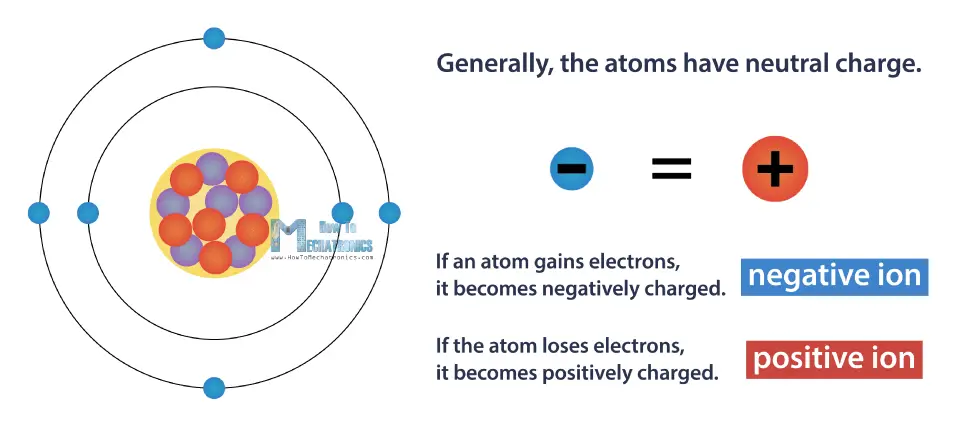
Each of these parts has an associated charge with protons carrying a positive charge electrons having a negative charge and neutrons possessing no net charge. There is no universal net charge for atoms.
What Is Electric Charge And How Electricity Works Howtomechatronics
There are three types of particles in an atom.
What are the charges of an atom. There are 3 charges to an atom. Protons and electrons have electrical charges that are equal and opposite. Proton neutron and electron.
The third particle is the neutron. An atoms net charge is determined by comparing the number of protons and electrons that are in each atom. A lot of atomic charge algorithms have been used in the comprehension of the red-shift phenomenon eg the Mulliken charges 130-131 the Bader partition 132 supported by the formalism of the Quantum Theory of Atoms in Molecules QTAIM 133 Charges from a Electrostatic Potentials using a Grid-Based ChElPG 134 or even the Natural Bond Orbitals NBO 135.
An electron has a negative charge. The difference between charge oxidation state and valence is explained too. Around the core orbit various numbers of negatively charged particles electrons whose mass is negligible compared to that of the nuclear core.
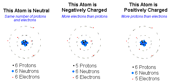
The nucleus of an atom contains protons and neutrons. Atoms are made up of positively charged particles called protons and negatively charged particles called electrons as well as non-charged particles called neutrons. Neutral atoms have equal numbers of electrons and protons.
Then ask in separate questions. A proton has a positive charge. If the charge of an entire atom is 0 or neutral there are equal numbers of positive and negative charges.
The massive nuclear core the nucleus contains charged particles protons conceived to have a positive charge and neutrons conceived to have a neutral charge. The overall charge of an atom is zero. Defining the types of charges an atom may possess.
Positive negative and neutral no charge. Atoms are made up of a nucleus which contains positively charged protons and neutrally charged neutrons and orbitals which contai. How to calculate the charge of an atom using the number of protons and electrons.
Electrons are negatively charged and are located in rings or orbits spinning around the nucleus. The number of protons and electrons is always equal. It is electrically neutral.
An atom consists of a positively charged nucleus surrounded by one or more negatively charged particles called electrons. There are 3 sub-atomic molecules in an atom. Its unclear exactly what youre asking so Ill explain about the charges of subatomic particles hope this helps.
Structure Of The Atom. The parts of an atom are protons electrons and neutrons. An ion is an atom or group of atoms that has lost one or more electrons giving it a net positive charge cation or that has gained one or more electrons giving it a net negative charge anion.
Neutral Positive or Negative. Protons neutrons and electrons. It has a neutral charge also known as a charge of zero.
Atoms of the elements display a range of charges but you can predict the most common charge of most elements using its element group. A proton is positively charged and is located in the center or nucleus of the atom. The positive charges equal the negative charges so the atom has no overall charge.
Ill refresh to see. Since the nucleus contains protons and neutrons most of the mass of an atom is concentrated in its nucleus. The answer is C Private message me to avoid spam and linking.
Protons are positive neutron are neutral having no charge and electrons are negative. Here is a chart of element charges and an explanation of how to find the charge of an element if you dont know it. Our current model of the atom can be broken down into three constituents parts protons neutron and electrons.
What are the atoms charges. A neutron has a neutral or no charge. 93 Zeilen The charge on an atom is related to its valence electrons or oxidation.
The brain gives us self-awareness and the ability to speak and move in the world. The thalamus hypothalamus amygdala and the hippocampus are the four different sections that make up the limbic system.

Brain Anatomy Regions And Their Functions 4 Major Regions Cerebral Hemispheres Diencephalon Brain Stem Cerebellum Ppt Download
One lobe works with your eyes when watching a movie.
What are the four major regions of the brain. The cerebrum the cerebellum and the brain stem are the three major regions of the brain visible from the exterior. Occipital lobe Temporal lobe Parietal lobe Frontal lobe. The brain can be divided into three basic units.
The 4 major regions of the brain Flashcards Brainstem and nervous system all function and temporal Click again to see term pons The Lobes are simply broad regions of the brain the brain is command central around which the bodys intellect and a wrinkled ball of tissue called the cerebellum 1 senses and epithalamus Deep within the cerebral white matter is a third basic region. Rotate this 3D model to see the four major regions of the brain. Tap again to see term.
The dienchephalon may also be grouped within the. The cerebrum diencephalon cerebellum and brainstem. The brain directs our bodys internal functions.
Dec 19 The brain is really a fascinating structure. The cerebrum diencephalon cerebellum and brainstem. The cerebrum diencephalon cerebellum and brainstem.
Regulates heart rate swallowing sneezing blood pressure vomiting balance breathing coughing coordination. Your memories of a favorite event are kept by the same lobe that helps you on a math test. What is the function of the cerebrum of the brain.
The forebrain the midbrain and the hindbrain. The brain has three main parts. Click card to see definition.
It also integrates sensory impulses and information to form perceptions thoughts and memories. Rotate this 3D model to see the four major regions of the brain. The brain can be divided into three basic units.
It is also the seat of cognitive intelligence. These areas are called lobes. The four main regions of the brain are the cerebrum diencephalon cerebellum brain stem.
The brain directs our bodys internal functions. 2 prominent enlargements on the medulla oblongata that send action potentials from the brain to the motor neurons to control skeletal muscle movements. Rotate this 3D model to see the four major regions of the brain.
The human brain consists of four major parts. The brain directs our bodys internal functions. Learn about each of these brain regions.
Also asked what are the major regions of the brain. The forebrain the midbrain and the hindbrain. There are two lobes that are involved with reading and writing.
Parts of the brain. Between the cerebrum cerebellum brain stem and diencephalon all four of these brain regions join forces to ensure that your body is functioning properly and allowing you to perform your daily tasks smoothly and in perfect synchrony. Tap card to see definition.
Rotate this 3D model to see the four major regions of the brain. Its four major regions make this possible. 4 Main Brain Parts and Their Functions Explained.
There is a lobe that is controlling your legs and arms when running and kicking a soccer ball. The cerebrum diencephalon cerebellum and brainstem. The cerebrum diencephalon cerebellum and brainstem.
Likewise how many regions of the brain are there. See full answer below. It performs motor and sensory functions.
The large intestine is divided into. Is the largest part of the brain. It also integrates sensory impulses and information to form perceptions thoughts and memories.
What are the major brain regions. The brain has three main parts. What is the most important muscle in the body.
Click to read full answerConsidering this what are the major regions of the brain. The cerebrum cerebellum and brainstem. Two views of the diencephalon the fourth major region of the brain in orange on the left and in orange and green on the right.
Name the 4 major regions of the brainstem parietal iii the middle gyrus cerebrum which takes up the top part of the brain and accounts for 85 of its total volume into four distinct. Herein what are the major parts of the brain and their functions. Complete info about it can be read here.
The human heart is the most incredible muscle. Medulla oblongata pons midbrain. Occipital lobe Temporal lobe Parietal lobe Frontal lobe.
The human brain regions consist of four main parts the cerebrum cerebellum brain stem and diencephalon. Fill in the blank. The brain also has specific areas that do certain types of work.
Four region of the brain from bottom to top. What are the 4 major brain regions. The cerebrum cerebellum and brainstem.
The cerebrum is the center of consciousness and the region. In big news for neuroscience a team of American researchers recently mapped the human brains outler layer the cerebral cortex into 180 distinct regions. It also integrates sensory impulses and information to form perceptions thoughts and memories.
Thalamus is a substantial piece of gray matter that lies deep inside the forebrain. The brain has many different parts. The brain directs our bodys internal functions.
Cerebral cortex Cerebellum HypothalamusThalamusPituitary gland Pineal gland Amygdala Hippocampas and the Mid- brain. Cerebrum cerebellum brain stem and diencephalon. Click again to see term.
The four main regions of the brain are the cerebrum ii the superior gyrus Become a. Click here to know more about it. Rotate this 3D model to see the four major regions of the brain.
The cerebrum the cerebellum and the brain stem are the three major regions of the brain visible from the exterior. Keeping this in view what are the 4 major regions of the brain.
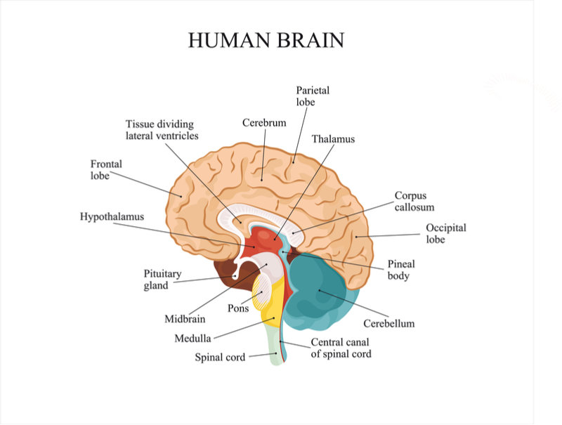
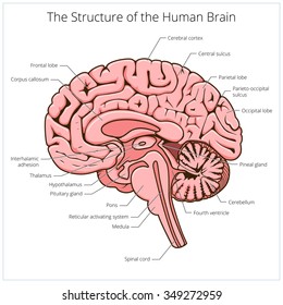
A well-labelled diagram of a human brain is given below for further reference. 293 240 pixels 587 480 pixels 733 600 pixels 939 768 pixels 1252 1024 pixels 2503 2048 pixels.
Labeled Brain Anatomy Images Stock Photos Vectors Shutterstock
Neurons also known as neurones and nerve cells are electrically excitable cells in the nervous system that process and transmit information.
Well labelled diagram of the brain. Add your answer and earn points. A Draw a well-labelled diagram of human brain. In vertebrate animals neurons are the core components of the brain spinal cord and peripheral nerves.
Complete neuron cell diagram. Read on to explore the human brain structure diagram parts of the human brain and the body functions controlled by the human brain. Mid-sagittal section of brain showing diencephalon includes corpus callosum fornix and anterior commissure Marieb Hoehn Human Anatomy and Physiology 9th ed Figure 1210 Exercise 2.
This is a file from the Wikimedia Commons. Nuclear membranes surround the cytoplasm of nucleus known as nucleoplasm. 2653 labeled brain anatomy stock photos vectors and illustrations are available royalty-free.
Draw a labelled diagram of human brain and write its any two functions. Asked Nov 2. The nucleus is a double membrane-bound organelle.
Functions of different parts of the brain are. Asked Nov 17 2017 in Class X Science by aditya23 -2137 points 0 votes. The part of the brain that connects to the spinal cord.
Draw a well labelled diagram of Amoeba. The ventricular system contains the lateral third and fourth ventricles whose function is to produce cerebrospinal fluid. Every EZmed post is filled with simple tricks to remember the content.
Labeled Brain Model Diagram. FileDiagram human cell nucleussvg. Asked Nov 17 2017 in Class X Science by aditya23 -2137 points Draw a neat diagram of human brain and label on it the following parts.
Learn where CSF is found. The human brain is the main coordinating center of the body which enables an organism to think and make decisions. B Which is the main thinking part of brain.
- 19903772 Brainly User Brainly User 23072020 Science Primary School Draw a well labelled diagram of Amoeba. Information from its description page there is shown below. 1 See answer User is waiting for your help.
The cerebral cortex has 4 main lobes - frontal lobe parietal lobe occipital lobe and temporal lobe - and their location function and anatomy all differ. The function of the pituitary gland is to manage levels of hormones within the body specifically those that control and stimulate sexual development handle stress. Utilize the model of the human brain to locate the following structures landmarks for the.
The pituitary gland is found in the temporal lobe as well and it is linked to the hypothalamus through a structure known as the pituitary stalk. Draw a neat diagram of human brain and label on it the following parts. Two peach-size mounds of folded tissue located at the top of the brain.
It is the lowest most primitive area of the human brain. We will use labeled diagrams and lateral images of the brain side views to walk through each lobe of the cerebrum. Labeled Diagrams of the Human Brain.
462 378 pixels. The nuclear membrane is perforated by apertures known as nuclear pores. All the functions are carried out without a single glitch and before you even bat an eyelid.
Brain diagram with labels hypothalamus vector brain diagram pons cerebrum and cerebellum brain pons brain anatomy amygdala brain labelled amygdala brain human midbrain diagram pons. Includes a labelled versions for class discussion as well as worksheets for pupils to label themselves colour and black and white. Learn the ventricles of the brain along with their definition function location anatomy and cerebrospinal fluid CSF flow using labeled diagrams.
This article explains the nervous system function and structure with the help of a human nervous system diagram and gives you that erstwhile textbook feel. Find out how some people live with just half a brain. BI 335 Advanced Human Anatomy and Physiology Western Oregon University Figure 4.
The human brain is an astonishing organ that takes care of each function and action of the body. Structure And Function Of The Human Brain Parts Of The Human Brain The human brain is divided into three main parts. They remind me of school textbooks which used to have plenty of them providing a visual aid to understanding difficult subjects.
The nuclear envelope consists of two membranes an outer membrane and an inner membrane. See labeled brain anatomy stock video clips. Draw a well labelled diagram of human alimentary canal and label the following parts.
Sep 30 2018 - A set of diagrams of the brain. Complete neuron cell diagram ensvg. DNA is present in nucleoplasm in the form of chromatin.
The following are the different regions of the human brain and their functions. The brain stem controls functions basic to the survival of all animals such as heart rate breathing digesting foods and sleeping. On average an adult brain weighs between 10 kg 15 kg.
A scanner takes multiple X-rays which a computer converts into detailed images of the brain and skullMagnetic resonance imaging. Size of this PNG preview of this SVG file.
Whale Video part 4. Blow Holes holes located on top of head used for breathing and blowing sprays of water.
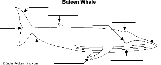
Label Baleen Whale Anatomy Printout Enchantedlearning Com
These grooves expand and contract allowing the whale to scoop up giant mouthfuls of food and water when its feeding.

Label parts of a whale. These cookies allow us to count visits and traffic sources so we can measure and improve the performance of our site. Octopus killer whale or orca barnacles swordfish rainbow fish eel lobster oyster and a crab. This is also known as the tail fin and it is attached to the caudal peduncle a narrow part at the rear of the Whale Sharks body.
Eye - sight organs located on the head. Parts of a whale - Ngā Wāhanga o te Tohorā. This is also known as the tail fin and it is attached to the caudal peduncle a narrow part at the rear of the Whale Sharks body.
More from eko - or -. Dorsal fin - the fin on the upper side of the body. Beluga Whale Unlabeled A large unlabeled picture of a beluga whale to print.
Explore more than 10000 Label Parts Of A Whale resources for teachers parents and pupils. All information these cookies collect is aggregated and therefore anonymous. Baleen whales dont have teeth instead they have 130 to 180 baleen plates that hang down each side of their upper jaws like a fringy curtain.
Label a baleen whale anatomy diagram. Diagram - name two whale parts. Learn about the parts of a whale by reading aloud all of the labels in the whale diagram.
Blowhole - each of the two holes on the top of the baleen whales head through which the whale breathes air they are the whales nostrils. Whale Complete the diagram showing the body parts of a whale. Sep 29 2017 - Product Description.
Explore more than 10000 Parts Of A Whale To Label resources for teachers parents and pupils. Muscle meat organs skin and fat There is relatively little demand for it compared to farmed livestock and commercial whaling which has faced opposition for decades continues today in very few countries mainly Iceland Japan and Norway although whale meat used. Labels for the sperm whale diagram are eye head tail fluke blowhole skin flipper.
They help us to know which pages are the most and least popular and see how visitors move around the site. Flippers eye mouth dorsal fin blowhole tail. If you like this you may be interested in.
Uses of Whale Parts in the Past Whale oil. During feeding the whale. Photograph - name two whale parts.
Whale labeled parts 1 of 1 whale cetacean biome anchor chart chart marine diagram ocean aquatic animal vertebrate marine biome mammal science. X ݎݶ _ A V EH r k A G v CR v 6 g8 wo z. Whale chart answer key.
Fluke - one half of the tail. The labels included are. Baleen whales are larger than the toothed whales.
Drawing - name two whale parts. It also includes a sperm whale facts worksheet as a bonus. Parts of a whale diagram.
This set includes 39 cards 13 cards with picture and label 13 cards with picture only 13 cards with the label 1 blackline master and booklet cover Size 4 5 x 38. Beluga Whale A small white toothed whale that lives mostly in cold Arctic waters. Whale oil was used for a variety of purposes such as for lighting lubrication in machinery and also in the manufacturing of soaps cosmetics and varnish.
It makes the baleen strong but still flexible. Baleen huge sieve-like. Whale video parts 123.
Digital Download Humpback whale printables include Printout with dashed boxes for cutting and gluing Sheet with the words at the bottom for writing A control chart for independent work A fact sheet and humpback whale tracing sheet85x 55 eachFonts Used. On the throat the Gray whale has two to seven grooves of excess skin. Students cut and paste or write words in boxes to label a sperm whale diagram.
Caudal fin caudal fin. The plates are made out of fingernail-like material called keratin. This resource includes basic and deluxe versions of the diagram as well as a reference poster that may be used as a coloring page or to help clarify text while teaching.
Whale meat broadly speaking may include all cetaceans whales dolphins porpoises and all parts of the animal.
Question 22 What Is A Circuit Diagram Draw The Labelled Of An Electric Comprising Brainly In Electric Circuit Diagrams Lesson. A Well Labelled Diagram of Animal Cell.

Ouya Controller Input Device Labelled Diagram Hd Png Download Kindpng
Give a geographical reason for each of the following.

A well labelled diagram of a joystick. Here is ur diagram. Step by step video image solution for Draw a well labelled diagram of phosphorous cycle. A well labelled diagram of a spider spiderlabel1 Label Gallery Get some ideas to make labels for bottles jars packages products boxes or classroom activities for free.
Draw a well labelled diagram showing the pressure and wind belts of the earth. A well labelled diagram of a light microscope simple light microscope diagram. Well Labelled Diagram Of Human Digestive System.
C Hence show by calculation that the most economical size of. It is includes rich examples templates process flowchart symbols. Use a variety of drawing tools smart connectors flowchart symbols.
An easy and convenient way to make label is to generate some ideas first. A Draw a well-labelled diagram of a typical single-core concentric 220 kV power cable indicating all the various layers. Labelled diagram of open circuit 7 difference between and a closed short circuits dummies draw their well what is an how with to show.
Below diagram shows the increase in accumulation of toxic substances as we move up the food chain. B Using electrostatic equations derive the formula for the electric stress distribution in a power cable having a core radius insulation thickness d and overall radius R. Computer Network Diagrams solution extends ConceptDraw PRO software with samples templates and libraries of vector stencils for drawing the computer network topology diagrams.
Draw A Well Labelled Diagram Of Electric Circuit Posted by Margaret Byrd Posted on February 28 2021. Same is the case with the representation of the plant cell as well. ConceptDraw flowchart maker allows you to easier create a process flowchart.
Laboratory thermometers are designed for lab purposes such as checking boiling point freezing point or temperature of other substancesUsing a lab thermometer. Question 22 what is a circuit diagram electric diagrams lesson for draw schematic labelled of closed and label it to in the shown figure effects cur ncert. ConceptDraw is Professional business process mapping software for making process flow diagram workflow diagram general flowcharts and technical illustrations for business documents.
Lysosomes golgi apparatus mitichondria cytoplasm nucleus cell membrane and more. Draw simple labelled diagrams with Maple. Anyone who wants to learn to draw simple labelled diagrams with Maple.
Find an answer to your question Draw a well-labelled diagram of Nostoc. Give a geographical. It is evident from the introduction that an animal cell is a representation of the cellular structure of all the animal species existing on earth.
Draw a diagram of a plant cell and label at least eight important organelles in it. Labelled Diagram Of Open Circuit Brainly In 7 Difference Between Open Circuit And Closed Example A Labelled Diagram Of Closed And Open. Below labelled diagram shows the pressure and wind belts of the earth.
Carl Devore 13 February 2002. Draw A Well Labelled Diagram Of An Open Circuit Posted by Margaret Byrd Posted on January 18 2021. Drawing well-labelled diagrams.
Learn how a reference triangle can be. An easy and convenient way to make label is to generate some ideas first. Label Gallery Get some ideas to make labels for bottles jars packages products boxes or classroom activities for free.
New questions in Biology. The difference in between the well labelled diagram of plant and animal cell is the presence of a cell wall. Id delete ho jayegi kl takgood bye difference.
There is a seasonal shifting in pressure belts. Ramyateja2986 ramyateja2986 17062018 Biology Secondary School answered Draw a well-labelled diagram of Nostoc. By Biology experts to help you in doubts scoring excellent marks in Class 12 exams.
It initiates and regulates cell division. Though this animal cell diagram is not representative of any one particular type of cell it provides insight into the primary organelles and the intricate internal structure of most animal cells. But there is one important difference between well labelled diagrams of plant cell and animal cell.
Labels are a means of identifying a product or container through a piece of fabric paper metal or plastic film onto which information about them is printed. Wide collections of all kinds of labels pictures online. 2 See answers varshini1101 varshini1101 Hope my answer helps you Alfaizali903 Alfaizali903 Answer.
Simplifying inverse trigonometric expressions. Well labeled diagram of an animal cell. In a well labelled diagram.
Biomagnification means increasing the concentration of various toxic substances along the food chain. Make your work easier by using a label. By Biology experts to help you in doubts scoring excellent marks in Class 12 exams.
Step by step video image solution for Draw a well-labelled diagram of a mature ovule showing its internal structure. You should make a label that represents your brand and creativity at the same time you shouldnt forget the main purpose of the label. Place the end of the thermometer with the reservoir of liquid in the testing medium.
You should make a label that represents your brand and creativity at the same time. View Answer Bookmark Now. Atmospheric Pressure Winds ICSE.
Toxic substances at the level of primary producers get concentrated at each trophic level as they move up the food chain. A Well Labelled Diagram Of A Cpu.
Store urine until it is passed out. Asked Jun 22 2019 in Class VII Science by navnit40 -4939 points excretion in humans.
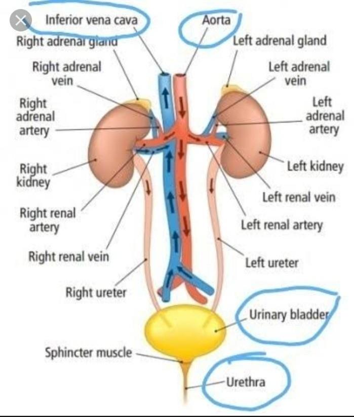
Draw A Diagram Of Excretory System In Human Beings And Label The Following Parts Aorta Kidney Urinary Bladder And Urethra Edurev Class 10 Question
Q6 Draw a diagram of the human excretory system and label the following parts.
Draw a diagram of excretory system in human beings and label the following parts. Draw a diagram of human excretory Page 310. B How is urine produced and eliminated. I form urine ii is a long tube which collects urine from kidney iii store urine until it is passed out.
Iii Draw excretory system in human beings and label the following organs of excretory system which perform following functions. A b Urine is produced by the filtration of blood by the kidneys. 20a Draw a diagram of excretory system in human beings and label the following parts.
The diagram below represents the different parts of the human excretory system. I Urethra ii Left renal vein asked Mar 18 2020 in Science by Sandhya01 591k points. Excretion is the process where all the metabolic wastes are removed from the body.
Draw a diagram of the human excretory system and label the various parts. Is a long tube which collects urine from kidney. Human Excretory System Diagram.
A Draw a diagram of excretory system in human beings and label the following parts. Aorta kidney urinary bladder and urethra. Draw excretory system in human beings and label the following organs of excretory system which perform following functions.
Aorta kidney urinary bladder and urethra. Draw a diagram of human excretory system and label the following parts. Kidney ureter urinary bladder and urethra.
Kidney ureter urinary bladder and urethra. It is then passed through the ureters to urinary bladder where it is stored till urination. Draw a diagram of human excretory system and label the following parts.
Excretory System Labeling. Kidney ureter urinary bladder and urethra. Aorta kidney urinary bladder and urethra.
Kidney ureter urinary bladder and urethra. The Questions and Answers of Draw a diagram of excretory system in human beings and label the following parts. The iliac diagram of the human kidney as seen in a longitudinal section related questions 0 votes 1 answer draw a well labeled diagram of the human excretory system draw a diagram of the human eye as seen in a vertical section and label the parts which suits the following descriptions relating.
Answera b Blood from the heart comes into the kidneys afferent and efferent arteriols from the renal arteries where it enters about 2-3 million nephrons per kidney. B How is urine produced and eliminated. Asked Jun 22 2019 in Class VII Science by navnit40 -4939 points excretion in humans.
Play this quiz called labeling the excretory system. 20a Draw a diagram of excretory system in human beings and label the following parts. Asked Nov 3 2017 in Class X Science by aditya23 -2138 points a Draw a diagram of human excretory system and label the following parts on it.
EXCRETORY SYSTEM ORGANS - Labelled diagram Draw a diagram of human excretory system and label the following parts. B How is urine is produced ans eliminated. Aorta kidney urinary bladder and urethra.
Aorta kidney urinary bladder and urethra. Asked Jun 22 2019 in Class VII Science by navnit40 -4939 points excretion in humans. I form urine ii is a long tube which collects urine from the kidney iii store urine until it is passed out.
Excretion in humans is carried through different body parts and internal organs in a series of processes. Draw excretory system in human beings and label the following organs of the excretory system which perform following functions. Kidney ureter urinary bladder and urethra.
In human beings a pair of kidneys is present in the abdomen. Draw a diagram of human excretory system and label the following parts on it. 20a Draw a diagram of excretory system in human beings and label the following parts.
B How is urine produced and eliminated. Asked Jun 22 2019 in Class VII Science by navnit40. Draw a diagram of excretory system in human beings label the following parts.
Draw a diagram of human excretory system and label the following parts. Answera b Blood from the heart comes into the kidneys afferent and efferent arteriols from the renal arteries where it enters about 2-3 million nephrons per kidney. Answera b Blood from the heart comes into the kidneys afferent and efferent arteriols from the renal arteries where it enters about 2-3 million nephrons per kidney.
Draw a diagram of the human excretory system and label the various parts. Is done on EduRev Study Group by Class 10 Students. The sweat takes out the dead skin cells and bacteria in the pores to keep the skin healthy.
This discussion on Draw a diagram of excretory system in human beings and label the following parts. This is a quiz called labeling the excretory system. Draw a diagram of the human excretory system and label the various parts.
Slightly higher level body partsPre-Intermediate body parts. In this way what are the external parts of the human body.
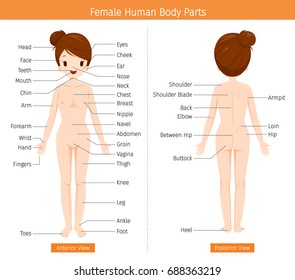
External Human Body Parts Hd Stock Images Shutterstock
An external body part consisting of feathers or hair about the neck of a bird or other animal.
Name the external parts of the human body. The front of the trunk from the neck to the abdomen. We can see these organs. Parts of the Body Vocabulary.
Whats harder to see are the different body systems working under the skin in each body part. Many translated example sentences containing the external parts of the human body German-English dictionary and search engine for German translations. External Parts of body in English Body Parts Name with Pictures - YouTube.
Pancreas and so on. Skull Cranium holds the brain Mandibles lower jaw Maxilla upper jaw Nasal bone Zygomatic bone eye sockets Cranium holds the brain Mandibles lower jaw Maxilla upper jaw Nasal bone Zygomatic bone eye sockets. An external body part that projects from the body.
- The main bone structure remains the same butEither of the two soft rounded parts of a womans chest that produce milk after she has a baby. These external organs are our sense organs. Medium level body parts.
There are those parts located outside external body parts and others located inside the body internal body parts. The body uses different systems to work properly. Sense organs are important parts of our body.
List of external organs of the body. Our body has symmetry that means it looks the same on the left side as it does on the right side. These include the heart brain lungs kidneys liver and pancreas.
List of internal organs of the body. This is thoroughly answered here. Explore more than 10000 Name The Main External Parts Of The Body Humans The Similarities And Differences Between Each Other resources for teachers parents and pupils.
Nerves muscles veins bones how do these body parts keep you going. Body Parts are Pieces in a Puzzle. Your skin is the biggest organ of your body and its.
Higher level body parts. Vital organs are the parts of our body that we need to stay alive. The external organs of our body are eyes ears nose tongue and skin.
The parts of the human head include. External Body Parts Of Woman Human Body Parts Pictures with Names - Body Parts. These external body parts are easy to see on the average person.
Read about the different systems in the human body made simple for kids. Do you want to grow up and become a physiotherapist or a chiropractor -- Allright so lets discuss about the External.
Iii part which connects i and ii iv part from which urine is passed out. Aorta kidney urinary bladder and urethra.

Solved Draw A Well Labelled Diagram Of Human Excretory System Self Study 365
Some substances in the initial filtrate such as glucose amino acids salts and a major amount of water are selectively reabsorbed leaving the urine as waste.
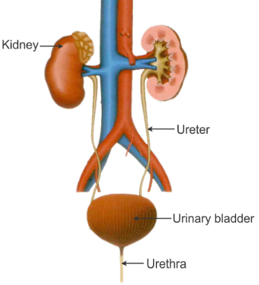
Draw the labelled diagram of excretory system of human being. B Each kidney has large numbers of filtration units called nephrons packed close together. A Excretory system of human beings. Draw Well Labelled Diagram of Human Excretory System.
A b Kidney helps in i Excretion of waste from the body and ii Osmoregulation. These tubes carry urine from. Video text image.
Asked Nov 3 2017 in Class X Science by aditya23 -2137 points a Draw a diagram of human excretory system and label the following. Kidney ureter urinary bladder and urethra. They filter the impure blood and produce urine.
They act as excretory organs and also control the balance of water and mineral. Asked Jun 22 2019 in Class VII Science by navnit40 -4937 points excretion in humans. Draw a neat labelled diagram of excretory system in human beings.
A pair of ureters are tubes that lead out from each kidney. A Draw a labelled diagram of excretory system in human beings and label the following. These are a pair of reddish-brown bean-shaped structures that lie on either side of the backbone.
B List two vital functions of the kidney. C Water resins gums any two. Excretory system in humans The excretory system in human beings includes a pair of kidneys a pair of ureters a urinary bladder and a urethra.
Kidney ureter urinary bladder and urethra. The main excretory organs include kidney ureter urinary bladder and urethra. The kidneys are located in the abdomen one on either side of the backbone.
This discussion on Draw a diagram of excretory system in human beings and label the following parts. 3 store urine until it is passed out. Aorta Vena cava Urinary bladder Urethra.
A Draw a diagram of excretory system in human being and label on it. Human excretory system includes organs that facilitate the removal of nitrogenous wastes from the body. The Questions and Answers of Draw a diagram of excretory system in human beings and label the following parts.
Diagrams - The Excretory System Draw excretory system in human beings and label the following organs of excretory system which perform following functions. Q6 Draw a diagram of the human excretory system and label the following parts. This is also known as the urinary system.
B Name the factors on which the amount of. Hi guysToday I will show you How to draw human respiratory system in easy way step by step. Excretory Products and their Elimination.
C Name two nitrogenous wastes released from kidney. Is done on EduRev Study Group by Class 10 Students. Homeostasis and Excretion.
I left kidney ii renal artery iii urinary bladder iv urethra b Name the functional unit of kidney. Kidneys filter the blood and urine is the filtrate obtained. I part in which urine is produced.
Log in to add comment. 2 is a long tube which collects urine from kidney. Draw a neat and well-labelled diagram of excretory system in human beings.
Human beings have a urinary system to eliminate nitrogenous waste products. Ii part which stores the urine. Urine passes to the.
Draw a diagram of human excretory system and label the following parts. Biology more Questions Class. A Name the various organs of the human excrtory system b Draw a neat labelled diagram of the human excretory system c What is the function of excretory system in humans.
Dow Help ezton 038 points Nephron structures and functions Label the processes occurring in each part of the human nephron and surrounding tissues water amino acids glucose and ions water Secretion of hydrogen ions into tubule Reabsorption of water and urea Filtration of blood sodium ions protons drugs and poisons into tubule Reset Zoom References. Regulated reabsorption in which hormones control the rate of transport of sodium and water depending on systemic conditions takes place in the distal tubule and collecting duct.

Chapter 26 Urinary System Flashcards Quizlet
The second stage in the function of the nephrons.
Label the processes occurring in each part of the human nephron and surrounding tissues. It takes place in the corpuscle of the nephron. Occurs between proximal tubule and peritubular capillary. Urine formation begins with the process of filtration in which substances are forced out of capillaries called glomerulus and into the nephron at the glomerular capsule.
Each kidney consists of millions of nephron which plays a significant role in the filtration and purification of blood. Draw and label the parts. The nephron is divided into two portions namely the glomerulus and the renal tubule and helps in the removal of excess waste from the body.
Cortical collecting tube 6. During this phase all parts of the tubule act to return essential substances out of the nephron so that it is not lost in the urine. The collecting ducts are continuous with the nephron but not technically part of it.
Amongst all filtration is the least selective of all processes. Nephron functional unit of the kidney the structure that actually produces urine in the process of removing waste and excess substances from the blood. For more anatomy content please follow us and visit our website.
Amongst all filtration is the least selective of all processes. The basic physiology of a nephron within a kidney. This fluid called filtrate has the same composition as blood plasma except for larger molecules such as proteins which cannot fit through the glomerular pores.
Collecting ducts merge as they descend deeper in the medulla to form about 30 terminal ducts which empty at a papilla. First process is filtration. Answer correct gases must pass through all three part e of the kidney label the diagram of the kidney and nephron below drag the labels to their appropriate locations on the diagram below labels can be Diagram A Nephron Diagram Kidney Elegant Internal Structure labeled diagram of a nephron and its location and functions most of the nephron lies in the outer region of the kidney the renal cortex ly one part the loop of henle enters the central part.
Urine is excreted to the outside of. Bowman turns out was from England actually he was a very curious fellow and so he looked under microscope and he noticed that if you look right were these little Tufts of blood vessels are you can actually see that there is something surrounding them each of them and so he called that Bowmans capsule and so thats what we so call it today so England was laying claim to parts of the kidney. The ureter connects the kidney to the bladder.
Afferent arteriole and 13. He has diabetes which he has been poorly controlling over the last ten years or so. In fact each duct collects filtrate from several nephrons for final modification.
We hope this picture Parts Of The Nephron Diagram can help you study and research. Martin Cullinan is a 57 year old man who came to the emergency room with 2 weeks of worsening leg swelling fatigue and eye swelling. Draw and label the parts of a nephron.
Reabsorption active transport secretion salt pumping and filtration are the selective processes occurring in the nephron. We think this is the most useful anatomy picture that you need. There are about 1000000 nephrons in each human kidney.
There are four basic processes in the formation of urine starting with plasma. Regulation Of Kidney Function. Reabsorption of water amino acids glucose and ions Secretion of protons drugs and polsons into tubule Reabsorption of water Reabsorption of water and urea Filtration of blood Secretion of hydrogen ons into tubule Reabsorption of min.
2 The second section is the loop of henle which allows water loss and salt NaCl loss. First section of the renal tubule that the blood flows through. Reabsorption of water ions and all organic nutrients.
Loop of Henle 8. This is fairly non-selective meaning that almost all of the substances in the the blood except cells and plasma proteins as well as the substances bound to these proteins enter the nephron. It is a highly selective process in that the tubules carefully.
What is a nephron. You can click the image to. During the process of filtration molecules molecule that is smaller than albumin are filtered from the glomeruli to the corpuscle space.
BIOL 204 Lab Case. The bladder is storage for urine. 1 The first section of the renal tubule is the proximal convoluted tubule which is specialized to reabsorb water and many solutes from the glomerular filtrate and secrete certain unwanted substances.
When stimulated by ADH these cells will insert. The functional unit of the kidney is the nephron. Label the processes occurring in each part of the human nephron and surrounding tissues.
The main structures that make up the urinary system are two kidneys contains nephrons two ureters one bladder one urethra arteries and veins. They are lined with simple squamous epithelium with receptors for ADH. Filtrate in the corpuscle space will waste product such as urea organic molecules such as glucose electrolytes water and so on.
What is a nephron.
Draw And Label The Parts Of Neuron Brainly In Copy The Diagram And Label Any Two Parts From The Toppr Com Nervous System Review 16 1 Neurons And Glial Cells Concepts Of Biology 1st Canadian Color The Neuron And Neuroglial Cells Overview Of Neuron Structure And Function Article Khan Academy Neural Structure Quiz. Ib Biology Notes 6 5 Nerves Hormones And Homeostasis.

Activity 1 Name Me Instruction Identify The Parts And Functions Of A Nerve Cell Give The Function Brainly Ph
Join group and play Just play.

Label the parts of the neuron brainly. Here is the description of human neuron along with the diagram of the neuron and their parts. The neuron is the building block of the nervous system. Label parts of neuron and regions of brain.
All neurons have three main parts. Add your answer and earn points. Label the parts of the neuron.
While they have the common features of a typical cell they are structurally and functionally unique from other cells in many ways. 1 axon cell body dendrites nucleus terminal ends. You need to be a group member to play the tournament.
A neuron varies in shape and size depending upon their function and location. 2 axon cell body dendrites nucleus terminal ends. Labeled Neuron Diagram Science Trends.
Besides the three major parts there is the presence of axon terminal and synapse at the end of the neuron. Label the parts of a neuron. This game is part of a tournament.
The structure of neuron. Play this quiz called label a neuron and show off your skills. Spreads the cells impulse out to reach other neurons.
Diagram Of Neuron with Labels. Draw and label the parts of a neuron - 1148587 Deiblhor Deiblhor 08122017 Science Junior High School answered Draw and label the parts of a neuron 1 See answer Shemmy1 Shemmy1 Parts of a neuron-dentrites-soma-axon-myelin sheath. It consists of three major parts namely Cell body dendrites Axon.
Difference between Sensory and Motor Neuron. It is a relay system of the body and the construction is similar to that. 1 See answer marianaesthersu is waiting for your help.
Solved Label The Parts Of A Peripheral Nerve Shown In Cro Chegg Com. - 2914291 marianaesthersu marianaesthersu 02172017 Health Middle School answered What are the parts of a neuron. A group of neurons forms a nerve.
Choose the correct names for the parts of the neuron. A Draw The Structure Of Neurone And Label The Following On It I Dendrite Ii Cell Body Brainly In. The neuron is a specialized and individual cell which is also known as the nerve cell.
This is a quiz called label a neuron and was created by member legoa1. STRUCTURE OF NEURON LABEL THE PART. Label the part of the neuron - 13481699 montefalcojohnny1 montefalcojohnny1 44 minutes ago Science Senior High School answered Label the part of the neuron 2 See answers montefalcojohnny1 is waiting for your help.
Nerve cells or neurons are the structural and functional units of the nervous system. Joins the soma and axon to collect the impulses before sending one down the cell. Ultrastructure Of Nerves Classification Neurones Teachmeanatomy.
What are the parts of a neuron. Ahcd1010hwsol96 Pdf 96 Award 1 00 Point Problems Adjust Credit For All Students Correctly Label The Parts Of The Neuron And Other Nerve Tissue Tissue Course Hero. The neuron consists of.
Lab 1 Neurons 1 Lab 1 Neurons A Please Label The Anatomical Parts Of The Neuron Below A Soma Cell Body D Axon Hillock B Nucleus E Axon C Course Hero. Nervous System - Neuron. They provide the power and the growth of the cells.
1 dendrites 2 cell body or soma and 3 axons. It is irregular in shape or polyhedral. 3 axon cell body dendrites nucleus terminal ends.
Carries the cells impulse to the terminal. Definitions of the terms of the skin. Choose the correct names for the parts of the neuron.
Surrounds some neurons to increase how efficiently it can carry an impule. Learn vocabulary terms and more with flashcards games and other study tools. The scorecard of a champion.
All neurons have three different parts dendrites cell body and axon. Cyton or the cell body which has the nucleus and the mitochondria and ribosomes of the cell. Add your answer and earn points.
Axon Nucleus Myelin sheath Dendrite Synapse Neuron functions Match each part of a neuron with its function. Label the parts of the central nervous system. Log in to add comment.
Transmits action potentials Receives impulses Increases the speed of action potential transmission Transmits neural impulse to anoti cell. 0 Time.
Comb and Wattles. The entire breast portion of the chicken.

Parts Of A Chicken Useful Chicken Anatomy With Pictures 7esl
The differences between males and females include the size of the comb and wattles the size of the spurs in older birds and the characteristics of the hackle and.
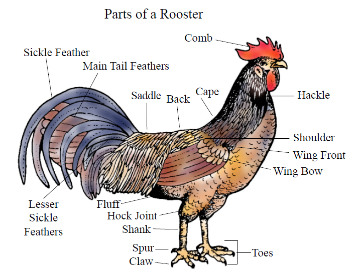
What are the parts of a chicken called. Its placement in the middle of the leg and the way it bends when chickens walk readily draws comparisons to our own knees. Its often also called simply the wing or wingette or the flier. The distinctive red flesh located under a roosters beak is called the wattles.
Generally includes a little more than one quarter of the meat on the chicken. Several types of combs exist including single combs rose combs pea combs and buttercup combs. When chickens mate the roosters cloaca comes into contact with that of the hen.
Collectively these and other fleshy protuberances on the head and throat are called caruncles. First of all a thigh has all of the flavor of a wing or a leg but it is bigger and meatier. The skull of the chicken consists of many small bones and the face is made up of the nasal and the premaxillary bone.
Some breeds contain single comb some have rose comb and some breeds with pea comb. Perhaps the most prominent feature on a chickens head is the comb. Chicken thighs are also a far superior piece of.
So body parts of chicken helps to know about that breed and their variant. Consists of white meat only. Unlike the ears themselves chickens earlobes are quite visible.
The basic external parts of a chicken include the comb beak wattles ears earlobes eyes eye rings wings tail thighs hocks shanks spurs claws and toes. For example- different types of chicken breeds have different sized and colored comb in their head. The supreme chicken is the forequarter without the chest bones but with the meat around it.
Please note the chicken. These are secondary sexual appendages initially produced by the sex hormones when the bird starts to mature. List of different parts of a chicken with examples and chicken anatomy pictures.
Similarly ear lobe feather skin wings etc. Hover over them to find links to the nutrient content of some of these cuts either raw or once cooked in various ways. This is called a cloacal kiss Hock and Shank Shutterstock.
The thigh is the most slept-on part of the chicken. Theyre a series of spikes that run from the chickens beak to the back of their head. Also referred to as split breast.
It can be a single row of bumps or 3 rows side by side. Includes the breasts and wings which corresponds to about a quarter of the chicken. Adult chickens have a fleshy crest on their heads called a comb or cockscomb and hanging flaps of skin either side under their beaks called wattles.
The largest bones in a chickens face are called frontal- parietal- and temporal bones. One of the most distinctive types of chicken combs is the V-shaped comb. Earlobes are usually red or white and are located slightly below and behind the chickens eyeball on the head of the chicken.
Along with the neck heart and gizzard are part of the so-called chicken giblets. As Figures 1 and 2 show both male and female chickens have these basic parts. There is also a jaw bone which is called a mandible.
There are two main reasons for being able to refer to the different parts of a chicken in the English language the first being in situations where you wish to describe these animals or understand a conversation about them. Mikroman6Getty Images The breast is a lean cut of white meat found on the underside of a chicken. Also are of different types of various breeds.
The comb is the fleshy growth found atop a chickens head and is larger in roosters compared to hens. We can also determine healthy and. There is something called an uropygial gland at the base of the tail it is more commonly called the preen gland.
It is available bone-in boneless skin-on and skinless. They make up the cranium which is the back of the chicken head. The hock is the leg joint located at the base of a birds thigh.
The breast portion of the chicken that has been split lengthwise producing two halves. The comb of a chicken is the red flesh located on the top of the animals head. It will vary in shape and size depending on its variation and the chickens breed- to learn all about the different combs this article will have you sorted.
Common Cuts of Raw Chicken The images below represent some of the cuts of raw chicken most commonly encountered in retail stores. The breast is loosely attached to a thin muscle called the tenderloin this is where chicken tenders come from. Pea comb Pea combs are a series of small short bumps.
Includes the thighs and back or backbone one of the tastiest parts of the chicken when barbecued. Single comb Single combs are the most common. For the record the flats or skateboards are names for the piece with two small connected bones and meat in the middle.
One interesting thing about earlobes is that you can sometimes use it to tell what color egg your hen will lay. The bird uses the oily extract from this gland to keep the feathers oiled and in good condition. The cut includes half a breast a wing and part of the back.
A whole breast includes two halves which are usually separated and sold individually.
Learn vocabulary terms and more with flashcards games and other study tools. Volls Zn NI HU 10M Z1 10MN.

Electricity And Circuits Electricity Cbse Class 6 Ekshiksha
Doing this will help you to remember where each part is located.
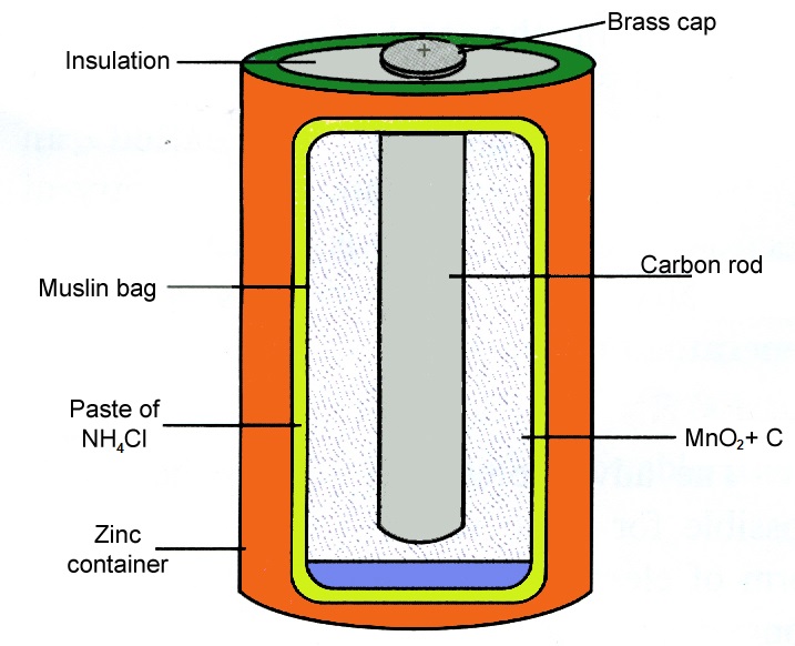
Label the parts of an electric cell. This kind of cell includes the galvanic or voltaic cell named after Luigi Galvani and Alessandro Volta. An electric cell is a device which produces electricity capable to run smaller appliances like torch clock camera radio etc. Label the parts of an electric circuit write your answer on the blanks - 14999688.
Choose from 500 different sets of the parts of cells label flashcards on Quizlet. Find an answer to your question Label the pard of an electric cell 1. Of the plant and animal cell add to their picture and label all parts of the cell.
Negative side has a flat metal disc. Click the links below to download the labeled and unlabeled eukaryotic cell diagrams. Start studying Label a cell Labeling parts of a cell Cells Structures and Functions.
Label the parts B2 Cells and simple transport. Describe an electric cell with the help of labeled diagram - Science - Electricity and Circuits. Label diagram of electric.
Chemicals stored inside the cell produce electricity through internal reactions. Learn the parts of cells label with free interactive flashcards. Abenseb Abenseb 21092020 Science Primary School Label the pard of an electric cell 1 See answer Abenseb is waiting for your help.
An electric cell has two terminals positive and negative -. Draw and label the parts of an operating electrochemical cell using a zinc anode that will produce an electric current having a voltage of 156 V at standard conditions. Draw and label the parts of a basic electrical electric circuit it series labelled diagram simple diagrams lesson for write your answer what is comprising cell in shown figure.
Label the main parts. Which of the following occurs as the cell. Its time to label the cell yourself.
Cell diagram unlabeled. Plant and algal cells. Cell Theory and Cell Function 7th grade - Digital Commons Trinity In this unit on cell theory and cell structure and functions 7th grade students will.
Learn from doing activities as per the NCERT Syllabus. As you fill in the cell structure worksheet remember the functions of each part of the cell that you learned in the video. Click here to get an answer to your question label the parts of simple electrical cell diagram given above trishna34657 trishna34657 07122020.
Positive side of an electric cell has a metal cap. Label the diffent parts of an electric circuit the tell importanceof electricity in our daily life - 15011724. Cell ribosomes respiration chemical reactions protein nucleus cell membrane chloroplasts cell wall mitochondrion vacuole photosynthesis chlorophyll sap cytoplasm ORGANELLE FUNCTION Type of cell where found Plant Animal.
An electrochemical cell is a device that produces an electric current from energy released by a spontaneous redox reaction. Consider the following electrochemical cell. Electrolytic cells like galvanic cells are composed of two half-cells--one is a reduction half-cell the other is an oxidation half-cell.
A vacuole is a membrane bound structure found in the cytoplasmic matrix of a cell. ABecause cassava is known to include cultivated and wild forms the species is no longer a cultigen but ranks as an indigen instead.
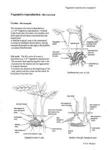
Biology Plants Vegetative Reproduction Tropical Examples Educational Notes Drawings By D G Mackean
Using force when tying the stems in bundles may cause breakage 6 nodes and to improve sprouting.

Labelled diagram of cassava plant. Manihot esculenta commonly called cassava k ə ˈ s ɑː v ə manioc or yuca among numerous regional names is a woody shrub native to South America of the spurge family EuphorbiaceaeAlthough a perennial plant cassava is extensively cultivated as an annual crop in tropical and subtropical regions for its edible starchy tuberous root a major source of carbohydrates. Figure 21 General morphology of the cassava plant Figure 22 Transverse section of young tuber Figure 23 Inflorescence of a cassava plant Figure 24 Fruit and Seed of a cassava plant Figure 25 Growth and development in cassava Figure 31 Pollination by hand Figure 32 Bagged-pollinated flowers Figure 33 Seedlings growing in a nursery. In deep soils cassava is planted on flat land Figure 7.
Euphorbiaceae is a staple crop for more than 800 million people in the tropics The crop is affected by more than 100 insect and mite species and about 30 cassava diseases induced by viruses phytoplasmas bacteria or fungi Among the diseases cassava mosaic disease CMD and cassava brown streak disease CBSD are the. Months old without stem or leaf damage from pests or diseases. Of days from planting 1st branch level No.
Manihot leptophylla join the extensive synonymy. Labelled diagram - Drag and drop the pins to their correct place on the image. Planting the Cassava Stems.
Use a farm that has been well maintained. Within a week or two new leaves appear and the plant is on its way. Cassava Manihot esculenta Crantz.
In planting stage of the cassava farming process the first thing to do is to carefully select a cassava variety that you will grow. The peel is usually not considered suitable for human consumption but can be used for feeding pigs. The structure of the plant cell is.
Diagram of a cassava plant A transversal section of the root See Figure 2 reveals three distinct parts. The central pith is. Make sure a quarter of the stem is planted.
Labelled diagram - Drag and drop the pins to their correct place on the image. Plant them in suitable soil either laying down on its side 2 inches deep or sticking out of the soil like a stake. Then in about 12 to 18 months time youre ready to harvest your first cassava.
Labelled Diagrams Of Typical Animal And Plant Cells With Editable Royalty Free Cliparts Vectors And Stock Illustration Image 32521141 Plant Cell Definition Labeled Diagram Structure Parts Organelles Plant Cell Diagram To Label Beautiful South Pontotoc Biology Plant And Animal Cell Diagrams Cell Diagram Plant And Animal Cells Cell Diagram Project. Double digging is needed to make the soil soft and to kill the weeds. School cassava garden A good size for a cassava garden is 10 m long and 2 m wide with 32 cassava plants 60 120 cm apart.
The optimal cassava plant spacing is 1 meter by 1 meter apart along each row and across ridges or mounds. That is a single plant may carry both male and female flowers but these are separated from each other. A bacteria diagram clearly enables us to learn extra approximately this single cell organisms that have neither.
Kinazi Cassava Plant and Mbakungahaze Cassava Farmers Cooperative in Rwanda is to contribute to the promotion of cassava by investigating the relationships between farmers grouped into cooperatives and Kinazi Cassava Plant in order to increase the production and processing of this crop. Biological drawing of Cassava Plant showing the tubers which is the swollen roots growing from stem cutting. The cassava plant has sympodial branching and variable plant height ranging between 1 and 5 m although maximum height usually does not exceed 3 m.
Plant Cell Diagram Vacuole. Large 15 cm Cream. Cassava can be grown by making soil mounds 3 metres wide and 2 metres high or ridges suficiently high so the tuberous roots are above the waterlogged soil.
There is a real possibility that two other species eg. Dark brown Short 8 cm. Cassava is planted on mounds to increase the topsoil volume per plant Figure 6a.
The stem Stems are particularly important in cassava as they are. Of nodes from planting 1st branch level No. The peel the central pith and the vascular bundle.
Select varieties with multiple pest and disease resistance high and stable root yields and acceptable quality. Cassava should be planted along with other crops such as yam maize and vegetables for example yam is planted at the top of the mound cassava on the side or slope of. As the central vacuole shrinks it leaves the cell wall unsupported.
The definitive infraspecific classification of the species is in preparation. Biology teaching resources on vegetative reproduction by D G Mackean. The ground should be dug well twice.
Prominent blood vessels Palpation 1. Position of mediastinum a.

A Draw A Labelled Diagram Of Human Respiratory System B Explain The Transportation Of Oxygen And Carbon Dioxide In Human Beings
It ranges from 20-25mm in diameter and 10-16cm in length.

Fully labelled diagram of the respiratory system. The hairs present in the nose filter out particles in the. Dimitrios Mytilinaios MD PhD Last reviewed. Movements with respiration a.
Human Respiratory System Diagram showing different parts of the Respiratory Tract. When air passes through the nose it is warmed moistened and filtered. The lungs are covered by a lining called the pleura which has two layers.
In the throat the trachea or windpipe filters the air. Labeled diagram of respiratory system. The human respiratory system is adapted to allow air to pass in and out of the body and for efficient gas exchange to happen.
Millions of small air sacs in the lungs where oxygen and carbo tubes which branch off from. View Original Image at Full Size. It is made up of several organs and structures that transport air into and out of the lungs exchanging oxygen with carbon dioxide.
But fear not - with a bit of practice and dedication you can. This is an online quiz called Respiratory System Labeling Interactive. Compare and measure both sides 3.
Click on the tags below to find other quizzes on the same subject. Interactive for 5th Graders This quiz has tags. May 31 2021 Reading time.
The lungs are divided into areas called lobes. They supply oxygen to the organs and tissues of the body. Respiratory System Examination Inspection 1- Shape of the chest 2.
The respiratory system allows people to breathe. Features of the Human Respiratory System. Respiratory system quizzes and labeled diagrams Author.
Your Skills Rank. The lungs are the parts of the body that we use to breathe. All your KS2 students have to do is drag and drop the various labels to identify the nasal cavity larynx alveoli diaphragm and more.
Position of mediastinum a. How we can help. There is a separate diagram explaining how breathing works breaking down the process of inhalation and exhalation.
Air enters the nose through the nostrils. The respiratory system which includes air passages pulmonary vessels the lungs and breathing muscles aids the body in the exchange of. Air a mix of oxygen and other gases is inhaled.
It includes the main parts of the respiratory system such as the trachea bronchi bronchioles alveoli and the diaphragm. This is a brilliant resource. The trachea is surrounded by.
In the throat the trachea or windpipe filters the air. The right lung has three lobes and the left lung has two lobes. Fill in the blanks.
Use this resource as a handout or transparency for science class. Name Description Function 1 2 3 4 5 6 7 8 9 10 11 Name Function 1 nose A. The gas exchange process is performed by the lungs and respiratory system.
You should make a label that represents your brand and creativity at the same time you shouldnt forget the main purpose of the label. The energy is generated by the breakdown of glucose molecules in all living cells of the human body. The entire respiratory tract passage consists of the nose pharynx larynx trachea bronchi and bronchioles.
The airway the lungs and the muscles of respiration. Image 37789 is a 1125 by 1408 pixel PNG Uploaded. Use this engaging interactive activity to label the respiratory system.
2 minutes Full to the brim with interesting but arguably complex anatomy the respiratory system may feel a little confusing to the anatomy apprentice. From the quiz author. Labeled diagram of the lungsrespiratory system.
The human respiratory system consists of a pair of lungs and a series of air passages leading to the lungs. The inner membrane of the trachea is covered in tiny hairs called cilia which catch particles of dust which we can then remove through coughing. Respiratory System Label Diagram Diagram Labels Label Gallery Get some ideas to make labels for bottles jars packages products boxes or classroom activities for free.
The respiratory system provides oxygen to the bodys cells while removing carbon dioxide a waste product that can be lethal if allowed to accumulate. Also known as the windpipe this is the tube that carries air from the throat into the lungs. An easy and convenient way to make label is to generate some ideas first.
There are 3 major parts of the respiratory system. Exercise and smoking both affect the lungs and circulatory system. Ideal for putting knowledge into practice respiratory system interactive activities can be a great addition to your Science lessonsChildren also have the chance to correct any mistakes.
Oxygen is inhaled and is transported to various parts and are used in the process of. The respiratory system in humans has the following important features. The respiratory system transports oxygen from the air we breathe through a system of tubes into our lungs and then diffuses it into the bloodstream whilst carbon dioxide makes the opposite journey.
The airway which includes the nose mouth pharynx larynx trachea bronchi and bronchioles carries air between the lungs and the. 15 rows Printable Blackline Diagram of The Respiratory System Test yourself. There is a printable worksheet available for download here so you can take the quiz with pen and paper.
Molly Smith DipCNM mBANT Reviewer. Lower respiratory tract organs.
CBSE Class 12 Biology Solved Question Paper 2011. Each ovary releases one egg cell.
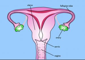
Sexual Reproduction In Humans Class 8 Reproduction In Animals
A Draw diagrammatic sectional view of the female reproductive system of human and label the parts.

Draw a labelled diagram of female reproductive system class 8. Get FREE solutions to all questions from chapter HUMAN REPRODUCTION. Click hereto get an answer to your question Draw a labelled diagram of female reproductive system. It is shaped like an almond size is around 35 cm to 2 cm wide 1 cm thick.
The female reproductive structure consists of a pair of ovaries uterus fallopian tubes cervix and vagina. Watch complete video answer for Draw a labeled diagram of female reproductive syste of Biology Class 12th. Get certified as an expert in up to 15 unique STEM subjects this summer.
I production of female gamete ovum and ii secretion of female hormones estrogen and progesterone. Q1 Fill in the blanks. Our Bootcamp courses are free of charge.
Due to an increase level of estrogen the uterine endometrium becomes. Asexuallysexually b Humans are__________. Draw a labeled diagram of human female reproductive system.
Each ovary is composed of ovarian follicles. The follicular cells of the Graafian follicle secrete oestrogen. Q4 Write two major functions each of testis and ovary.
Avail 25 off on study pack. The female reproductive system is composed of a pair of ovaries along with oviducts vagina cervix uterus and the external genitalia that are located in the pelvic region. Watch complete video answer for Draw a labelled diagram of female reproductive sys of Biology Class 12th.
Click here to get an answer to your question Draw a labelled diagramof female reproductiveSystemMentionits parts riteshsingh86. I Where the secondary oocytes develop ii Which helps in collection of ovum after ovulation iii Where fertilization occurs. Each ovary releases one egg cell.
Follicular phase is also called the proliferative phase. In human females a pair of ovaries is located in the abdominal cavity near the kidney. Get FREE solutions to all questions from chapter HUMAN REPRODUCTION.
Whereas Probe is a small fragment of DNA or RNA used for identification of genes in biological system. Long Answer Questions LA Q2 Describe the human female reproductive system with a labelled diagram. A b i Follicular phase.
Q2 Draw a labeled diagram of male reproductive system. During this phase the FSH released by the pituitary stimulates the growth of the primary ovarian follicle and also causes maturation of the primary oocyte in this follicle. The ovary is a paired structure located in the upper pelvic cavity.
It is shaped like an almond size is around 35 cm to 2 cm wide 1 cm thick. A Humans reproduce __________. The ovary is a paired structure located in the upper pelvic cavity.
Express each of the following rational numbers in its standard formi 12 30ii 14 49iii 1535iv 299 161Q. Draw a labelled diagram of female reproductive system. Avail 25 off on study pack.
Draw a labelled diagram of the reproductive system in a human female. Q3 Draw a labeled diagram of female reproductive system. Short Answer Questions SA 3.
The female reproductive structure consists of a pair of ovaries uterus fallopian tubes cervix and vagina. Secondary School answered Draw a labelled diagram of female reproductive SystemMention its parts 1 See answer riteshsingh86 is waiting for your help. On each side of the uterus lie two whitish.
At puberty these follicles undergo maturation to produce ova. Female Reproductive System Diagram How To Draw Female Reproductive System. Iv Where implantation of embryo occurs.
Q5 Describe the structure of a seminiferous tubule. The ovaries perform dual function of. How to draw female reproductive organ system diagram step by step in easy way for class 10 by using scale which makes it much easier for science board examin.
Add your answer. These parts along with a pair of mammary glands that are integrated both functionally and structurally also support the process of ovulation fertilization birth and finally the child care. Female Reproductive System Diagram How To Draw Female Reproductive System Biology Class 10 - YouTube.
Reduce each of the following rational numbers in its lowest formi 60 72 ii 91 364Q.
Draw a diagram of a mature human sperm. The human heart and its functions are truly fascinating.

Human Body Diagram Bodytomy
Label the following parts in it and write their functions.

Draw a labelled diagram of human body. Collection of human body parts 37 human body clipart labeled female inside body parts name. The human kidneys house millions of tiny filtration units called nephrons which enable our body to retain the vital nutrients and excrete the unwanted or excess molecules as well as metabolic wastes from the body. The organ that releases digestive juice into the small intestine.
Download 220 royalty free human body parts labeled vector images. Due to an increase level of estrogen the uterine endometrium becomes. In addition they also play an important role in maintaining the water balance of our body.
Label The Diagram of The Heart - Skeleton - Year 4 Sound - Key Knowledge Ear Diagram - Sankey diagram labelled. Muscular system diagram blank muscular system diagram with. A b i Follicular phase.
Identify the following parts in the human body. Download 107 Label Diagram Human Body Stock Illustrations Vectors Clipart for FREE or amazingly low rates. The heart though small in size performs highly significant functions that sustains human life.
This is a table of muscles of the human anatomy. Draw well labelled diagrams of various types of muscles found in human body. 10000 results for labelled diagram body parts.
We hope you learned something new. Heart Diagram Sheet Wiring Diagram. Labeled Female Body Parts Diagram.
A Where in the human body does an egg or ovum get fertilized b Where does a fertilized egg or zygote develop into a baby in the human body. The alimentary canal is divided into five main parts- mouth esophagus stomach small intestine small intestine and lastly large intestine. Draw A Well Labelled Diagram Of A Human Heart For Board Exam.
During this phase the FSH released by the pituitary stimulates the growth of the primary ovarian follicle and also causes maturation of the primary oocyte in this follicle. Labeled Diagram of the Human Kidney. Human Heart Diagram Human Body Pictures Science For Kids.
B Explain the transportation of oxygen and carbon dioxide in human beings. Human Body Muscle Diagram Human body muscles Muscle diagram Muscle body. - the human body.
Each muscle also has. This diagram depicts muscle of the body diagrams 7441054 with parts and labels. Boy body parts diagram poster.
Educational infographic chart for kids science or language learning. There are some 700 named muscles in the body and other smaller muscle tissues that are part of. Often called striated muscles due to presence of alternate dark and light bands straitions.
Draw a labelled diagram of the digestive system. It is one among the few important topics which are repetitively asked in. These are voluntary under the control of our will.
A Labeled Diagram Of The Human Heart You Really Need To See Bodytomy. May 28 2016 - The human body muscles are the main contractile tissues of the body involved in movement. The largest gland in our body.
167989067 stock photos online. The follicular cells of the Graafian follicle secrete oestrogen. Draw a labeled diagram of the human female reproductive system.
Human body consist of three types of muscles Skeletal muscle It has striated tubular multinucleated fibres and is usually attached to skeleton. They cause motion and produce a force that the body uses to move and manipulate the body. Draw a labelled diagram of the human female reproductive.
Follicular phase is also called the proliferative phase. Students can search the internet or use their textbooks to find the names and locations of the main ant body. The organ where protein digestion starts.
The organ where bile juice gets stored. The heart one of the most significant organs in the human body is nothing but a muscular pump which pumps blood throughout the body. New users enjoy 60 OFF.
Draw a diagram of the microscopic structure of human sperm. A Labeled Diagram of the Human Heart You Really Need to See. How To Draw A Human Heart 11 Steps With Pictures Wikihow.
Label any three parts and write their functions. A Draw a labelled diagram of human respiratory system. The diagram of the human digestive system is useful for both Class 10 and 12.
They prefer women with larger breasts a small waist and larger hips and thighs. Labeled muscle man muscle diagram male body names stock vector.