Microscope diagram with labels and functions. Through the eyepiece you can visualize the object being studied.

Microscope Diagram Labeled Unlabeled And Blank Parts Of A Microscope
This part rotates to change which objective lens is active.

Labelled diagram of a microscope with functions. 872018 3 Compound Microscope. Compound Microscope Types Parts Diagram Functions And Uses. You should always carry a microscope with.
By Uncategorized 0 Comments. Its magnification capacity ranges between 10 and 15 times. It is the topmost part of the microscope.
Objects that are not visible to the human eyes. Bottom base of the microscope that houses the illumination supports the compound microscope. Labelled Diagram Of A Microscope And Their Functions - Fun for my own blog on this occasion I will explain to you in connection with Labelled Diagram Of A Microscope And Their Functions.
Electron microscope parts and functions. In a book a photomicrograph of the cell measured 100 mm. Give A Well Labelled Diagram Of Compound Microscope Using Of.
Microscopy is the science of investigating small objects and structures using. Label the microscope worksheet activities par. To calculate the magnification.
So if you want to get great shots related to Labelled Diagram Of A Microscope And Their Functions just click on the save icon to save the photo to your computer. Microscope Parts and Functions Microscope One or more lenses that makes an enlarged image of an object. The parts of a microscope labeled printable this diagram labels and explains the function of each part of a microscope.
Its actually not a toy microscope its. Microscope diagram with labels and functions. The Microscope Image courtesy of.
Parts of the optical parts are as follows. You should always carry a microscope with two hands one on the arm and the other under the base. Supports the microscope head and attaches it to the base.
Ncert Class 9 Science Lab Manual Slide Of Onion Peel And Cheek. This particular image Parts Of A Compound Microscope With Diagram And Functions with Diagram Of The Microscope Parts earlier mentioned is actually labelled along with. Its actually not a toy microscope its.
Text magnification frac text 100. 872018 2 Simple Compound Stereoscopic Electron Simple Microscope Similar to a magnifying glass and has only one lens. Published by simply CARPNY TEAM at May 7 2016.
Mirror A simple microscope has a plano-convex mirror and its primary function is to focus the surrounding light on the object being examined. You should always start on the lowest power objective lens and should always leave the microscope on the low power lens. The real size of the cell shown above is 005 mm 50 μm.
Diagram of microscope parts and function diagram of the microscope parts labeled diagram of microscope parts. Microbiology 3510l Dustman Flashcards Lecture 1. Microscopy is the science of investigating small objects and structures using.
Parts And Components Of Light Microscopes Light Microscope. Labelled diagram of compound microscope. Weve gathered our favorite ideas for Microscope Slide Diagram Explore our list of popular images of Microscope Slide Diagram Photos Collection with high resolution.
A Study Of The Microscope And Its Functions With A Labeled Diagram. Parts Of The Microscope And Their Functions. Microscope Labelled Diagram And Functions Written By JupiterZ Tuesday December 18 2018 Add Comment Edit.
What Is The Function Of The Pointer On A Microscope. Electron microscope parts and functions. Holds the objective lenses attaches them to the microscope head.
Labelled Diagram Of A Microscope Optics Amp Binoculars Exe Microscope Side Vector Drawing With Parts Labelled Free Svg. Parts Of A Compound Microscope With Diagram And Functions Microscope Labeling News Information Microscope Labeling Biology. Compound Microscope Diagram Labelled.
Let us take a look at the different parts of microscopes and their respective functions. Lens The biconvex lens is placed above the stage and its function is. Shop compound microscopes compound microscope definitions for.
Microscope labeled diagram 1.
Here at the Bosque Village we waste none of the rabbit. In nature rabbits are an integral part of ecosystems throughout the world.
Meat Rabbits Truths No Homesteading Article Tells You Northwest Edible Life

They make a great replacement for eating chicken since they are easy to raise and easier to process than chicken.

What parts of a rabbit are edible. The head can be crushed and fed to the chickens the blood bones and meat is considered good for the laying hen and blood mixed in the mash can be used for the same purpose. Closely related to rodents they exhibit physical. The brains can also be used for brain tanning the pelt.
The hindmost part of the rabbit that forms a distinct flexible appendage to the trunk of the body. In Europe rabbits are sold with the head on this is cooked or used for soup stock. Acokanthera Acokanthera-fruit flowers very poisonous.
Its is a straggling medium annaual plant with very small white flowers. O Choke Cherry Seeds. If no parts are listed assume that the whole plant is poisonous and should not be in fed to your rabbit.
From the vast Sonoran desert of North America to the Arctic tundra of Greenland they have adapted to nearly every climate that the Earth has to offer. To see whether something is edible a rabbit will touch the object with their sensitive top lip. Do rabbits eat cauliflower leaves.
This includes the florets the leaves and the stems too. By using all the meat and raisin. Dandelions are definitely safe for your rabbit to eat.
Apple is a good example. Rabbits are fantastically efficient and fun. The joint of the hindquarters that attaches the hind legs to the trunk of the body.
Dandelions are very nutritious and contain even more beta-carotene that carrots more iron. You will find this plant in hedgerows fens disturbed and waste griounds. Photo courtesy of Woodstock Farm Sanctuary.
O Cherry tree all parts but fruit. It flowers from May to Sept. The rounded upper portion of the hindquarters.
Whilst you might think that only the head of a cauliflower should be eaten actually the whole thing is edible. Isabel Leah and Sherman. Read the complete listing of the plant to get details regarding which parts to avoid.
Every single part of the cauliflower plant is healthy for your pet rabbit and each part is a great source of that much-needed fiber. O Christmas Candle Sap. In difficult conditions rabbits will eat almost any plants but in normal circumstances there are some plants that rabbits find so tasty that they are drawn to landscapes containing them.
You can feed bunny all parts of the plant it has an aquired tastebut onvce bunny gets used to it they eat it with relish. Rabbits also have big eyes. The seeds are poisonous but the fruit is perfectly fine for rabbits.
A rabbits eyes protrude from the side of its head which gives them near 360ú vision - the one area they cant see is right in front of their own nose. The top part of the rabbits shoulders loin and hindquarters.
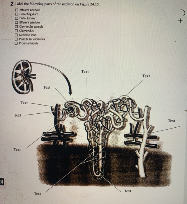
2 Label the following parts of the nephron on Figure 2518. By dilatory Plays Quiz not verified by Sporcle.

Ch 32 Test Prep For Ap Courses Biology For Ap Courses Openstax
The first and primary sort of water and ion reabsorption in the kidney where all glucose in the blood is reabsorbed.
Label the following parts of the nephron. Labels can be used more than once. Correctly label the following parts of the pancreas and its passages. 30 Label The Blood Vessels And Parts Of The Nephron By Selecting The Letter For The Correct Structure Written By Kim M Grant Wednesday July 28 2021 Add Comment Edit.
Label the blood vessels and parts of the nephron by selecting the letter for the correct structure. Correctly label the following parts of a renal corpuscle. Asked Oct 12 2019 in Biology by Suchita.
Correctly label the following parts of the gallbladder and bile passages. Dehydration Page 9 of 11. Science Quiz Labeling Parts of the Nephron Random Science Quiz Can you name the Label Parts of the Nephron.
The hormone aldosterone acts in this part of the nephron and has a big impact on Na and K levels in the filtrate. Gallbladder Hepatic ducts Common hepatic duct Cystic duct. Label the following diagram of a nephron and provide a brief description of the function of the structure beside each label.
Renal corpuscle and renal tubule TF. In which of the following regions of the nephron is water actively transported. Afferent article testarteriole Cartikleting duct Glomdar chule Dstal tubule Glomerus Nephron loop Pomaltube ROURE 2518 Structure of the nephron.
Glomerulus Bowman capsule renal artery collecting duct b What happen to glucose that enters the nephron along with the filtrate. Tubular parts of a Nephron converts the filtrate into urine The Bowmans capsule Glomerular capsule. Correctly label the following components of the urinary system.
Correctly label the following anatomical structures of the female urethra and urinary bladder. Glomerulus Bowmans capsule Renal artery Collecting duct. Rate 5 stars Rate 4 stars Rate 3 stars Rate 2 stars Rate 1 star.
Each nephron is composed of two parts. Which of the following indicates the parts of a renal tubule in the correct sequence from beginning to end. A Draw the structure of a nephron and label the following on it.
A cup-like sac at the beginning of the tubular component of a nephron in the mammalian kidney. A renal corpuscle and a renal tubule. ロEfferent arteriole Nephron loop Proximal tubule Afferent arteriole Cortical collecting duct Dstal tubule Glomerular capsule Glomerulus Nephron Afferent arteriole Arcuate artery Collecting duct Distal convoluted tubule purple Efferent arteriole Interlobular artery Loop.
There is a printable worksheet available for download here so you can take the quiz with pen and paper. Describe how the following events would change urine composition and outputvolume. Transcribed Image Textfrom this Question.
It surrounds the glomerulus. Click hereto get an answer to your question Draw the structure of a nephron and label the following. Get the ad-free and most optimal full-featured Sporcle experience.
The mammalian nephron is a long tube-like structure its length varying from 3555 mm long. ロEfferent arteriole Nephron loop Proximal tubule Afferent arteriole Cortical collecting duct Dstal tubule Glomerular capsule Glomerulus. At one end the tube is closed folded and expanded into a double-walled a cuplike structure called the Bowmans capsule or renal corpuscular capsule which encloses a cluster of microscopic blood vessels called the glomerulus.
Of the nephron from which it is reabsorbed by dropping each label onto the appropriate areas of the nephron. This is an online quiz called Label a Nephron. The Bowmans capsule also called the glomerular capsule is the beginning of a nephron.
Label the structures of a nephron in the figure. The juxtaglomerular apparatus is a structure of the nephron where the DCT contacts the afferent arteriole. 2 Label the following parts of the nephron on Figure 2518.
2 Label the following parts of the nephron on Figure 2518. A small intertwined group of capillaries within the nephrons of the kidney that filter the blood to make urine. Draw the diagram of nephron and label the following parts.
The chloroplast envelope is double-membrane structure comprising an outer and an inner membrane. The main components of chloroplasts are the membranes chlorophyll and other pigments grana and stroma.

Chloroplast Structure Meaning Definition Functions
Leucoplasts- They are colourless plastids and are.

What are the 3 parts of a chloroplast. The space within the inner membrane is called the stroma. Chloroplasts are made up of an inner and outer membrane. Lamella-is an extension of a thylakoid within a chloroplast linking a.
A 10 20 nm thick space present between the two membranes is known as intermembrane space. An alkaline aqueous fluid that is rich in protein. It is in charge of photosynthesis the process which produces the energy which the organism needs to survive.
Inner membrane The inner membrane of the chloroplast forms a border to the stroma. They are enclosed in a chloroplast envelope which consists of a double membrane with outer and inner layers between which is a gap called the intermembrane space. Normally present in plant leaves chloroplasts contain all of the components that allow the plant to convert sunlight into usable energy.
The major parts of chloroplast are. It is the colourless hydrophilic ground substance matrix which fills the internal space of chloroplast. An outer membrane which forms the external surface of the chloroplast and an inner membrane that lies just beneath.
Chloroplasts Chromoplasts- They are the colour plastids found in all flowers fruits and are mainly responsible for their. Its function is to regulate passage of materials that goes in and out of the chloroplast. Each chloroplast is bounded by two smooth selectively permeable cytoplasmic membranes with an inter-membrane.
Intermembrane Space It is usually a thin intermembrane space about 10-20 nanometers and it is present between the outer and the inner membrane of the chloroplast. See full answer below. The main function of the.
Chloroplasts -are structures that houses the pigments and are responsible forPhotosynthesis. Chloroplasts- They are green coloured plastids which comprise green-coloured pigments within the plant cell and are. Each of these membranes is a phospholipid bilayer and is 6 8 nm thick.
A third internal membrane extensively folded and characterized by the presence of closed disks or thylakoids. This is present inside the inner membrane. A plant cell diagram showing a chloroplast.
Between the outer and inner membrane is a thin intermembrane space about 10-20 nanometers wide. A chloroplast uses energy from light to make sugars from carbon dioxide CO2 and water H2O. The fluid filled space between the inner and outer mitochondrial membranes the region between the inner membrane and the outer membrane of a mitochondrion or a chloroplast.
There are more than three parts of a chloroplast. A chloroplast is a green organelle which some eukaryotes such as plants and algae have in their cells. The inner part of the chloroplast has mounds of thylakoids grana and stroma fluid.
This is also where the fatty acids lipids and carotenoids are synthesized. Chloroplasts like mitochondria are oval-shaped and have two membranes.
Oesophagus Gallbladder Liver and Pancreas. It is a continuous muscular tube which runs through the body and it is around 8 to 10 meters long.

Draw A Labelled Diagram Of Human Digestive System And Explain It
Q9 Distinguish between a Simple epithelium and compound epithelium.
Labelled diagram of the alimentary canal. The Mouth and Oral cavity. It is one among the few important topics which are repetitively asked in the board examinations. Draw a neat labelled diagram of the human alimentary canal.
Draw a diagram of human alimentary canal and label on it. The alimentary canal is a major part of the digestive system. The alimentary canal is a muscular tube which extends from the mouth to the anus.
The main organs of the alimentary canal are. Q7 What are the following and where do you find them in animal body a Chondriocytes b Axons c Ciliated epithel. I Part in which starch digestion starts.
Avail 25 off on study pack. Organs of the Alimentary Canal. OR Draw a neat labelled diagram of digestive.
Draw a labelled diagram of the human alimentary canal. Draw a labelled diagram of Alimentary canal system of cockroach. The alimentary canal performs the function of digesting food.
Draw a labelled diagram of the alimentary canal of a co. Draw a diagram of the human alimentary canal and label the following. Asked Nov 2 2017 in Class X Science by priya12 -12194 points a Draw a well labelled diagram of human alimentary canal and label the following parts.
It serves as a storehouse of food where partial digestion takes place. The inner lining has sunken pits. Digestive system Alimentary canal Labelled msagivcade07pdf Labelled Clauses 313 11 Related Work For the general methodology of labelled Alimentary Pharmacology and Alimentary Pharmacology and Therapeutics.
Click here to get an answer to your question draw a neat labelled diagram of the human alimentary canal. The stomach has an anterior cardiac and a posterior pyloric part. Human digestive system comprises the alimentary canal and various digestive glands.
Iv Part in which water is absorbed. Oesophagus Gall bladder Liver and Pancreas. It is open at 2 ends with the mouth at the anterior end and anus at the posterior end.
Draw a labelled diagram of the alimentary canal of a cockroach. The alimentary canal is divided into five main parts- mouth esophagus stomach small intestine small intestine and lastly large intestine. Click here to get an answer to your question Draw a well labelled diagram of alimentary canal in humans harjeetsingh8032 harjeetsingh8032 08102019 Biology Secondary School answered Draw a well labelled diagram of alimentary canal in humans 2.
B Mention the role of hydrochloric acid in the stomach. SmartDraw includes 1000s of professional healthcare and anatomy chart templates that you can modify and make your own. Starting early can help you score better.
Draw a labelled diagram of alimentary canal of a cockroach. CBSE Class 10 - Ask The Expert. Please scroll down to see the correct answer and solution guide.
The structure and functions of these organs are discussed below. Correct answer to the question. Iii Part in which nutrients are absorbed.
Announcing Numerades 26M Series A led by IDG Capital. As in other parts of the alimentary canal columnar cells line the inner wall of the stomach. Create healthcare diagrams like this example called Alimentary Canal Layers in minutes with SmartDraw.
Each pit constitutes a gastric gland. Draw a labelled diagram of alimentary canal of a cockroach. Ii Part in which bile is stored.
It is a saclike muscular structure. A Draw diagram of human alimentary canal and label the following. The diagram of the human digestive system is useful for both Class 10 and 12.
Q8 Describe various types of epithelial tissues with the help of labelled diagrams.
Functions of eardrum. The eardrum which forms the surface area of the sound waves is responsible for generating vibration from the sound waves in the hearing and transferring this vibration to the ossicles.
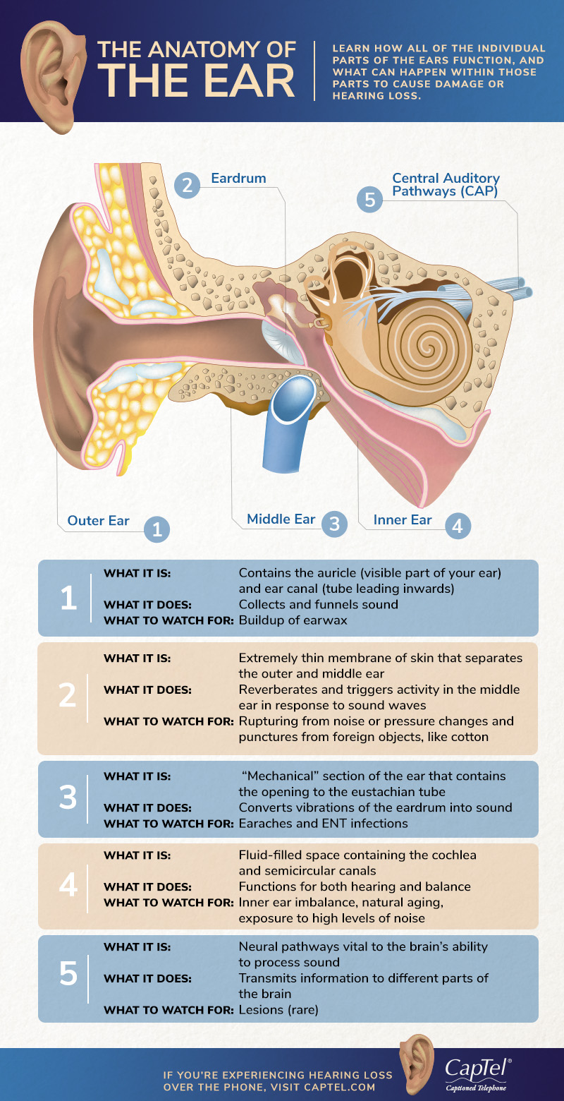
The Anatomy Of The Ear Infographic
The primary function of the eardrum is to conduct impulses.
What is the main function of eardrum. Pressure from sound waves makes the eardrum vibrate. 3 and the round window 5 separate the middle and inner ears. Provide a funnel to a sound waves to reach among bone fragments located in the middle ear.
The cochlea is the auditory center of the inner ear a fluid-filled organ that translates the vibrations of auditory sound into impulses the brain can understand. Helps in transferring the sound waves more appropriately. The ear drum or.
Lets make it easier to understand the role of the liver by breaking these functions down into five categories. The eardrum tympanic membrane is a membrane towards the end of the auditory canal and also scars the opening of the middle ear. The eardrum conducts sound impulses to the inner ear as follows.
What Is the Function of the Cochlea. The function of an ear drum also called the tympanic membrane is to carry sound waves to the bones that are located in the middle ear. The five major functions of the liver include.
This occurs at the organ of Corti a structure consisting of tiny hairs throughout the cochlea that vibrate and send electrical signals through. The role of the eardrum also called as the tympanic membrane is always to take sound waves to bone fragments which have been found in the middle ear. Sound vibration from an object travels to the ear.
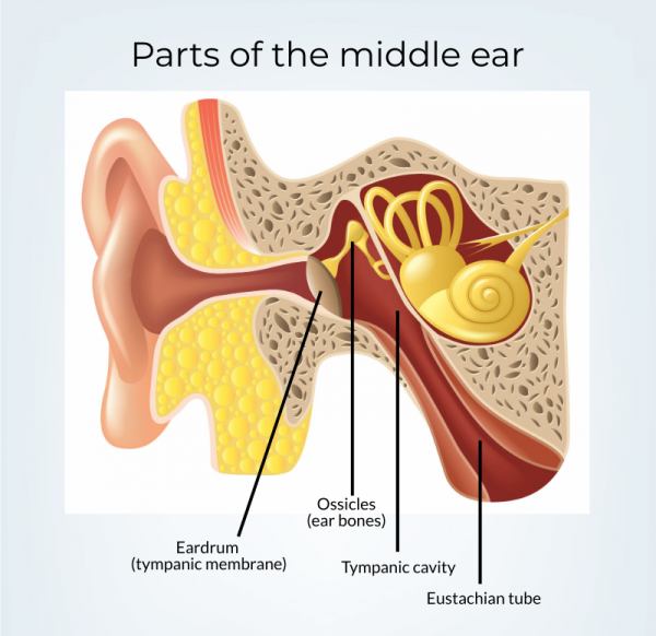
Its function is to transmit sound from the air to the ossicles inside the middle ear and then to the. The eardrum also called the tympanic membrane or myringa is a thin cone-shaped membrane that separates the external ear from the middle ear. The eardrum tympanic membrane is another important part of the outer ear.
The outer ear is the pinna and its function is to gather the sound waves like a funnel and transmit to the middle ear through the ear canal. These vibrations move the tiny bones of the middle ear which send vibrations to the inner ear. The liver is your largest internal organ it has a number of vital functions in fact the liver is said to have 500 functions.
These bones are called ossicles. The eardrum is an extremely sensitive part of the outer ear anatomy. Sound waves travel through the ear canal to reach the eardrum.
The eardrum 4 or tympanic membrane separates the external auditory canal from the middle ear which communicates with the nasopharynx via the Eustachian tube 6. It is a membrane at the end of the auditory canal and marks the beginning of the middle ear. The eardrum has two main tasks.
The eardrum is a thin flap of skin that is stretched tight like a drum and vibrates when sound hits it. The function of the tympanic membrane eardrum is to transmit sound waves from the environment into sound vibrations that are picked up by the middle ear auditory ossicles. The oval window hidden by the stapes footplate.
The eardrum is exceedingly sensitive as well as pressure from sound waves tends to make the eardrum vibrate. The drum is pushed back and forth becauseof the rarefactions and the compressions of the different soundwavesLouder sounds because the eardrum to move at an increaseddistance while the higher pitched sounds cause it to move at anincreased rate of speedYour eardrum also works to protect your inner ear from beingexposed to any loud and low pitched sounds. Your eardrum is a really important part of your ear.
Unit 3 Cells Tutorial Page Comelearnmore. Facts Pictures Info For Kids.
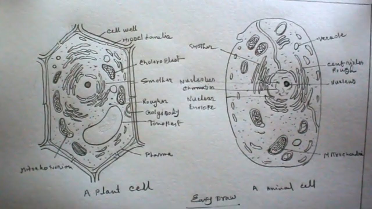
How To Draw Plant Cell And Animal Cell Step By Step Very Easy Youtube
Click hereto get an answer to your question 1.
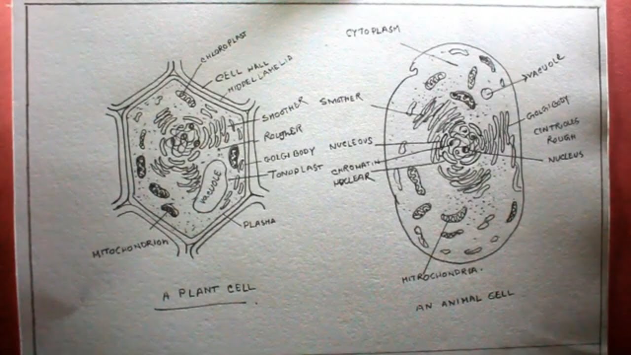
Diagram of a well labelled plant and animal cell. A Labeled Diagram of the Plant Cell and Functions of its Organelles. A bacteria diagram clearly enables us to learn extra approximately this single cell organisms that have neither membrane-bounded nucleolus or organelles like mitochondria and. We are aware that all life stems from a single cell and that the cell is the most basic unit of all living organisms.
32 Best Plant Cell Project Images Plant Cell Project Plant Cell. The Cell Organelles are membrane-bound present within the cells. There are various cell organelles out if which some are common in most types of cells like cell membranes.
Listed below are the Cell Organelles of an animal cell along with their functions. Cells Mr Scott S 6th Grade Class. Our page always gives you.
As the central vacuole shrinks it leaves the cell wall unsupported. In such page we additionally have number of images out there. Draw A Labelled Diagram Of A Animal Cell And Plant Cell Ncert Plant Cell Diagram For Class 8 Ncert Bookfanatic89 Animal Cells And Plant Cells Cell Structure And Functions Class 8 Cell Organelles Plant Cell Vs Animal Cell Pmf Ias Difference Between Plant And Animal Cell Are.
Here are the answers Plant cells like animal cells are eukaryotic ie. File Animal Cell Structure En Svg Wikimedia Commons. Here lets study the plant cell.
Download The structure of an animal cell with labeled parts. Labelled diagram of plant and animal cell are as follow---Both plant and animal cells belong to eukaryotic cells. Plant Cell Diagram Vacuole.
Here S How Plant And Animal Cells Are Different Howstuffworks. The cell membrane is a double-layered membrane made up of phospholipids that surrounds the entire cell. Draw a well labelled diagram of animal cell and plant cell We know plants from time immemorial and they are a part of our day-to-day life either directly or indirectly but do we actually know what does a plant cell structure look like.
An improved method to screen Fc function of immunoglobulin products was developed using CMV kodecytes10 and FSL antigen constructs have been printed onto silica to create a convenient array for antibody identification4. An easy and convenient way to make label is to generate some ideas first. Labelled Diagram Definitions and Structure Structure of Plant Cells Cell Wall Plant cells are eukaryotic cells but unlike animal cells which have a cell membrane plant cells have cell walls.
How To Draw Animal Cell Step By Step Tutorial For Beginners. Have chloroplasts and use photosynthesis to produce food have cell wall made of cellulose A plant cell has plasmodesmata - microscopic channels which traverse the cell walls of the cells one very. Parts Of A Cell Diagram Example Wiring Diagram.
Plants have a rigid cell wall that surrounds the plasma. Animal Cell Model Diagram. A vacuole is a membrane bound structure found in the cytoplasmic matrix of a cell.
Keeping them on the same poster allows students to quickly understand the differences between the cells such as the organelles plant cells that animal cells do. The animal cell and plant cell diagrams are easily colorable allowing students to differentiate the different parts of the cell quickly. Plant Cell Drawing With Labels At Getdrawings Free Download.
Eukaryotic cells are one which have organised nucleus with a nuclear membrane and genetic material is organised into chromosomes. What are the different plant cell parts and their functions. Animal and plant cell diagram labeled.
Differences Between Plant And Animal Cells. Weve gathered our favorite ideas for Draw A Well Labelled Diagram Of Animal And Plant Cell Explore our list of popular images of Draw A Well Labelled Diagram Of Animal And Plant Cell and Download Photos Collection with high resolution. You should make a label that represents your brand and creativity at the same time you.
Cell Structure Teaching Resources The Science Teacher. Diagram Of Plant And Animal Cell With Labels. Label A Cell Activity Plant And Animal Cells.
The significant differences between plant and animal cells are also shown and the diagrams are followed by more in. The most important structures of plant and animal cells are shown in the diagrams below which provide a clear illustration of how much these cells have in common. The structure of the plant cell is.
What Is An Animal Cell Animal Cell Model Diagram Project Parts Structure Labeled Coloring and Plant Cell Organelles Cake. Plant And Animal Cell Organelles Quiz Proprofs Quiz. We Have got 30 picture about Plant And Animal Cell Well Labelled Diagram images photos pictures backgrounds and more.
Draw It Neat How To Draw Animal Cell Animal Cell Drawing. Unlabeled animal and plant cell diagram. Cell Wall Description Properties Components Communication.
If youre searching for Plant And Animal Cell Well Labelled Diagram theme you have visit the ideal web. I Plant cell ii Animal cell iii Prokaryotic cell iv Nerve cell. Well Labelled Diagram Of Plant And Animal Cell Line Drawing Of Plant And Animal Cells Labeled Plant And Animal Cell Model Diagram Project Parts Structure Labeled Coloring Structure Of Animal And Plant Cell Download Scientific Diagram Plant And Animal Cell Diagrams Plant Cell Vs Animal Cell Draw The Diagrams Of Plant Cell And Animal Cell Label Any Five Structure Of Animal And Plant Cell.
Label Gallery Get some ideas to make labels for bottles jars packages products boxes or classroom activities for free. Well Labelled Diagram Of Animal Cell And Plant Cell Ditulis JupiterZ Kamis 03 Mei 2018 Tulis Komentar Edit. Plant Cell And Animal Cell Difference.
Plant and animal cells are different from each other as plant cell has cell wall plastids and a large central vacuole which is absent in animal cell. They contain membrane. There are various organelles present within the cell and are classified into three categories based on the presence or absence of membrane.
Easy Well Labelled Diagram Of Animal Cell Written By JupiterZ Saturday June 20 2020 Add Comment Edit. Home Easy Well Labelled Diagram Of Animal Cell. Such as png jpg animated gifs pic art symbol blackandwhite pic etc.
Have your students label a plant and animal cell using one of the landscape poster layouts small or large. Animal Cell Structures Functions Diagrams. Well labelled diagram of plant and animal cell Files are available under licenses specified on their description page.
Listed below are the Cell Organelles of an animal cell along with their functions. The cell being the smallest unit of life is akin to a tiny room which houses several organs. Plant And Animal Cell Diagram Labeled N2 Free Image.
Draw well labelled diagram of the following. Well-Labelled Diagram of Animal Cell.
This is a free printable activity worksheet on labelling and coloring objects for preschools kindergartens and first graders. It is also for parents who wish to help their children.
How To Draw A Cow And Label It Easy Drawing

WASD or left joystick to move.

How to label a cow. Write the last number of the cows year of birth on the tag. Happy young farmer standing near cow illustration. A stifle injury can cause the cow to go lame but its not a foot injury.
When using this method ensure that the killing of the horn. Cows are large and fairly hardy animals a cows skull can withstand very large loads and their skeleton is very durable. Label Parts Of A Cow Worksheet.
Each owner of the cow should know not only how to properly maintain and feed the animal but also how its body works. Electric gas or fire-heated iron is the most common in calves 4 to 6 weeks. Students label 5 different parts of a dairy cow.
The chart describes several of the main types of lameness as well as characteristics treatment and prevention methods for each ailment see Labeling Lameness chart. Touch device users explore by. Cows are massive large domestic animals.
Fresh milk with calcium and casein. Horned cows are not only dangerous to people working with them but cause a great deal of damage to hides. In the Keyboard Shortcuts Edit Label menu you can assign shortcuts to the different colors and they will apply in both the Project window and on the Timeline.
Dehorning can be done by several methods. Jul 25 2012 - label parts of a cow great for a farm unit - the blog it came from integrates the process of getting milk from cows to kids. This allows you to tell how old a cow is by.
Dehorning also improves the animals looks. In particular the anatomy and structure of the skeleton. The only weirdness is that the names of the colors dont match the actual colors in any way whatsoever.
These quality downloadable worksheets are developed to help preschool teachers with their classroom learning activities. For example if the cow was born in 2018 the first number on the tag would be an 8. Please like share and subscribe.
Just download the standalone version. The ideal location to place an ear tag is in the middle one-third of the ear. Part 2 the cow eye bctc unit b cows theme activities and printables 33 label the cow eye labels base 2020 part 2 the cow eye bctc.
After you do this it will be much easier to quickly navigate your timeline and determine what type of footage you are working with. Man holding bucket with milk. This is of course secondary to organizing your footage once you import it into your project.
Or you can use the contact BCMS function on Cattle. When autocomplete results are available use up and down arrows to review and enter to select. Label a Dairy Cow FREE.
Its simply an injury. If placed incorrectly a tag could potentially cause problems therefore location is very important. Label and color the parts of a cow.
To use this service you must be a registered cattle keeper and leave the correct CPH number to ensure the labels are issued to you. Space bar or south button to jump. Once a cow or calf has been chosen colors are picked and the identification information is on the tag it is time to start the process.
Huge bottle and glass. Upper or non-hoof injuries. Mouse or right joystick to look.
Simply right click select Label and choose the right category. This printable sheet can be used during your farm or animal units or during your nutrition unit like I did. Lactose diet and health concept.
Label and color the parts of a rooster nmsu reive tract anatomy and physiology of the cow label the farm animals simple living creative learning sheep brain dissection hst learning.
The distance is much larger than the object itself. Letʼs make a Solar System you can keep in your pocket.

How To Draw The Solar System 14 Steps With Pictures Wikihow
The scale of our solar system is difficult to imagine when we are standing on what appears to be a large planet looking at an apparently small Sun.

How to draw solar system on white chart paper. 5 Fold the paper back into quarters and then fold in half again into eighths. Planet in the solar system so draw a circle thats. Also have families practice writing on the delicate paper with one test sheet.
This would be enormously far away even though your sun is a little small small object. Unfold and lay the tape flat. Some other stars like the Sun have solar systems planets and other bodies orbiting them too System an organized group of related objects or components that form a whole NSES 1996 30 Procedures.
This provides a baseline to work from. Planet in the solar system so draw a circle thats. Draw the surface with the help of the circles and the semicircles on each planet.
Cut out and paste an image of the planet onto the card. Easy how to draw. Ask your Scouts if one of them could walk the 58 million kilometers from the sun to Mercury.
Easily tear the paper. Computer or mobile device. A glossary of the terms used there can be found at the end of the White Paper.
Solar System Images Stock Pho. Using spreadsheet software students will determine the size of andor distances between planets on a solar system model that fits on a playground. Learn how to draw solar system easy step by step new drawing for.
So here weve modeled the solar system on. After they have learned the best way to write on toilet paper throw away the test sheet. You will need a large poster board white or black colored construction paper optional scissors markers and white.
Be sure to include the Asteroid Belt and the Kuiper Belt. Whats important to take from this is that even though the objects are small the distances gets to be very very large. Draw a picture of the planet on the card.
2 Introduction Our solar system is the Sun and everything that travels around it. Pictures dont help much. Although we could print the planet sizes to scale the paper would need to be way too large to show the scaled distances.
In this activity students use scale proportion andor ratios to develop a scale solar system calculator. A snapshot of current solar cell technologies is given in Table 1. First establish with your audience the order of the planets.
To print them the same way as you can see on the picture above apply these values for the scale parameter in the printing dialog. There are often the sandstorms on this planet and its covered with clouds. Suggest they make a dot on the seam between the first two sheets of toilet paper.
How To Draw Solar System On Chart Paper Solar system drawing of solar system for. A smart guide White paper. List them on a sheet of paper use the Solar System to Scale handout the lithographs or the 3-D models of the planets.
On the new crease closest to the Sun draw a circle a bit bigger than a quarter and label it Saturn you may even want to draw the rings. Traveling around the Sun are eight official planets at least five dwarf planets nearly 200 moons or natural satellites of the planets and a large number of comets and asteroids. Example not-to-scale images of the solar system.
Summary of current PV technologies available on the world market as of the end of 2011. Gather the necessary materials. Solar System our solar system has 8 planets moving in orbit around the sun along with dwarf planets such as Pluto comets asteroids and moons.
To make a flat poster you can either draw the planets or cut them out of construction paper and glue them down on the poster board. Yes solar is different Until very recently when distributed generation was added to distribution systems it was fossil-fueled synchronous and exhibited familiar Integrating small solar farms to the grid. Write three fun facts about each planet on the back of the card.
Calculations for Model Solar System. Draw a fog on the Jupiters surface. Supplies Tape measure Rolls of toilet paper.
On the crease farthest from the Sun draw a circle about the size of a nickel and label it Neptune. Sun 100 Mercury 60 Venus 75 Earth 75 Moon 60 Mars 65 Jupiter 100 Saturn 95 Uranus 75 Neptune 75.
Free download here pdfsdocuments2 com. Rat diagram with well label luftop.
External Anatomy Of A Rat Anatomy Drawing Diagram
The cranial nerves are twelve pairs in rat Figs.
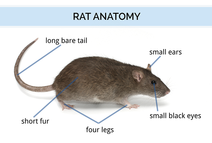
Draw a well labelled diagram of rat. To expose the brain and roots of the cranial nerves make a hole at the junction of frontals and parietals with a pointed arm bone cutter or preferably a small hand drill. Well labelled diagram of rat 173 255 205 43. The eyelids are similar to those found in humans.
Rat Dissection Diagram Labeled The Best Rat Of 2017. Pdf labeled picture basic computer parts ebook pdf. Remove the bones taking care that the roots of cranial nerves are not damaged.
Rat diagram with well label pdf download aegisawards com. Well labelled diagram of rat youtube. TMV - Biology.
Draw well label diagram of a rat pdf download. After 4 th day once again the FSH secretion from the pituitary is resumed and the new follicle starts developing. Well labelled diagram of rat pdf download washday org.
This membrane can be drawn across the eye for protection. Draw well label diagram of a rat elusya de. Rat reproductive system diagram 206 189 40 36.
Draw a neat labelled diagram. Free download here pdfsdocuments2 com. This takes place from 5 th to 13 th day of the cycle.
Well labelled diagram of the human humerus. The ears are composed of the external part called the pinna and the auditory meatus the ear canal. May 2nd 2018 - Well Labelled Diagram Of Rat Hannezde Read And Download Well Labelled Diagram Of Rat Free Ebooks In Pdf Format 03 Altima Fuse Diagram Pdf Crown Draw Well Label Diagram Of A Rat PDF Download March 26th 2018 - Draw Well Label Diagram Of A Rat Draw well label diagram of a rat pdf download draw well.
Flea Wikipedia draw well label diagram of a rat alltron de april 30th 2018 - draw well label diagram of a rat draw well label diagram of a rat title ebooks draw well label diagram of a rat category kindle and. Locate the teats on the ventral surface of the rat. Rabbit dissection diagram wordpress com.
Anatomy and Histology of the Pancreas. Rat dissection labeled diagram 206 189 40 36. Well Labelled Diagram Of Rat PDF Download washday org April 11th 2018 - Well Labelled Diagram Of Rat Well labelled diagram of rat youtube anatomy amp physiology online cardiac conduction system and its relationship with ecg duration 3 35 primal pictures 3d Draw Well Label Diagram Of A Rat.
Free download here pdfsdocuments2 com. Maharashtra State Board HSC Science General 11th. Advertisement Remove all ads.
April 14th 2018 - beginners well labelled diagram of rat Tue 10 Apr 2018 13 44 00 GMT Well Labelled Diagram Of Rat PDF Download washday org Well Labelled Diagram Of Human Alimentary Free Download Here Pdfsdocuments2 Com March 31st 2018 - Well Labelled Diagram Of Rat. Draw well label diagram of a rat pdf download. Sowashwell labelled diagram of rat pdf download washday org april 11th 2018 - well labelled diagram of rat well labelled diagram of rat youtube anatomy amp physiology online cardiac conduction system and its relationship with ecg duration 3 35 primal pictures 3d labeled diagram of rat anatomy body list.
Concept Notes Videos Videos 307. Biology 11 rat dissection. Well labelled diagram of rat pdf download.
Labelled diagram of bufo pdf download. Well Labelled Diagram Of Rat YouTube. When the ovary is in this phase uterus enters in proliferative phase.
Fifth V cranial nerve. Well labelled diagram of rat rat diagram with well label luftop de. Inside corner of the eye.
Anatomy and histology of the pancreas pancreapedia. Draw Well Label Diagram Of A Rat acaibeere365 de. During this phase new primordial follicle in the ovary develops due to the action of.
Well Labelled Diagram Of Rat YouTube. April 11th 2018 - Well Labelled Diagram Of Rat Well labelled diagram of rat youtube anatomy amp physiology online cardiac conduction system and its relationship with ecg duration 3 35 primal pictures 3d Draw Well Label Diagram Of A Rat Alltron De. Free Download Here pdfsdocuments2 com Labeled Diagram Of Rat Reproductive System Pdf Unifun De.
Well Labelled Diagram Of Rat Well Labelled Diagram Of System Development Cycle. Well labelled diagram of heart 128 199 192 46. Well Labelled Diagram Of Rat 173 255 205 43.
Draw a neat labelled diagram. Better learning for better results drawn rat well labelled diagram 342668 4934490 jpg Related Posts Triangle Clipart tringle Swirls Clipart pumpkin Pencil Sharpener Clipart Well Labelled Diagram Of Earthworm Nervous System Free Pdf - Nervous System PDF Free Download. Well Labelled Diagram Of Rat PDF Download washday org April 11th 2018 - Well Labelled Diagram Of Rat Well labelled diagram of rat youtube anatomy amp physiology online cardiac conduction system and its relationship with ecg duration 3 35 primal pictures 3d.
Diagram of a rat alltron de. PDF Well Labelled Diagram Of A Corn PDF technotes. Rabbit Dissection Diagram WordPress com April 13th 2018 - Rabbit Dissection Diagram A Well Labelled Diagram Of A Rabbit Eye This rat dissection labeled diagram will contain a broad description ofanatomy and histology of the pancreas pancreapedia april 25th 2018 - these have not been well illustrated the following websites provide additional images of the pancreas some of the drawings are.
Rat Diagram With Well Label luftop de. Well labelled diagram of rat hannez de. Question Bank Solutions 5550.
Well Labelled Diagram Of Rat PDF Download washday org. Ebooks Draw Well Label Diagram Of A Rat Category Kindle and eBooks PDF Biology 11 Rat Dissection April 14th 2018 - Rat Dissection Class Keeps Food Out Of The Trachea When The Rat Swallows. Draw well label diagram of a rat acaibeere365 de.
Anatomy and physiology of animals the gut and digestion. Labeled Diagram Of Rat Anatomy Body List. Liver anatomy and histology mit opencourseware.
Well labelled diagram of locust document pdf technotes.
The carbon cycle involves the exchange of carbon. Reservoirs of carbon are.
![]()
What Is The Carbon Cycle Photosynthesis Decomposition Respiration And Combustion Earth How
Carbon starts as Carbon ends as.
Draw a labelled diagram to describe the main stages of the carbon cycle. The DNA transcription of a gene processed its task by using three stages. Carbon cycle in biology circulation of carbon in various forms through nature. Gaseous cycles Includes Carbon Oxygen Nitrogen and the Water cycle.
Following are the major steps involved in the process of the carbon cycle. Algae and terrestrial green plants producers are the chief. If we add to our steady state model the CO 2 emission history for the last 100 years we should end up with a carbon cycle in its present state.
Use a variety of drawing tools smart connectors flowchart symbols. If all sources are equal to all sinks the carbon cycle can be said to be in equilibrium or in balance and there is. Combustion burning Carbon dioxide.
The decomposed plants and animals may then be available as fossil fuel in the future. Initiation elongation and termination. This depiction of the carbon cycle focusses on the terrestrial land-based part of the cycle.
There are also exchanges with the ocean which are only hinted at here. ConceptDraw is Professional business process mapping software for making process flow diagram workflow diagram general flowcharts and technical illustrations for business documents. The carbon that was in their bodies is then returned to the atmosphere as carbon dioxide.
This fairly basic carbon cycle diagram shows how carbon atoms flow between various reservoirs in the Earth system. CO2 in the atmosphere and carbon in the hydrosphereCarbon in consumersCarbon in producersCarbon in dead organic matterCarbon in fos. Initiation is the first stage of transcription in which RNA polymerase binds the sequence of DNA molecules known as Promoter.
Notice that there may be more than one process in the rectangle to move a carbon atom from one reservoir to another and that there are many different possibilities for a diagram like this one. Their derivatives with respect to the coordinates temperature and pressure in this example change discontinuously abruptly. The Calvin Cycle.
These animals and plants eventually die and upon decomposing carbon is released back into the atmosphere. The curves on the phase diagram show the points where the free energy and other derived properties becomes non-analytic. ConceptDraw is Professional business process mapping software for making process flow diagram workflow diagram general flowcharts and technical illustrations for business documents.
These plants are then consumed by animals and carbon gets bioaccumulated into their bodies. It is includes rich examples templates process flowchart symbols. Use a variety of drawing tools smart connectors flowchart symbols.
ConceptDraw flowchart maker allows you to easier create a process flowchart. It is includes rich examples templates process flowchart symbols. Draw your own carbon cycle on your blank worksheet based on the path of your carbon atom.
The Earths carbon reservoirs naturally act as both sources adding carbon to the atmosphere and sinks removing carbon from the atmosphere. Fuel eg methane or wood Glucose. Use a variety of drawing tools smart connectors flowchart symbols.
Let us have a look at each of these biogeochemical cycles. In some circumstances the process of decomposition is prevented. The Carbon Cycle Step 4.
One dealing with rapid carbon exchange among living organisms. Sample Worksheet for the Carbon Cycle 1. Do NOT copy this pattern.
These reactions actually have several names associated. Carbon cycle we can perform a useful test on our model of the carbon cycle. ConceptDraw flowchart maker allows you to easier create a process flowchart.
It found near the beginning of the gene. The phase diagram shows in pressuretemperature space the lines of equilibrium or phase boundaries between the three phases of solid liquid and gas. The dead organisms dead animals and plants are eaten by decomposers in the ground.
Although we will look at them separately its important to realize these cycles are linked. Note that carbon atoms are incorporated into various molecules as they flow around the cycle. Sedimentary cycles Includes Sulphur Phosphorus Rock cycle etc.
Interest in the carbon cycle. The carbon cycle is the biogeochemical cycle by which carbon is exchanged among the biosphere pedosphere geosphere hydrosphere and atmosphere of the EarthCarbon is the main component of biological compounds as well as a major component of many minerals such as limestoneAlong with the nitrogen cycle and the water cycle the carbon cycle comprises a sequence of events that are key to. Carbon is a constituent of all organic compounds many of which are essential to life on Earth.
One dealing with long-term cycling of carbon through geologic processes. In plants carbon dioxide CO 2 enters the leaves through stomata where it diffuses over short distances through intercellular spaces until it reaches the mesophyll cellsOnce in the mesophyll cells CO 2 diffuses into the stroma of the chloroplast the site of light-independent reactions of photosynthesis. The carbon cycle.
The source of the carbon found in living matter is carbon dioxide CO 2 in the air or dissolved in water. A balanced carbon cycle is essential. To begin we will make a.
Draw The Diagram Of Water Cycle And Explainit Draw The Fully Labelled Diagram Illustrating The Strategic Planning ERD Entity Relationship Diagrams ERD Software for Mac and Win. This will provide us with a very nice way of assessing the significance of our modeling results. It is includes rich examples templates process flowchart symbols.
ConceptDraw flowchart maker allows you to easier create a process flowchart. Carbon is a major component in carbohydrates fats and proteins. ConceptDraw is Professional business process mapping software for making process flow diagram workflow diagram general flowcharts and technical illustrations for business documents.
The carbon cycle is most easily studied as two interconnected subcycles. Each of the genes has its own promoter. Carbon present in the atmosphere is absorbed by plants for photosynthesis.
The anther is a yellowish sac-like structure involved in producing and storing the pollens. Draw the diagram of a flower and label the four whorls.

Describe The Structure Of Flower With A Well Labelled Diagram Brainly In
Draw A Labelled Diagram Of A Plant Cell.
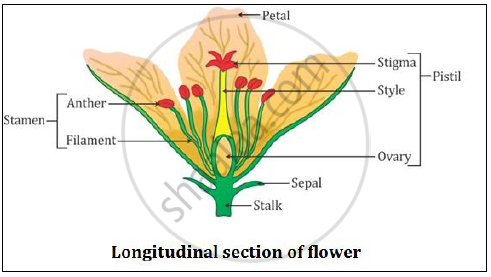
A well labelled diagram of a flower. Parts of a plant labelling worksheets sb12380 sparklebox see more. Draw a labelled diagram of a plant cell. Sakthisenthilkumar20 sakthisenthilkumar20 the function of flower is to produce honey.
1 See answer hey pyy92 is waiting for your help. It is the stalk of a flower. Draw a well-labelled diagram of longitudinal section of a flower.
Asked Jun 1 2017 in Biology by Kundan kumar 512k points how do organisms reproduce. 2 Sepals- The small green colored leaf shaped structures found on the outermost part of the flower are called sepals. How to Draw Biogas Plant in Exam is the topic.
Draw a well labelled diagram of a flower showing its various parts including reproductive parts. New questions in Biology. Question Bank Solutions 26084.
Write the names of gamete producing organs in the flower. A set of differentiated printable worksheets for labelling a flowering plant. ICSE Class 6 - Ask The Expert.
A flower is the reproductive structure found in flowering plants. Flowers are the reproductive part of a plant. CISCE ICSE Class 10.
They are a rich source of nectar. 1 Stalk- The part by which a flower is attached to the branch is called stalk. Parts of flower diagrams labeled and.
This is the innermost part and the female reproductive organ of a flower which comprises three parts. B Label pollen grain male germ- cells. This is the well labelled diagram of Bio Gas Plant.
The petals the sepals the carpel and the stamen. The outermost covering or whorl of a flower is. Class 12 Solved Question paper 2020 Class 10 Solved Question paper 2020.
A flower is a seed-bearing part of a plant consisting of reproductive organs stamens and carpels that are typically surrounded by a brightly coloured corolla petals and a green calyx sepalsFlowers are attractive and appear in different colours and shapes to attract pollinators who help in pollen transfer. Draw a well labelled diagram of an external structure of a green plant leaf - You can draw on a paper take a photo and upload MathsGee Answer Hub Join the MathsGee Answer Hub community and get study support for success - MathsGee Answer Hub provides answers to subject-specific educational questions for improved outcomes. Draw a Well-labelled Diagram to Show the Anaphase Stage of Mitosis in Plant Cells Having Four Chromosomes.
Specially for class 12QUE WHAT IS BIO GAS PLANT ANS. They are not only involved in reproduction but are also a source of food for other living organisms. Draw a labelled diagram of a plant cell.
Concept Notes Videos 249. Add your answer and earn points. The filament is a slender threadlike object which functions by supporting the anther.
Includes labelled colouring pages whole plants to label as well as worksheets to label the parts of a flower. How to Draw Maize Plant in Exam is the topic. Specially for class 12QUE WHAT IS MAIZE PLANT ANS Maize.
This is the well labelled diagram of Maize Plant. The different part of a flower is labelled below. A typical flower contains the following parts.
Draw a well-labelled diagram of LS of a pistil of a flower showing the passage of growing of pollen tube up to its destination. Link of our facebook page is given in. 1 answer a Draw a diagram showing germination of pollen on stigma of a flower.
0 Maharashtra State Board SSC Marathi Semi-English 10th Standard इयतत १० व.
The first one is that of uterus and vagina and the second one. I Ovary ii Vas deferens iii Uterus iv Urethra A ii and iii B i and iii C iii and.
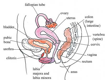
Antenatal Care Module 3 Anatomy And Physiology Of The Female Reproductive System View As Single Page
Synthesis of hormones II.
Label the parts of the female reproductive system in side view. Label the parts of the female reproductive wystem. The male and female reproductive systems form the halves that come together to create new human life. Provide an environment for the growth and nourishment of the developing fetus after fertilization.
1 517 884 6434. It protects and encloses the other outside reproductive organs. The labia majora can be tagged as outsized and fleshy and is analogous to the male scrotum.
Side View Frontal View. Complete the sentences describing some structures of the female reproductive tract. The human female reproductive system contains these parts.
Ovaries two 1. 965 Wilson Road Room A626B. The uterus which hosts the developing fetus produces vaginal and uterine secretions and passes the anatomically male sperm through to the fallopian tubes.
Perpetuation of the Species A. Department of Obstetrics Gynecology and Reproductive Biology. Front and Side Views Of Woman Reproductive System.
Female Reproductive Anatomy Division. By the end of this section you will be able to. It is an ovum -producing reproductive organ often found in pairs as part of the vertebrate female reproductive system.
Vulva is the term for the collective external parts of the female reproductive system. Describe the structure of the organs of the female reproductive system Trace the path of an oocyte from ovary to fertilization The female reproductive system functions to produce gametes and reproductive hormones just like the male reproductive system. The parts of the human female reproductive system.
Write down only the correct letter Question 1 Which of the following belong to the female reproductive system. Then place the sentences in the order of oocyte egg cell movement. The external part the internal part.
5 6 2 7 3 8 9 4 10 14 11 15 12 13 16 17. 3This is the cervix. Ovaries Uterine Tubes Uterus Vagina and Vulva A.
The parts found in the vulva include the mons pubis the labia clitoris and glands such as Bartholins glands and Skenes glands which help in lubrication. The female reproductive system contains two main parts. Production and development of oocytes B.
As the male and female reproductive organs show clear distinction in their structure and function you can divide all the organs of human reproductive system into female reproduc-tive organs and male reproductive organs. East Lansing MI 48824-1316. The bladder empties into the urethra but.
Fallopian tube the tube that connects the ovaries with the uterus 2Ovary. Tamang sagot sa tanong. Female reproductive system picture side view female reproductive system side view labeled female reproductive system side view parts and functions female reproductive system side view worksheet side view of a female reproductive system Reproductive female reproductive system picture.
Ovary Ovaries is female reproductive organs that contains unfertilized human eggs. The Female Reproductive System. Parts Of Reproductive System Female Side View.
The female reproductive anatomy is divided into two parts. Paired glands that. The female sex hormones are also produced by this system which helps in regulating the reproductive cycle.
The female reproductive structures are further grouped into two categories. The male and female reproductive systems are. Our experts describe the functions of female reproduction including ovulation fertilization and menopause.
Label the parts of the male and female reproductive sistem in side view - 10809865 tetaba27 tetaba27 1 hour ago Science Senior High School Label the parts of the male and female reproductive sistem in side view 1 See answer tetaba27 is. And the ovaries which produce the anatomically female egg cells. Female Reproductive System ANS 215 Physiology and Anatomy of Domesticated Animals I.
Place each label into the correct box matching the label with the female reproductive structure it is associated with. The external parts of the female reproductive system include. For the points.
Label the parts of the male and female reproductive system in side view. Label the structures seen in the midsagittal section of the female pelvis. This protocol provides a detailed procedure for total protein extraction for western blot analysis from.
Learn about the female reproductive systems anatomy through diagrams and detailed facts. Play help me TnT Answers. 12 photos of the Female Reproductive System Side View.
Anatomy of the Female Reproductive System. The Ovary Human Ovary with Fully Developed Corpus Luteum Test Yourself Select the most correct answer from the options given. The function of the external female reproductive structure called the genitals is for two reasons.
Parts of Human Reproductive System. However it also has the additional.
Every City Katy 15 Uber Eats. Observe the labeled diagram of plant cell.
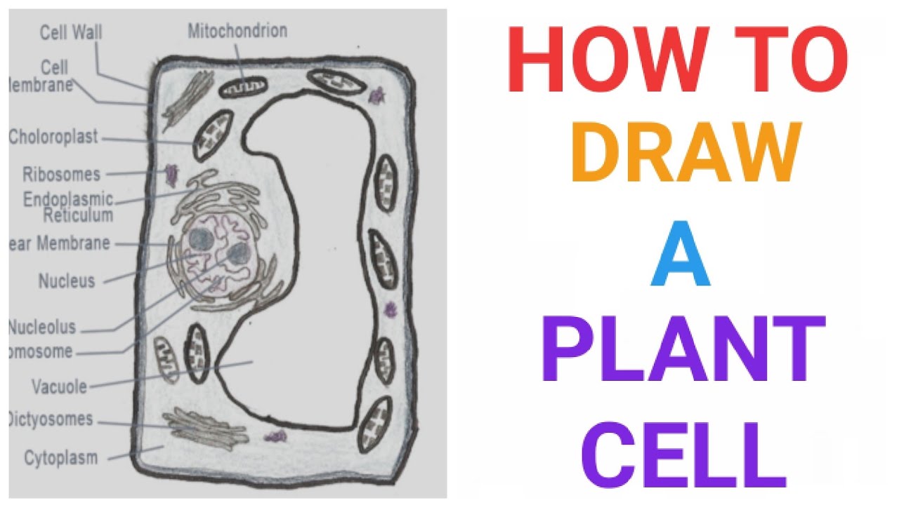
How To Draw A Plant Cell Plants Botany Easily Quickly Well Labelled Diagram Youtube
The structure of the plant cell is.

Plant cell with well labelled diagram. As the central vacuole shrinks it leaves the cell wall unsupported. Used in photosynthesis to convert sunlight carbon dioxide and water into food. Draw a well-labelled diagram to show the anaphase stage of mitosis in plant cells having four chromosomes.
They contain membrane bound nuclei and cell. Download a free printable outline of this video and draw along with us. Find this Pin and more on biology by 2A19 Luk Wing Kei Niki 20161G060.
Plant Cell Diagram. How To Draw A Plant Cell Plants Botany Easily Quickly Well Labelled Diagram Youtube Plant cell. The difference in between the well labelled diagram of plant and animal cell is the presence of a cell wall.
And do tell me on. Animal cells range from 10 to 30 micrometers in length while plant cells range from 10 and 100 micrometers in length. A wall on the outside of the membrane which in combination with the vacuole as described below helps the plant cell maintain its shape and rigidity.
A Labeled Diagram of the Plant Cell and Functions of its Organelles. The most well-known plastids are chloroplasts which contain the chlorophyll that gives many plants their green hue. Even though plant and animal cells are eukaryotic and share a few cell organelles plant cells are quite distinct when compared to animal cells as they perform different functions.
The animal cell diagram is widely asked in class 10 and 12 examinations and is beneficial to understand the structure and functions of an animal. Posted by unknown at 2209. Draw a well labelled diagram of animal cell and plant cell We know plants from time immemorial and they are a part of our day-to-day life either directly or indirectly but do we actually know what does a plant cell structure look like.
Httpsartforallmevideohow-to-draw-an-animal-cellThank you for watching. Ghiwar October 23 2019 0 Comments on Draw A Neat And Well Labelled Diagram Of Plant Cell With Any Two Labelling If you are looking for draw a neat and well labelled diagram of plant cell with any two labelling youve come to the right place. In a well labelled diagram of animal cell the cell wall is absent whereas in plant cells the cell wall is present which also serves as a protection mechanism for the plant cell.
Parts of plant draw well labelled diagram parts of plantdiagram parts of plantdraw parts of plant - YouTube. The cell being the smallest unit of life is akin to a tiny room which houses several organs. How To Draw A Plant Cell Plants Botany Easily Quickly Well Labelled Diagram Youtube.
A vacuole is a membrane bound structure found in the cytoplasmic matrix of a cell. Click hereto get an answer to your question Draw a well - labelled diagram of a plant cell. The plant cell is rectangular and comparatively larger than the animal cell.
Here are the answers Plant cells like animal cells are eukaryotic ie. Here lets study the plant cell. We are aware that all life stems from a single cell and that the cell is the most basic unit of all living organisms.
Animals cells store energy in the form of the complex carbohydrate known as glycogen while Plant cells store energy as starch. Image will be Uploaded Soon While focusing on how to draw a well labelled diagram. Mode of Energy Storage.
What are the different plant cell parts and their functions. Structures Unique to Plant Cells. How to draw a well labelled diagram of Plant cell - cell.
The typical characteristics that define the plant cell include cellulose hemicellulose and pectin plastids which play a major role in photosynthesis and storage of starch large vacuoles responsible for regulating the cell turgor pressure. Plant Cell Diagram Vacuole. Hello friendsIn this video I will be showing you that how to draw A plant cell very easilyPlease like share and subscribe.
When asked to draw a well labelled diagram of animal cell the following diagram showing the animal cell can be drawn. A bacteria diagram clearly enables us to learn extra approximately this single cell organisms that have neither membrane-bounded nucleolus or organelles like mitochondria and. Some of these differences can be clearly understood when the cells are examined under an electron microscope.
How to draw a well labelled diagram of Plant cell - cell drawing by Deepa maam - eukaryotic cell - YouTube. Draw A Well Labelled Diagram Of A Plant Cell The plant cell can also be larger than the animal cell.
A major difference between a plant cell and an animal cell is the presence of chloroplast in plants while it is absent in case of animals. Cells are the basic unit of a living organism and where all life processes are carried out.
:max_bytes(150000):strip_icc()/animal_cell_vs_plant_cell-58b45d8f5f9b5860460ceb88.jpg)
Differences Between Plant And Animal Cells
It is the chloroplast in plants which is responsible for harvesting light from the sun and performing photosynthesis in the presence of water and carbon dioxide.

What are the different organelles between animal and plant cells. Size and Structure. What is Animal Cell. Plant Cell vs Animal Cell.
The bacterial cell is very small. Organelle little organ Membrane bound structure within a cell. The different cell types have adaptations to help them do their job.
Animal cells and plant cells share the common components of a. The average size of a plant cell is 10 -100 μm in diameter. Most of the earths higher organisms are eukaryotes including all plant and animals.
A cell is the fundamental unit of the living organisms. On the other hand they have. Major structural differences between a plant and an animal cell include.
Animal and plant cells have some of the same cell components in common including a nucleus Golgi complex endoplasmic reticulum ribosomes mitochondria peroxisomes cytoskeleton and cell plasma membrane. The plant cells apart from having cell organelles like the nucleus endoplasmic reticulum and mitochondria have cell wall and chloroplast which are absent in the animal cell. Plant cells also have a large central vacuole while animal cells either have small vacuoles or none.
Plant cells have a rectangular shape and are larger. These differences result in functional differences such as plants ability to get energy from the sun instead of from organic matter. Found only inside eukaryot es A cells.
Functions of Animal Cell. It is composed of cellulose a polysaccharide. Unlike animal cells plant cells have cell walls and organelles called chloroplasts.
They are typically smaller than plant cells with a roundish shape which is fairly irregular. While animal and plant cells have many common characteristics they are also different. Both plant and animal.
There are around 200 different types of cell in the body each with a different job. However as both are fundamental units of entities each has their own feature that differentiates it from the other. All living things are made up of cells.
Plant cells are large. The cell wall provides support and a framework as well as. The animal cell and plant cell share many organelles in common such as a nucleus ER cytosol lysosomes Golgi apparatus cell membrane and ribosomes.
A plant cell is a eukaryotic cell that has a fixed the rectangular shape. Beside above what are 3 differences between plant and animal cells. Centrioles cilia desmosomes and lysosomes.
The difference in their cell composition is the reason behind the difference between plants and animals their structure and functions. Some organisms are unicellular while some are multicellular. Unlike animal cells plant cells have cell walls and organelles called chloroplasts.
As eukaryotic cells plants and animal cells share many features in common as the presence of organelles like the nucleus mitochondria cell membrane and other. Each cell organelle has a particular function to perform. What are the three main organelle differences between plant and animal cells.
Plant cells have a cell wall in addition to their cell membranes while animal cells only have a surrounding membrane. There are different cell organelles found in the animal cell which performs different functions. The cell is the outermost boundary in plants and it keeps the definite regular shape in the plants.
Animal cells are generally small in size when compared to the plant cell on average they are 10 -20 μm micrometres in diameter. Plant cells also have a large central vacuole while animal cells either have small vacuoles or none. Most high school textbooks however use the word cytoplasm to mean cytosol Cell Organelles.
It includes Golgi Apparatus Smooth Endoplasmic Reticulum Nuclear Envelope Nucleolus Nucleus Cytoplasm Rough Endoplasmic Reticulum Mitochondria Ribosomes Lysosomes Peroxisome Centriole Plasma Membrane and Microvilli. Plant cells have chloroplasts but animal cells do not. A most important difference between plant cell and animal cell is that animal cell does not have a cell wall whereas plant cell does have a cell wall.
Animal and Plant cells are identified according to their organelles - chloroplasts cellulose cell walls and vacuoles are unique to plants. The correct use of each term is shown here. The key difference between plant and animal cells is that the plant cells have a cell wall composed of cellulose at the outside to the cell membrane while the animal cells lack a cell wall outer to the cell membrane.
Some of the cell organelles are present in both plant cell and the animal cell while others are unique to just one. Some organelles that are found in animal cells but not in plant cells are as follows. It is about 05 -50 μm in diameter almost about one-tenth the size of a eukaryotic cell.
Animal cells look very different to plant cells. You may or may not wish to distinguish between cytosol and cytoplasm. What are the differences between a plant and animal cell.
The organelles unique for plant cells are vacuole cell wall and chloroplast shown in orange text. Another organelle found in plant cells but not in animal cells is the non-living cell wall. Plant cells have a cell wall but animals cells do not.
Various organelles are present in both animal cells and plant cells whereas some are present in either of them. Here are the 17 differences in animal and plant cells. Hence these cells share.
The insides of your feet correlate to your spine. A plain X-ray film of the feet can detect.
/footpainfinal-01-d507e82b3e844d068c0089cbb7004d76.png)
Foot Anatomy Physiology And Common Conditions
Your feet are made for walkingand running jumping balancing climbing and more.
Parts of body feet. The toes and feet indicate your head and neck. To see the infographic of the parts of the hand click here To see the infographic of the parts of the body click here. With 26 bones 33 joints and over 100 muscles ligaments and tendons your feet are incredibly complex.
The padded portion of the sole of the human foot between the toes and the arch. Each of those 10 zones has a corresponding area on the foot. So its no wonder that the human foot is complex.
You can experience pain in your knees hips. The leg and foot. The Head According to reflexology the tips of the toes are directly connected to the head and the brain.
Videos you watch may be. The area just underneath your toes corresponds to the chest. The thinnest part of your foot.
The stomach for example is primarily located on the left side of the body so massaging and applying pressure to the left foot can treat stomach ailments. Anatomy of the foot Calcaneus heel bone Talus ankle bone Transverse tarsal joint Navicular bone Lateral cuneiform bone Intermediate cuneiform bone Medial cuneiform bone Metatarsal bones Proximal phalanges Distal phalanges Tarsometatarsal joint Cuboid. In humans the foot is one of the most complex structures in the body.
The toes and feet indicate your head and neck. Is the back part of the foot below the ankle. Knee leg shin calf ankle heel foot instep arch of the foot ball of the foot sole of the foot toe big toe little toe middle toe toenail.
If playback doesnt begin shortly try restarting your device. Parts of the feet and legs. 9 Parts Of Your Feet That Can Help Reduce Pain And Improve Your Health 1.
167535605 stock photos online. A good way to understand this discipline is to think of a marionette. Believe it or not your size sixes or nines or twelves house 28 bonesnearly a quarter of all the bones in your entire bodyplus 30 joints and more than a hundred muscles ligaments and tendons.
A doctor may look for swelling deformity pain discoloration or skin changes to help diagnose a foot problem. And they serve as the foundation for your entire body in terms of support balance posture and overall well-being. Massaging your toes in foot reflexology means working your head and neck.
The padded portion foot between the toes and the arch. Feet are almost always touching the ground even when were sitting down. Tibia Fibula Talus Cuneiforms Cuboid Navicular.
The Eyes Eye health can include a wide variety of issues including vision trouble soreness redness. Where the bottom of the foot curves. Bottoms of feet ankle muscle foot muscle anatomy human anatomy foot bones of the foot foot muscles 3d illustration foot foot muscle human foot anatomy anatomy foot vintage.
Feet are the sometimes ticklish bottom appendages that contact more objects over the course of our lives than probably any other part of our body. The right foot is associated with the right part of the body and left foot is associated with the left side of the body. The sinuses are linked to the tips of each of the toes and the knee is linked to part of the outer border of the sole of the foot.
45699 foot anatomy stock photos vectors and illustrations are available royalty-free. Covers the end of the top of the toes. We walk around on our feet use them to jump balance stabilize ourselves and jog.
Each part of the body is represented on a certain part of one or both feet. The other bones of the foot that create the ankle and connecting bones include. New users enjoy 60 OFF.
Download 428 Body Parts Feet Stock Illustrations Vectors Clipart for FREE or amazingly low rates. Try these curated collections. Parts of the Body.
Reflexology divides the body into zones instead of acupressures meridians The 10 zones start at the top of the head and divide the body into equal sections down to the feet. See foot anatomy stock video clips. It is made up of over 100 moving parts bones muscles tendons and ligaments designed to allow the foot to balance the bodys weight on just two legs and support such diverse actions as.
Its not as simple as drawing a body on your foot instead the size position and scale is altered eg.
This brief tutorial covers the conduction system of the heart About Press Copyright Contact us Creators Advertise Developers Terms Privacy Policy Safety How YouTube works Test new features. Your heart is at the center of your circulatory system.

16 Conduction System Of The Heart Ideas Cardiac Nursing Conduction Cardiac
They have this inherent capability rhythmicity to contract by themselves unlike skeletal muscle that has to wait for an impulse from a nerve.
How does the conducting system of the heart work. This system is a network of blood vessels such as arteries veins and capillaries that carries blood to and from all areas of your body. Your heart muscle is made of tiny cells. The SAN is found in the top of the right atrium and sets the.
In order to be able to analyze a rhythm strip you must first learn how the electrical system of the heart works. The conduction system of the heart initiates and coordinates the electric signal that causes the rhythmic and synchronized contractions of the atria and ventricles. These special cells are able to generate an action potential on their own self-excitation and pass it on to other nearby cells conduction including cardiomyocytes.
Its a muscle about the size of your fist in the middle of your chest tilted slightly to the left. Your heart beats as a result of the generation and conduction of electrical impulses. The conducting system of the heart consists of cardiac muscle cells and conducting fibers not nervous tissue that are specialized for initiating impulses and conducting them rapidly through the.
These impulses cause the heart to contract and then relax. Conducting System of the Heart Heres the deal with cardiac muscle cells. The electrical system of the heart is the power source that makes this possible.
Each day your heart beats around 100000 times. Your hearts electrical system controls the timing of your heartbeat by sending an electrical signal through these cells. Two different types of cells in your heart enable the electrical signal to control your heartbeat.
An electrical stimulus is generated by the sinus node also called the sinoatrial node or SA node. Cardiac conduction system The heartbeat starts in the heart itself due to the sino-atrial node SAN. The atria and ventricles work together alternately contracting and relaxing to pump blood through your heart.
This is a small mass of specialized tissue located in the right upper chamber atria of the heart. Except for the very small part which penetrates through the AV fibrous tissue and has low conduction velocity the bundle of His is made up of purkinje fibers which possess maximum conduction velocity in the heart. Cardiac conduction is the rate at which the heart conducts electrical impulses.
The electrical conduction system of the heart is responsible for the EKG tracing you see on a patient. As Ive said many times before this was my least favorite thing learning. Purkinje fibers are very large fibers and they transmit action potentials at a velocity of 15 to 40 m.
The constant cycle of heart muscle contraction followed by relaxation causes blood to be pumped throughout the body. In higher vertebrates this system comprises the sinuatrial SAN and atrioventricular nodes AVN and the wiring of the ventricles. This article will look at the key structures involved in the generation and conduction of that electrical impulse.
How does the hearts electrical system work. The cardiac conduction system is a network of specialized cardiac muscle cells that initiate and transmit the electrical impulses responsible for the coordinated contractions of each cardiac cycle. It determines heart rate how fast the heart is beating and also coordinates and organizes the beating of the heart muscles so that the heart works efficiently with each heartbeat.
The electrical system of the heart is critical to how it functions. Your blood carries the oxygen and nutrients that your organs need to work properly. Cardiac muscle can if it had to just contract on its own.
Conducting cells carry your hearts electrical signal. From the AV node arises a special conducting pathway named the bundle of His. The hearts pumping action is regulated by an electrical conduction system that coordinates the contraction of the various chambers of the heart.
How your heart works The human heart works like a pump sending blood around your body to keep you alive. How does the heart beat. Electrical Conduction System of the Heart Explained.
The pumping action of the heart muscle is controlled by an spontaneous electrical impulse conducted around the heart by specialised cells.
Students label the parts of a volcano diagram using words provided in the word bank. This printable can be used as post-unit assessment for earth science or volcanoes and earthquakes or can be assigned as a take-home or independent completion activity.
Https Sacredheartliverpool School Wp Content Uploads 2020 07 Activity 8 Geography Pdf
Magma chamber - a magma chamber contains magma molten rock deep within the Earths crustcohduit - a conduit is a passage through which magma molten rock flows in a volcano.
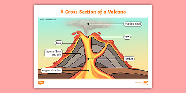
Label the diagram of volcano using the words inside the box. Conduit - a conduit is a passage through which magma molten rock flows in a volcano. Demonstrate your knowledge of volcanoes by labeling a diagram of a volcano. A word bank full of terms such as ash cloud crater and magma chamber will help them along the way.
The largest volcano in the entire world mauna loa in hawaii is a shield volcano. Look at this diagram of the inside of a volcano. Label the diagram of a volcano using the words inside the box Main Features of a Volcano Figure 3.
It also looks at different types of plate boundary and where volcanoes are located. Students label the parts of a volcano diagram using words provided in the word bank. In this topic we look at how movements in the earths crust can cause both volcanic eruptions and earthquakes.
Tamang sagot sa tanong. Label the diagram of a volcano using the words inside the box. They can also colour in the volcano diagram when the activity has been completed.
Resources like this one are great for use. Use Textables and arrows to label the major parts of the volcano and describe what they are. This illustrated science worksheet challenges students to label a colorful volcano diagram.
The first worksheet contains a volcano diagram for kids that includes the labels at the bottom of the page so students simply have to match up the words to the corresponding labels. After an eruption the tip or top of the volcano tends to get blown off leaving a small depression at the top of it. Students label the parts of a volcano diagram using words provided in the word bank.
It then goes on to look at the different features of volcanoes by looking at a cross-section of a volcano. Parts of a volcano labeling worksheet students label the parts of a volcano diagram using words provided in the word bank. Volcano Worksheet Definitions ash cloud an ash cloud is the cloud of ash magma chamber a magma chamber that forms in the air after some volcanic eruptions.
Students label the parts of a volcano diagram using words provided in the word bank. Label the diagram of a volcano using the words inside the box. These workbooks are ideal for the two kids and adults to make use of.
Parts of a volcano labeling worksheet teachervision students label the parts of a volcano diagram using words provided in the word bank this printable can be used as post unit assessment for earth science or volcanoes and earthquakes or can be assigned as a take home or independent pletion activity an answer key is provided volcano diagram printout enchantedlearning the largest volcano. Label the diagram with the missing words from the bottom of the sheet - 15448221. Printable Volcano Worksheets may be used by anybody at your home for educating and studying purpose.
Label our volcano diagram to understand the different. The composition of a volcano introduces a few complicated concepts to children so it can be quite handy for them to be able to visualise what the inside of a volcano looks like when learning about them. Label the Volcano Diagram Read the definitions then label the diagram below.
Thats what this parts of a volcano labelling activity aims to do with a vibrant illustration being presented so that children can get a clear image of what a volcano looks like making the learning process more effectiveTwinkl Go. Main Features of a Volcano Source. Volcano diagram to label.
1 on a question K to 12 BEC CG. Definitions ash cloud - an ash cloud is the cloud of ash that forms in the air after some volcanic eruptions. Printable Volcano Worksheets Printable Volcano Worksheets might help a trainer or student to find out and understand the lesson plan within a a lot quicker way.
An answer key is provided. Landforms Formed From Volcanic Activity Magma from the belly of kilauea in hawaii collects as it flows out to the pacific ocean. 1 question Direction.
Read the definitions then label the diagram below. Conduit a conduit is a passage through which magma molten rock flows in a volcano. On large volcanoes magma.
This sheet comes with answers which you can use to mark the students work. Label the Volcano Diagram. Search for volcano drag and drop the diagram into a cell.
Discover the different parts of an active volcano. Volcanoes form when magma that sits within the earths mantle magma reservoir and works its way up to the surface. SOES -IIIa25 SSES -IIIa27 AC5Direction.
If a volcano continues to erupt it will grow and grow into a larger volcano. Ash cloud - an ash cloud is the cloud of ash that forms in the air after some volcanic eruptions.
Larger cell does not divide and forms a conspicuous part of suspensor. By understanding the structure of dicot root and monocot root we can make comparisons between them and distinguish them by studying them under a microscope.
Plant Parts And Their Functions Pmf Ias
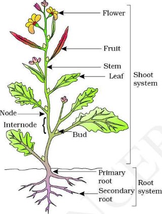
Labelled Diagram Of A Monocot Leaf.

Labelled diagram of a monocot plant. Labelled diagram of monocot and dicot seed. Development of Monocot Embryo. A carrot is an example of a dicot root.
Ts of dicot leaf monocot leaf grass draw a neat labelled diagram of t s ts of dicot leaf. Two epidermal layers are present one each on upper and lower surfaces. It consists of parenchymatous cells.
The embryonal cell forms suspensor and embryo. Monocots and dicots. Monocot and Dicot Stems.
Monocot Stem with Secondary Thickenings 3. Three cell stage is formed by transverse division of embryonal cell. Few big motor cells or bulliform cells are present in groups here and there in the furrows of upper epidermis.
External structure of a woody stem. Normal Dicotyledonous Stems 4. Of Monocot root.
Experiment to study the external features of plants with diagrams. Record your observations 10 following the same procedure study the stomata of other dicot and monocot plants observations sl no observation dicot monocot 1 number of stomata in the microscopic field 2 sep 28 2019 this pin was. In this diagram the parts of the monocot stem are labeled and usually consist of the vascular bundle the parenchyma the cortex the epidermis the xylem and the phloem.
Maharashtra State Board HSC Science General 11th. Root branches develop from the pericycle. In this upper cell is large and the lower cell is small and is called embryonal cell.
Root anatomy of monocot root anatomy plant tissue monocot root diagram botany preperation before practicals biology grade 9 docx biology 1409 chapter 25 understanding stems roots and labelled diagram of a leaf awesome simple leaf cell difference between dicot and monocot leaf with comparison monocotyledon wikipedia lab 6 notebook docx plant. Angiosperms flowering plants are divided into two groups. Monocot and dicot flowers edinburgh garden school.
Zygote first divides by transverse division. Dicots have two cotyledons. Plant Tissues and Anatomy - Exercise Page 96 Q 5.
Draw a well labelled diagram of monocot seed you have studied. In older roots the cells are thick-walled. A labeled monocot stem is a diagram that features the cross section of a monocot plant stem.
It is the outermost layer of the stele that lies below the endodermis. Dicot root cross section labeled diagrams. Monocot roots interestingly have their vascular bundles arranged in a ring.
Diagram illustrating the tissue layers and their organization within monocot and dicot roots. Freeman and Sinauer Associates used by permission. Monocotyledoneae EMorren ex Mez.
The following points highlight the top four types of monocot and dicot stems. Draw a neat labelled diagram. Dicot roots have their xylem in the center of the root and phloem outside the xylem.
Masuzi february 4 2020 uncategorized 0. Pericycle give rise to lateral roots cork cambium and part of vascular cambium 2. Dicots vs monocots monocot vs dicot flowers stems.
Monocotyledons ˌmɒnəˌkɒtəlˈiːdən commonly referred to as monocots Lilianae sensu Chase Reveal are grass and grass-like flowering plants angiosperms the seeds of which typically contain only one embryonic leaf or cotyledon. Both groups however have the same basic architecture of. Normal Monocot Stems 2.
It may be single-layered maize or multi-layered. Radial vascular bundle monocot root. Draw a well labelled diagram of the monocot seed you have studied.
Uniseriate upper and lower epidermal layers are composed of more or less oval cells. Monocot seeds have one seed leaf termed a cotyledon in fact monocot is a shortening of monocotyledon. Question Bank Solutions 5551.
In monocot roots the pericycle does not form cambium because there is no secondary growth in monocots.