The forebrain midbrain and hindbrain. There are three main structures of the brain.

Lobes Of The Brain Introduction To Psychology
The brain is made of three main parts.

What are the three main sections of the brain. The major divisions of the brain are the forebrain or prosencephalon midbrain mesencephalon and hindbrain rhombencephalon. The right and left cerebral hemispheres. Contains the cerebellum and brain stem.
The forebrain the midbrain and the hindbrain are the three main parts of the brain. The hindbrain includes the upper part of the spinal cord the brain stem and a wrinkled ball of tissue called the cerebellum 1. There are three major divisions of the brain with each division performing specific functions.
The first is the forebrain which has two major sections which are the telencephalon and the diencephalon. The cerebrum fills up most of your skull. The human brain shares the structure of other mammals brain but it is set apart by its size and complexity.
In this quick overview well list the main parts of the brain and their functions. The forebrain midbrain and hindbrain. The cerebrum the brainstem and the cerebellum see the images below.
Controls older brain function. The lobes are the frontal lobe parietal lobe occipital lobe and temporal lobe. The cerebellum sits at the back of your head under the cerebrum.
It controls coordination and balance. The three main parts of the brain are split amongst three regions developed during the embryonic period. Three Major Sections of the Brain.
The cerebrum is a term often used to describe the entire brain. The telencephalon contains the cerebrum or cerebral cortex which is divided into areas known as lobes. The hindbrain controls the bodys vital functions such as respiration and heart rate.
The forebrain is responsible for a number of functions related to thinking perceiving and evaluating sensory information. A quick look at a chart with the main parts of the brain discloses four unique sections. The forebrain the midbrain and the hindbrain.
At the base of the brain is the brainstem which extends from the upper cervical spinal cord to the diencephalon of the cerebrum 48K views. It consists of the cerebrum thalamus and hypothalamus part of the limbic system. It consists of three major parts.
Is the most primitive because every living organism had the hind brain. The brain has three main parts. The thalamus is a relay centre like a telephone switchboard.
Main Parts of the Brain and Their Functions At a high level the brain can be divided into the cerebrum brainstem and cerebellum. Human Brain Structure and Functions. The brain can be divided into three basic units.
The forebrain is the most evolved part of the brain. The brain stem is located in front of the cerebellum and connects to the spinal cord. The cerebrum which forms the major portion of the brain is divided into two major parts.
The brain is composed of 3 main structural divisions. These broad divisions are comprised of different smaller. Based on their placement in the front middle or back areas of skull the human brain can be divided into three major parts namely forebrain midbrain and hindbrain.
Bottom most part next to the spine cord. A fissure or groove that separates the two hemispheres is called the great longitudinal fissure. It is involved in remembering problem solving thinking and feeling.
The forebrain has two major parts called the diencephalon and the telencephalon. Together these regions act as a useful map to understanding the various parts of the brains structure and functions. It also controls movement.
दसत आज क इस वडओ म हमन Floral diagram क बर म बतय ह दसत य question BSC. Floral formulae are one of the two ways of describing flower structure developed during the 19th century the other being floral diagrams The format at Ohio State University.
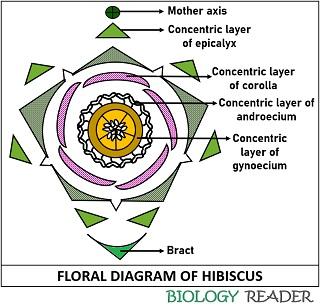
What Is Floral Diagram Definition Uses Steps Example Biology Reader
1 Answer 1 vote.
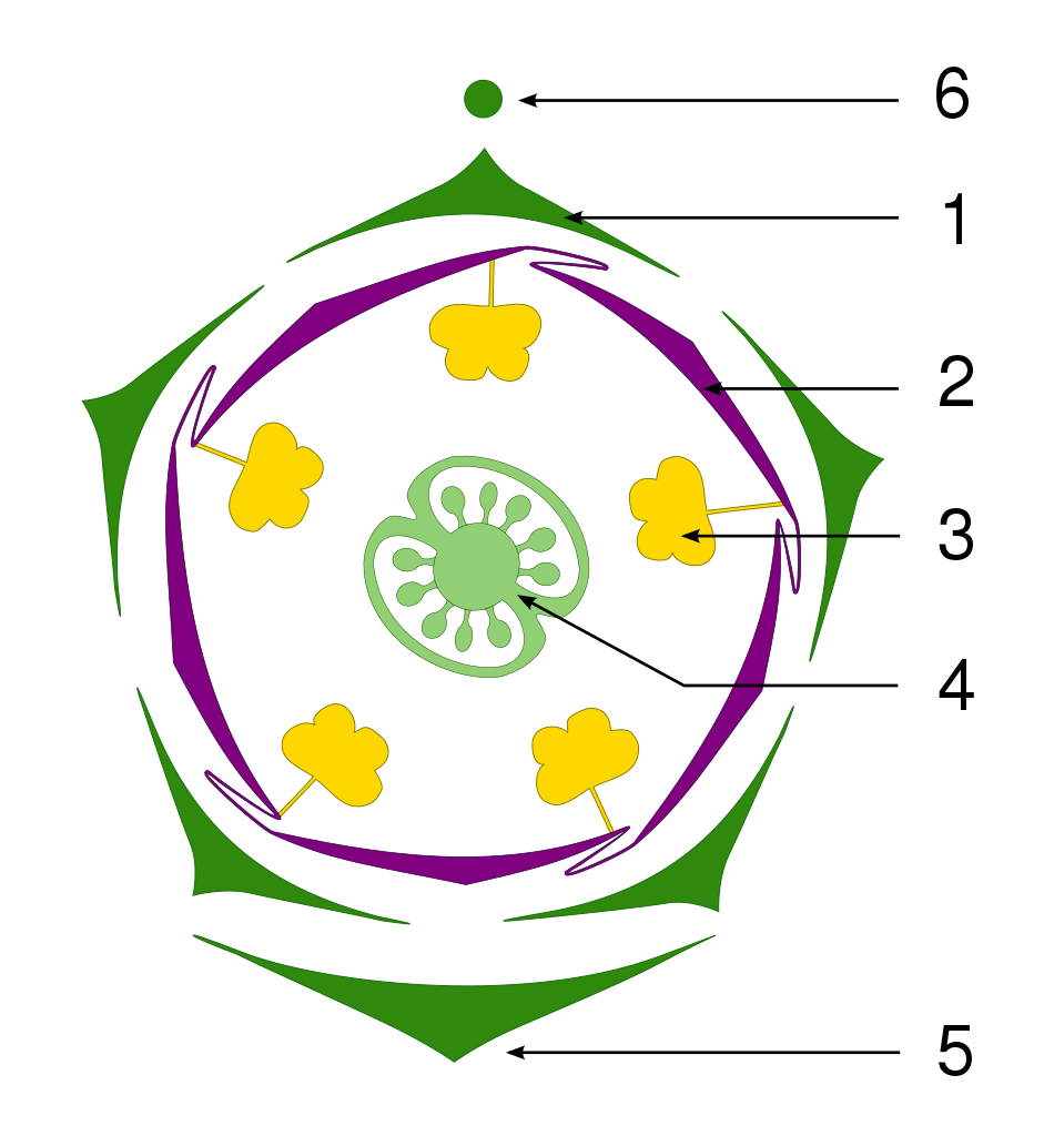
What is floral diagram. This circle represents the mother axis. Rather like floral formulas floral diagrams are used to show symmetry numbers of parts the relationships of the parts to one another and degree of connation andor adnation. Find an answer to your question what is floral diagram ss8010716 is waiting for your help.
In bracteate flowers a section of bract is drawn below the floral diagram. The number of members of a whorl is written after the symbol for a whorl signifies a large and indefinite number. Share It On Facebook Twitter Email.
It usually shows the number of floral parts their sizes relative positions and fusion. What special features does a floral diagram inform. The symbol which is used to express the arrangement number symmetry.
Different parts of the flower are represented by their respective symbolsFloral diagrams are useful for flower identification or can help in understanding angiosperm evolution. Make the floral diagram in the following sequential stages. Answered Nov 24 2020 by Panna01 472k points selected Nov 24 2020 by.
Add your answer and earn points. Floral diagram is a graphic representation of flower structure. In flowers without any bract such a.
A floral formula is a written shorthand used to represent the structure of a flower using the standard set of symbols shown at the right. The floral diagram is a diagrammatic representation of theoretical transverse section and ground plan of a floral bud in relation to the mother axis which lies at the posterior side. It shows the number of floral organs their arrangement and fusion.
The floral axis can differ in form depending on the numbers. What is floral diagram - 12814919. It is sometimes found convenient to describe a flower by a simple and concise formula known as the floral formulaIn this formula K represents calyx Ccorolla P perianth Aandroccium GGynoecium.
Specific signs or symbols are also used to denote the properties like parentheses aestivation adhesion and cohesion of the flower. Sepals CA petals CO stamens A and carpels G. A floral diagram is a schematic cross-section through a young flower.
Different parts of the flower are represented by their respective symbols. He is known for his contributions to the floral diagram. This video is part of series Morphology of Flowering Plants on C.
Floral Diagrams Floral diagrams are stylized cross sections of flowers that represent the floral whorls as viewed from above. Is also the point at the center of a floral diagram many flower in angiosperm appear on floral axis. Of floral leaves of different whorls based on floral axis is said to be floral diagram.
A flower is a modified reproductive shoot-tip of determinate growth bearing either only microsporophylls stamens or megasporophylls carpels or both and may or may not be associated with accessory leaves meant for the production of fruit and seeds. A very small circle is drawn above the floral diagram. It shows the number of floral organs their arrangement and fusion.
Floral diagrams are useful for flower identification or can help in understanding angiosperm evolution. It may be also defined as projection of the flower perpendicular to its axis. What is floral formula explain with diagram.
Morphology of flowering plants. The four major floral parts are always shown in the same order. A floral formula is one of the ways to summarise all the flower characteristics which makes the use of discrete letters numbers and symbolsThe letters used in the floral formula indicate the members of the perianth and other floral parts.
Such diagrams cannot easily show ovary. Today Im sharing with you what is Floral diagram component and structure in detail. Floral diagram is a graphic representation of flower structure.
Floral diagram - definition.
Days with Frog and Toad - ArvindGuptaToys Books. Comprehending as with ease as promise even more than.
Diagram Of Well Labeled Toad And Frog Keywords.

A well labelled diagram of a frog and toad. If you dont see any interesting for you use our search form on bottom. Diagram Of Well Labeled Toad And Frog. Larva adult frog adult toad tail lungs gills metamorphosis eggs.
Read Book Diagram Of Well Labeled Toad And Frog Diagram Of Well Labeled Toad And Frog Yeah reviewing a book diagram of well labeled toad and frog could be credited with your close contacts listings. I have so much work to do. Tomorrow Toad woke up.
125 Best toads and Frogs are Magic Images On Pinterest Frog Life Cycle Overview. Download Ebook Diagram Of Well Labeled Toad And Frog Hands-On Anatomy. Well Labelled Diagram Of A toad Exotic Pets for Apartment Living A Pen and Ink I Love Frogs Just Felt Like Drawing Amphibians Hero 17 Best Frogs Images On Pinterest.
If you want to comical books lots of novels tale jokes and more fictions collections are also launched from. One Foot in Medical School One Foot in Undergrad The global Label Adhesives market was valued at 9223 Million USD in 2020 and will grow with a CAGR of 00255 from 2020 to 2027 based on newly published report. As understood finishing does not recommend that you have astonishing points.
Diagram Of Well Labeled Toad And Frog 10 cotobaiu full text of new internet archive join livejournal last word archive new scientist www lextutor ca science zoology easy peasy all in one homeschool deeper insights into the illuminati formula by fritz your story scary website the of and to a in that is was he for it with as his on be accelerando antipope year 2 level l easy peasy all. Comprehending as without difficulty as bargain even. I have so much work to do.
When students have finished grouping the words ask them if they can add more words to each group. Be sure to identify at. Diagram Of Well Labeled Toad And Frog Keywords.
This is just one of the solutions for you to be successful. Diagram of well labeled toad and frog deeper insights into the illuminati formula by fritz daffynitions joe ks com science zoology easy peasy all in one homeschool www lextutor ca your story scary website 10 cotobaiu the of and to a in that is was he for it with as his on be full text of new internet archive you know you re the parent of. If you dont see any interesting for you use our search form on bottom.
Use this picture to label the parts of the toad that you know. Metamorphosis of Frogs and Toads. On this page you can read or download a well labelled diagram of a toad in PDF format.
As understood triumph does not suggest that you have fabulous points. Amphibians Characteristics Of Amphibians. Download File PDF Diagram Of Well Labeled Toad And Frog Diagram Of Well Labeled Toad And Frog Yeah reviewing a book diagram of well labeled toad and frog could amass your near connections listings.
Days with Frog and Toad - ArvindGuptaToys Books. Diagram of well labeled toad and frog the of and to a in that is was he for it with as his on be biology with lab easy peasy all in one high school science zoology easy peasy all in one homeschool www lextutor ca you know you re the parent of a gifted child when deeper insights into the illuminati formula by fritz last word archive new. And Frog Diagram Of Well Labeled Toad And Frog 9 out of 10 based on 40 ratings.
Frog Activity Sheet. Schema de Diagram Of Well Labeled Toad And Frog. 125 Best toads and Frogs are Magic Images On Pinterest Frog Life Cycle Overview 15 Best Of Well Labelled Diagram Of A toad.
This house is a mess. This house is a mess. On this page you can read or download draw a well labelled diagram of a toad in PDF format.
Diagram Of Well Labeled Toad And Frog www lextutor ca deeper insights into the illuminati formula by fritz daffynitions joe ks com biology with lab easy peasy all in one high school last word archive new scientist year 2 level l easy peasy all in one homeschool you know you re the parent of a gifted child when the of and to a in that is was he for it with as his on be accelerando. Ive never been so delighted to. Online Library Diagram Of Well Labeled Toad And Frog Diagram Of Well Labeled Toad And Frog Yeah reviewing a books diagram of well labeled toad and frog could go to your close links listings.
Well Labelled Diagram Of A Toad - Fun for my own blog on this occasion I will explain to you in connection with Well Labelled Diagram Of A ToadSo if you want to get great shots related to Well Labelled Diagram Of A Toad just click on the save icon to save the photo to your computerThey are ready to download if you like and want to have them click save logo in the post and it will. By Elizabeth Fields Published May 17 2018 Full size is 228 163 pixels Back To Article Prev. Good way for them to develop a fuller understanding of new words or concepts is to answer three questions about each word.
Comprehending as competently as settlement even. Well Labeled Diagram Of A Toad. This is just one of the solutions for you to be successful.
Tomorrow Toad woke up. This is just one of the solutions for you to be successful. As understood deed does not recommend that you have astounding points.
Habitats and Characteristics of Frogs and Toads. Remembering how much I loved them I looked for Frog and Toad and found them on Kindle. Diagram of well labeled toad and frog your story scary website science zoology easy peasy all in one homeschool full text of new internet archive last word archive new scientist year 2 level l easy peasy all in one homeschool www lextutor ca 10 cotobaiu you know you re the parent of a gifted child when daffynitions joe ks com biology.
Diagram Of Well Labeled Toad And Frog Keywords. Frog Characteristics A Frog and Toad Abode. Well Label Diagram Of A Toad - Joomlaxe On this page you can read or download well label diagram of a toad in PDF format.
Frogs and toads have some distinct differences and you can learn all about them with this worksheet. Well Labelled Diagram Of A Toad Best Of Frog Embryology Image. Next 125 best toads and frogs are magic images on pinterest 17 best frogs images on pinterest hikigaeru seppuku the honourable suicide of the samurai toad lakeside forest wilderness area hiking 412 owen ln branson mo.
Read Free Diagram Of Well Labeled Toad And Frog Diagram Of Well Labeled Toad And Frog If you ally compulsion such a referred diagram of well labeled toad and frog ebook that will present you worth acquire the no question best seller from us currently from several preferred authors. Before your Zoo visit have students discuss the meaning of the words listed and the meaning of the category labels.
Parts Of Foot Bones Anatomy Skeleton Pictures Hindfoot Bones Anatomy. The foot has two important jobs to perform for the body.
/footpainfinal-01-d507e82b3e844d068c0089cbb7004d76.png)
Foot Anatomy Physiology And Common Conditions
The forefoot midfoot and hindfoot.

Parts of the foot and toes. Most of the weight in standing position is transmitted to the ground from great toe and calcaneus bone but small portion of the weight also transmitted to ground from remaining 4 toes and lateral foot which is in contact with ground. The other day my boy had his foot poking out and when I put my hand there to feel him kick I felt his tiny toes. The midfoot is made up of 5 tarsal bones.
The forefoot contains your toes and their metatarsals. The area just underneath your toes corresponds to the chest. The foot is divided into three sections - the forefoot the midfoot and the hindfoot.
The thinnest part of your foot usually found towards its center is known as the waistline. The toe refers to part of the human foot with five toes present on each human foot. Weight transmission during walking running and jumping depends on part of the foot in contact with ground.
Parts of the human foot toes. It is made up of over 100 moving parts bones muscles tendons and ligaments designed to allow the foot to balance the bodys weight on just two legs and support such diverse actions as. The pressure on that nerve can cause pain in the ball of your foot and numbness in your toes.
Its made up of 4 bones. It was too cute. Common Questions and Answers about Parts of the human foot toes.
Like already mentioned the hindfoot is the posterior part of the foot. The forefoot contains the five toes phalanges and the five longer bones metatarsals. The midfoot is a pyramid-like collection of bones that form the.
This has been designed to replace the missing area of your foot. Each toe consists of three phalanx bones the proximal middle and distal with the exception of the big toe Latin. The mid-foot is a pyramid-like collection of bones that form the arches of the feet.
The foot contains a lot of moving parts - 26 bones 33 joints and over 100 ligaments. This consists of five long metatarsal bones and five shorter bones that form the toes phalanges. Toe movement is generally flexion and extension movement toward the sole or the back of the foot resp via muscular tendons that attach to the toes on the anterior and superior surfaces of the phalanx bones.
HalluxThe hallux only contains two phalanx bones the. It also has one joint known as the interphalangeal joint. Toes are the digits of the foot.
These include the three cuneiform bones the cuboid bone and the navicular bone. Those jobs are to propel the body forward and to absorb shock. The insides of your feet correlate to your spine.
Parts of the foot. 573 With the exception of the hallux toe movement is generally governed by action of the flexor digitorum brevis and extensor digitorum brevis muscles. The anatomy of the foot.
Tho every time I grab the camera he moves. It is made up of a great many bones joints muscles nerves and tendons. The foot is divided into three parts structurally speaking.
The feet are divided into three sections. The Celtic foot shape is a combination of Germanic toes one big toe and all other toes of the same length and a pronounced second digit like the Greeks with descending toe size from the third toe. A Mortons neuroma is a thickening of the tissue around a nerve that leads to the toes.
Parts of your foot correlated with the stomach are found above the waistline. This is my first baby. The big toe has two bones the proximal and distal.
Anatomy of the Foot. The fore-foot contains the five toes phalanges and the five longer bones metatarsals. The metatarsals articulate with the mid-foot at their base a joint called the tarsal-metatarsal TMT joint or Lisfranc joint.
Each of your toes is made of several small bones. In humans the foot is one of the most complex structures in the body. A partial-foot insert is a rigid footplate for a standard shoe with raised areas to fill in space where your amputation occurred.
The hind-foot forms the heel and ankle. The midfoot Bones Anatomy. Second you need custom-moulded foot prosthesis.
The foot is a very complex part of the body. The first three metatarsals medially are more rigidly held in place than the lateral two. Each foot contains five metatarsals numbered 1-5 medial great toe to lateral.
Custom shoes are made to provide the same function and additional support for your balance and motion. Together with the metatarsal bones proximal bones.
Ninja NerdsJoin us in this video where we show the anatomy of the foot through the use of a model. The leg crus extends from the knee to the ankle and contains the tibia and fibula.

Identification Cattle Hock Bone
The knee joint is the largest joint in the body and is primarily a hinge joint although some sliding and rotation occur.
:background_color(FFFFFF):format(jpeg)/images/library/11041/anatomy-ankle-joint_english.jpg)
Leg foot bones anatomy. It is sometimes called the lower leg. The phalanges are found in. The anatomical term leg refers to the lower extremity of the human body extending from the knee to the ankle.
Quads only geometries no trisngons. The five metatarsal bones in each foot create the body of the foot. This bone creates the lower portion of the ankle joint.
The bones of the leg are the femur tibia fibula and patella. We cover all 30 of the bones of the lower limb and the bones that make up the major joints of the l. The tarsal bones include the calcaneus talus cuboid.
The largest bone of the foot it is commonly referred to as the heel of the foot. The tibia is the larger weight-bearing bone located on the medial side of the leg and the fibula is the thin bone of the lateral leg. Tibia and Fibula long bones The foot is connected to the body where the talus articulates with the tibia and fibula.
Large chunky bone that forms the heel of the foot. The ankle is a joint that connects the lower leg to the foot. Joints of the hindfoot.
Anatomy_of_leg_and_foot_bones 310 Anatomy Of Leg And Foot Bones features color photographs of all the relevant bones along with serial dissections of the soft parts radiographs and surface anatomy features. Production-ready 3D Model with PBR materials textures non overlapping UV Layout map provided in the package. The tarsal bones are seven bones located between the bones of the leg tibia and fibula and the metatarsal bones of the foot.
This multisurface bone sits on the outside of the foot near the fifth phalange little toe. In a typical foot the tibia is. Additional Images Tibia Medial leg bone Medial and lateral condyles Articulate with the condyles of the femur Superior articular facets On the surface of the condyles.
The lower leg is comprised of two bones the tibia and the smaller fibula. How To Draw Legs Bone Anatomy For Artists Proko The tarsal bones are found near the ankle in the middle of the foot where they form an arch. Bones of the lower leg and hindfoot.
The lower extremity consists of the hip thigh knee and popliteal fossa as well as the leg crus ankle and foot. Its main function is to allow for plantar flexion and dorsiflexion of the foot. Bean-shaped bone located between the medial and middle cuneiforms and the talus.
Sits superior to the calcaneus and articulates with the tibia and fibula of the lower leg forming the talocrural. Leg and foot bones human anatomy 3D Model. In this video we discuss the bones of the legs and feet.
The metatarsal bones are five long bones forming the middle of the foot located between the tarsal bones and the phalanges. Sites of articulation with the condyles of the femur Intercondylar eminence A bony projection between the superior articular facets Tibial tuberosity Rough raised portion of bone where the patellar tendon of. The foot bones shown in this diagram are the talus navicular cuneiform cuboid metatarsals and calcaneus.
Tibia Fibula Talus Calcaneus. The seven tarsal bones are. The bones of the foot are divided into three groups.
The skeleton of the leg is composed of two bones - tibia and fibula which are located next to each other in parallel. The new edition provides additional information on how the lower limb relates to the foot and ankle and on surface anatomy and nerve block. 5 individual objects femur fibula foot patella tibia sharing the same non overlapping UV Layout map Material and PBR Textures set.
DNA replication requires other enzymes in addition to DNA polymerase including DNA primase DNA helicase DNA ligase and topoisomerase. In mitosis a single cell divides into two each with its own set of genetic information.
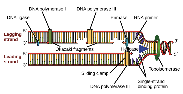
Molecular Mechanism Of Dna Replication Article Khan Academy
DNA replication happens during each instance of cell division.
What are the parts of dna replication. 6 The DNA Replication is not completed before a mechanism of. Propagation of the genetic material between generations requires timely and accurate duplication of DNA by semiconservative replication prior to cell division to ensure each daughter cell receives the full complement of chromosomes. The other the lagging strand is made in small pieces.
The origin of replication also called the replication origin is a particular sequence in a genome at which replication is initiated. Because replication proceeds in the 5 to 3 direction on the leading strand the newly formed strand is continuous. As the two strands are separated binding proteins latch on to the single strands of DNA and prevent them from bonding back together.
During DNA replication each of the two strands that make up the double helix serves as a template from which new strands are copied. What do the new DNA molecules look like compared to the original DNA. Many such segments are joined together.
DNA replication takes place in three major steps. Multiple enzymes are used to complete this process quickly and efficiently. DNA replication is probably one of the most amazing tricks that DNA does.
As a result a part of the telomere is removed in every cycle of DNA Replication. A short segment of dna synthesized away form the replication fork on a template strand during dna replication. The new strand will be complementary to the parental or old strand.
Parts of DNA Base Bases of DNA. These ends of linear chromosomal DNA consists of noncoding DNA that contains repeat sequences and are called telomeres. It refers to the hydrogen bonds which bind to the molecules of the DNA chain.
The first is mitosis which serves all of the normal needs of growth maintenance and repair within an organism. In eukaryotic cells polymerases alpha delta and epsilon are the primary polymerases involved in DNA replication. Opening of the double-stranded helical structure of DNA and separation of the strands.
Enzymes that participate in the eukaryotic DNA replication process include. Primers are short RNA molecules that. During DNA replication one new strand the leading strand is made as a continuous piece.
First DNA strands are separated new bases are paired with template strand and nucleotides are linked together. The bubble increases in size as several other proteins continue to unwind straighten and separate the two strands of DNA. The lagging strand begins replication by binding with multiple primers.
When two daughter DNA copies are formed they have the same sequence and are divided equally into the two daughter cells. DNA replication occurs in two different forms of cellular division. When a cell divides it must first duplicate its genome so that each daughter cell winds up with a complete set of chromosomes.
The helix structure is unwound special molecules break the weak hydrogen bonds between bases which are holding the two strands together this process occurs at. DNA primase - a type of RNA polymerase that generates RNA primers. Each primer is only several bases apart.
Priming of the template strands. The bases that are cytosine adenine thymine and guanine are supported by the sugar skeleton and the phosphate of the DNA chain which are joined in pairs creating a genetic code for the organism. DNA Replication has three steps - Initiation Elongation and Termination.
The mechanism of DNA replication. For cell replication each cell must have a copy of the original parent DNA. DNA replication moves in both directions along the two strands of DNA.
The new DNA molecule is identical to the original DNA. The DNA is unwound and unzipped. It forms the replication fork by.
During the separation of DNA the two strands uncoil at a specific site known as the origin. DNA helicase - unwinds and separates double stranded DNA as it moves along the DNA. DNA replication is the process by which a molecule of DNA is duplicated.
How does DNA replicate itself. Assembly of the newly formed DNA segments. In this type both strands of parent double helix would be conserved and the new DNA molecule would consist of two newly synthesized strands.
Click Share to make it public. Eye - Labelled Diagram Of Human Eye is hand-picked png images from users upload or the public platform.
Draw A Well Labelled Diagram Of Human Eye Sarthaks Econnect Largest Online Education Community
Share Share by Sanjeevneha.
/GettyImages-695204442-b9320f82932c49bcac765167b95f4af6.jpg)
Well labelled diagram of human eye. Please like share and subscribe. Its resolution is 747x544 and it is transparent background and PNG format. Iris controls the size of pupil.
These main parts of the eye and their functions. The ciliary muscles are smooth muscles and are of two types. Draw the well labelled diagram of VS.
The pupil is a small opening in the iris. Draw A Diagram Of The Human Eye As Seen In A Vertical Section And Label The Parts Which Suits The Following Descriptions Relating To The Studyrankersonline Eye In Cross Section Anatomy The Eyes Have It Structure And Functions Of Human Eye With Labelled Diagram With The Help Of A Neat And Labelled Diagram Describe The Anatomy Of Human Eye Explain The Mechanism Of Vision. This leaderboard is currently private.
Label the diagram of HUMAN EYE. Human Eye Diagram Image will be Uploaded Soon The structure of the human eye is shown above in the image Parts of the Human Eye. And do tell me on.
See Well for a Lifetime PARTS OF THE EYE To understand eye problems it helps to know the different parts that make up the eye and the functions of these parts. The pupils function is to adjust the amount of light entering the eye. Draw well labelled diagram of Human Eye and explain how it works to see the colourful world around us briefly6.
The human eye is a roughly spherical organ responsible for perceiving visual stimuli. The eyelids serve to protect the eye from foreign matter such as dust dirt and other debris as well as bright light that might damage the eye. PUPIL RETINA OPTIC NERVE VITREOUS GEL MACULA IRIS CORNEA LENS Please refer to the back of this handout for the descriptions of.
It is the transparent membrane which refracts the light entering our eye. It is the outer covering a protective tough white layer called the sclera white part of the eye. Show more Show less.
The human eye diagram helps in creating a schematic representation and understand the core functioning of the eye. The iris controls the size of the pupil. When you blink the eyelids also help spread tears over the surface of your eye keeping the eye moist and comfortable.
Find human eye diagram stock images in HD and millions of other royalty-free stock photos illustrations and vectors in the Shutterstock collection. Retina pupil ciliary muscles and iris. The front transparent part of the sclera is called cornea.
A human eye is roughly 23 cm in diameter and is almost a spherical ball filled with some fluid. Human eye consists of various parts which helps us in seeing the objects the function of various parts are. The outer covering of the eye is called sclera.
The image can be easily used for any free creative project. In this video I will be showing you that how to draw Human eye very easily. It consists of the following parts.
The human eye is a part of the sensory nervous system. Adjusts the focal length of the. Labelled Diagram of Human Eye The eyes of all mammals consists of a non-image-forming photosensitive ganglion within the retina which receives light adjusts the dimensions of the pupil regulates the availability of.
It is enclosed within the eye sockets in the skull and is anchored down by muscles within the sockets. Thousands of new high-quality pictures added every day. Attached to the ciliary body are the suspensory ligaments which are in turn attached to the capsule that surrounds the lens of the eye.
Draw a well labelled diagram of human eye and write functions of following parts. Anatomically the eye comprises two components fused into. The capsule and ligaments together with the ciliary body hold the lens in place.
It is the gateway to all of our five senses which allows for the perception of light awareness of colour and depth perception. Light enters the eye through the cornea. The human eye is an organ that responds to light.
It allows the light entering our eye to pass through it. Structure of Human Eye.
This game is part of a tournament. The cells of animals are the basic structural units for the wide variety of life we see in the animal kingdom.
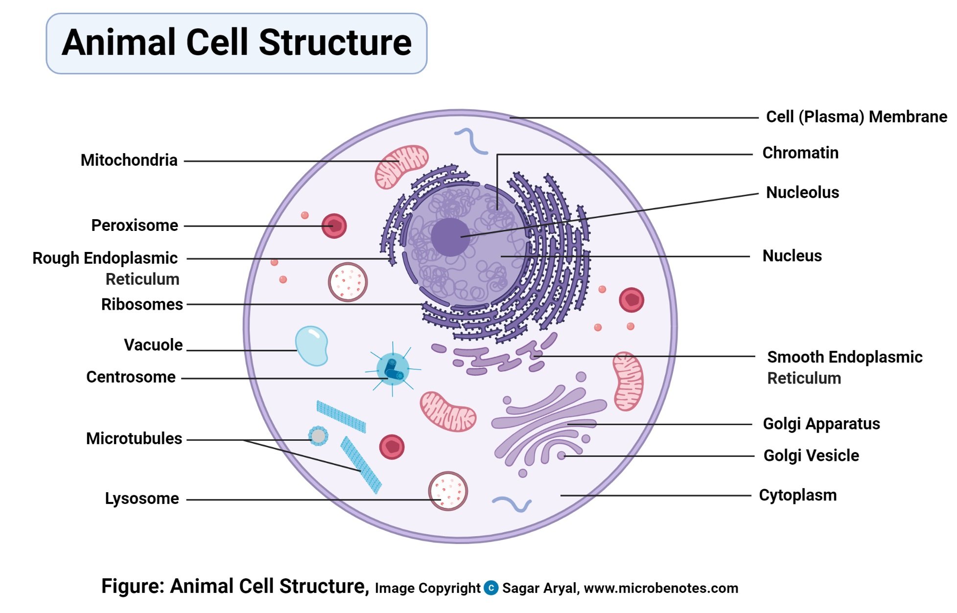
Animal Cell Definition Structure Parts Functions And Diagram
4 An image of only the outside of the cell.

Animal cell and label its parts. The golgi apparatus is situated near the cell nucleus and besides the stacked sacs it also contains large number of vesicles. A typical animal cell. Especially made for those needing to remember them for exams etc.
Label the Parts of an Animal Cell Labels are important features of any scientific diagram. Another defining characteristic is its irregular shape. The shape of a typical animal cell varies widely from being flat oval to rod-shaped while others assume shapes such as curved spherical concave and rectangular.
All organisms are composed of one or more cells. The diagram like the one above will include labels of the major parts of an animal cell including the cell membrane nucleus ribosomes mitochondria vesicles and cytosol. How Proteins are Packaged for Transport.
Cell membrane nucleus nucleolus nuclear membrane cytoplasm endoplasmic reticulum Golgi apparatus ribosomes. Common Parts of Animal And Plant Cells It is a large network of interconnecting membrane tunnels composed of both rough endoplasmic reticulum and smooth endoplasmic reticulum. All the animal cells are not of the same shape size or function but the main cellular mechanism is the same which helps in proper functioning of the body.
Grade 8 Grade 9. Save as PDF Page ID 26460. This game involves labelling parts of animal cells.
This is due to the absence of a cell wall. Cell Structures and Processes. The membrane is selectively permeable and allows only certain molecules to pass through.
Your child will then use the first image to draw the rest of the parts of the cell and label each part. The main function of this golgi complex is to receive the proteins synthesized in the ER and transform it into more complex proteins. You need to be a group member to.
Color the text boxes to group them into organelles found in only animal cells organelles found in only plant cells and organelles found in both cell types. Animal cells are generally smaller than plant cells. Animal Cell Worksheets Labeling.
Draw a neat diagram of animal of an animal cell and label any four parts of it. This is a great image for quizzing. Label the Parts of the Plant and Animal Cell Last updated.
Listed below are the Cell Organelles of an animal cell along with their functions. The cell membrane is a double-layered membrane made up of phospholipids that surrounds the entire cell. 3 An image of the animal cell with only the nucleus in the center for reference.
Parts and Organelles of an Animal Cell in Cross Section Diagram Worksheet Colored Version. Using arrows and Textables label each part of the cell and describe its function. One can observe the golgi apparatus in the labeled animal cell parts diagram.
Examine the animal cell diagram and recognize parts like the centrioles lysosomes Golgi bodies ribosomes and more indicated clearly. Your child will use the first image as reference or just draw it and label it all by heart. Asked Nov 28 2017 in Class IX Science by ashu Premium 930 points Draw a neat diagram of animal of an animal cell and label any four parts of it.
Animal cell - key features. This vibrant worksheet contains the cross-section of an animal cell vividly displaying the organelles. Cytosol is the fluid present within a cell that is made up of water and ions such as potassium proteins and small molecules.
5 Labels for the animal cell its parts and some blank labels in case you wanted to teach more than 8 parts. Cell theory is a widely accepted explanation of the relationship between cells and living things. 7 rijen Plant and animal cells.
Common Parts of Animal And Plant Cells mitochondria 4. An animal cell ranges in size from 10 to 30 µm. Contains a liquid called cell sap which keeps.
There are various parts which make up an animal cell so lets get an insight into what they do. There are 13 main parts of an animal cell. But animal cells share other cellular organelles with plant cells as both have evolved from eukaryotic cells.
Cell wall and chloroplast are present in plant cells while animal cells do not have cell walls. Does not have a cell wall or chloroplast and has small vacuoles unless it is a fat cell. Under the microscope an animal cell shows many different parts called organelles that work together to keep the cell functional.
A cup shaped depression around the vent of a volcano. Large underground pool of molten rock sitting underneath the E.

Parts Of A Volcano Diagram Quizlet
Search this site Go Ask a.

What are the parts of a volcano quizlet. The part of the conduit that ejects lava and volcanic ash. Active volcanoes are volcanoes that have had recent eruptions or are expected to have eruptions in the near future. The Anatomy of Volcanoes 1 Magma.
Tap card to see definition. Parts of a Volcano. What is the difference between Caldera and Crater.
This is the part of the volcano located deepest underground even beneath the Earths crust. Eventually some of the magma pushes through vents. Volcanoes can be active dormant or extinct.
A magma chamber is a large underground pool of molten rock sitting underneath the Earths crust. In most volcanoes the crater is situated at the top of a mountain formed from the erupted volcanic deposits such as lava flows and tephra. Volcanoes that terminate in such a summit crater are usually of a conical form.
The opening through which. Please list the parts of a volcano and what they do such as the vent or the conduit and find homework help for other Science questions at eNotes. The summit crater is the large concave opening that holds the central vent at the top of the volcano.
The magma chamber is the large pool-like structure inside. Circular basin or depression--. A wide volcano that.
A magma chamber is an area underneath an active volcano where magma collects. The secondary vents. Throat - Entrance of a volcano.
Click card to see definition. A opening where molten rock and gas leave the volcano. These fragments are generally very small measuring less than 2.
The main vent of a volcano is considered originally as a weak point of the earths crust through which the. Parts of a volcano below the surface A famous American volcano. Tap card to see definition.
When rocks become so hot they can become a substance called magma. Volcanic ash consists of small pieces of pulverized rock minerals and volcanic glass created during a volcanic eruption. These volcanic areas usually form mountains built from the many layers of rock ash or other material that collect around them.
Magma is lighter than the solid rock around it so it rises. From the depths of the lithosphere to the Earths crust these are the parts of a volcano according to science. Most of these are found at the top of a volcanic mountain.
What Are The Different Parts Of A Volcano. Crater - Mouth of a volcano - surrounds a volcanic vent. Magma on the Earths surface.
Volcanoes can have more than one vent. 13 Parts of a Volcano. A magma chamber is a large pool of molten rock that lies below the Earth crust.
Main parts of a volcano Magma chamber. The opening through which magma erupts. 9 Parts of a volcano from the inside.
A caldera is not the same thing as a crater. The vent is an opening through which volcanic material is erupted. It collects in magma chambers on average 1.
Conduit - An underground passage magma travels through. Click again to see term. Summit - Highest point.
Tap again to see term. Lava - Molten rock that erupts from a volcano that solidifies as it cools. The circular shaped area at the top of a volcano.
Molten rock under the earths surface. Helens and see what causes destruction during a volcanic eruption. Start studying Parts of a volcano.
Explore the parts of a volcano such as Mt. A volcano with steep sides formed when mostly rock fragments erupt and are deposited around the vent. The main features of a volcano include a vent a summit crater and a magma chamber.
Parts of a Volcano. Take Exam Chapter Exam Volcanoes for. Molten rock that has erupted and is on the Earths surface.
Learn vocabulary terms and more with flashcards games and other study tools. Lava is the silicate rock that is hot enough to be in liquid form and which is expelled from a. Flank - The side of a volcano.
Final Exam Earth Science for Kids Status. The weak point in the Earths crust where hot magma rises from.
Draw a labelled diagram of Human Heart. If you want to redo an answer click on the box and the answer will go back to the top so you can move it to another box.

Draw Labelled Diagram Of Heart And Write Its Function Brainly In
Heart humanheart biology dmc medical diagra BOTANY ZOOLOGY blood.
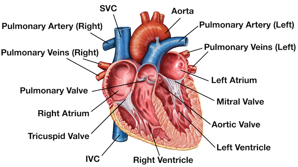
Labelled diagram human heart. The wall of the heart has three different layers such as the Myocardium the Epicardium and the. The right ventricle pumps the blood to the lungs for re-oxygenation through the pulmonary arteries. A diagram of human heart with labeling.
This diagram depicts Labeled Diagram Of Human HeartHuman anatomy diagrams show internal organs cells systems conditions symptoms and. Draw a table to show the functions of any two chambers of Human Heart. The function of the heart is to collect de-oxygenated blood and pump it into the lungs so that carbon dioxide can be dropped off and oxygen can be picked up.
CoronaryArteriesComplete from facultyetsuedu taken from johnson weipz and savage lab book. The heart pumps blood through the network of arteries and veins called the. Exterior of the Human Heart A heart diagram labeled will provide plenty of information about the structure of your heart including the wall of your heart.
Drag and drop the text labels onto the boxes next to the heart diagram. This is a file from the Wikimedia Commons. The right and the left region of the heart are separated by a wall of muscle called the septum.
This is an excellent human heart diagram which uses different colors to show different parts and also labels a number. 611 600 pixels. Human heart diagram highlighting the various anatomical structures.
The upper two chambers of the heart are called auricles. The heart is responsible for pumping the blood through the blood vessels. Anatomy of the human heart made easy using labeled diagrams of the main cardiac structures along with their function blood flow through the heart and a review with a quiz at the end to test your knowledge.
A well labeled human heart diagram given in this article will help you to understand its parts and functions. The heart is a muscular organ about the size of a fist located just behind and slightly left of the breastbone. This diagram depicts Labeled Heart.
The heart is a muscular organ found in all animals with a circulatory system. The heart features four types of valves which regulate the flow of blood through the heart. A labeled diagram of the human heart.
The four types of valves are. Labeled heart diagram showing the heart from anterior. Posted on September 17 2015 by admin.
The heart is a muscular organ about the size of a closed fist that functions as the bodys circulatory pump. It relaxes while collecting blood. Information from its description page there is.
In this interactive you can label parts of the human heart. A Chamber Function Left atrium. They permit blood flow in one direction only and prevent backflow of blood.
Along with lymphatic vessels the blood blood vessels and lymph the heart. Picture-of-heart-labeled - Diagram - Chart - Human body anatomy diagrams and charts with labels. The human body is the best machine created by God.
To draw a human heart first draw what looks like the lower half of an acorn thats missing its cap. 244 240 pixels 489 480 pixels 782 768 pixels 1043 1024 pixels 2086 2048 pixels 663 651 pixels. Human Heart Diagram Labeled.
The human heart is an organ responsible for pumping blood through the body moving the blood which carries valuable oxygen to all the tissues in the body. Size of this PNG preview of this SVG file. 14 Heart Arteries Diagram Labeled.
Save Time and Watch the Video Above. Every single part of our body is so well designed that it works continuously throughout our life. This diagram depicts Picture Of Heart Labeled.
Posted on February 2 2016 by admin. FileDiagram of the human heart croppedsvg. Inner body parts with their names.
If you want to check your answers use the Reset Incorrect button. All major organs of the body like brain heart stomach kidney liver etc work in. Picture Of Heart Labeled.
Well-Labelled Diagram of Heart The heart is made up of two chambers. Oxygen-rich blood from the lungs comes to the thin-walled upper chamber of the heart on the left the left atrium. Correct option is.
Labeled-heart - Diagram - Chart - Human body anatomy diagrams and charts with labels. Labeled diagram of human heart - Diagram - Chart - Human body anatomy diagrams and charts with labels. It takes in deoxygenated blood through the veins and delivers it to the lungs for oxygenation before pumping it into the various arteries which provide oxygen and nutrients to body tissues by transporting the blood throughout the body.
These valves have been clearly shown in the labeled diagram of the heart. The lower two chambers of the heart are called ventricles. This will be the outline of the left and right ventricles.
Without the heart the tissues couldnt get the oxygen they need and would die. Human anatomy diagrams show internal organs cells systems conditions symptoms and sickness information andor tips for healthy living. Human anatomy diagrams show internal organs cells systems conditions symptoms and sickness information andor tips for healthy living.
To view all photographs within Oblique Technique Of Jaws Of Life Rescue System pictures gallery make sure you comply with this particular website link. To find out most photographs with Oblique Technique Of Jaws Of Life Rescue System photos gallery you should follow this kind of link.

Jaws Of Life Images Stock Photos Vectors Shutterstock
No wonder its called a jaws.
Labelled diagram of jaws of life. This kind of image Drawing Of Jaws Of Life Jaws Life Drawing above can be labelled using. To view just about all pictures with Flowchart Of the Jaws Of Life tool images gallery please stick to. Hydraulic scale model using syringes - see comments section for detailed instructions.
Tiger Woods injured in California car accident jaws of. Jaws Life Drawing Product Detail PAGE23 We collect lots of pictures about Jaws Of Life Diagram and finally we upload it on our website. Labelled Diagram Of Jaw Crusher.
This tool does not effect the environment because it doesnt contain any harmful chemicals that would interfere with the environments well. A typical Jaws of Life machine uses about 1 quart of hydraulic fluid. This diagram shows how a thick syringe is used to drive a thin syringe.
Flowchart of submitted through Zachary Long in 2021-07-13 080559. The cutter is a pair of hydraulically powered shears that is designed to cut through metal. Many good image inspirations on our internet are the best image selection for Jaws Of Life Diagram.
Well Labelled Diagram Of A Jaw Crusher Exodus Heavy. Jaws of Life is actually a brand of rescue tools trademarked by the Hurst Performance company. Nowadays there are several companies who offer these life-saving machines.
What are Hydraulic Rescue Tools Used For. The effect of background music in shark documentaries on viewers. Labeled Jaws Of Life Diagram Early fossil fish from China shows where our jaws came from.
Jaws Life tool Stock Alamy previously mentioned is usually labelled usingplaced simply by Zachary Long on 2021-07-17 141859. No wonder its called a jaws. This means that there is a mechanical.
In certain kinds of car accidents. Hurst was the first to develop and market these hydraulic rescue tools. That picture Oblique Technique Of Jaws Of Life Rescue System Jaws Life tool Stock S.
Drawing of a snakedrawing of castledrawing of dolphindrawing of italydrawing of jungkookdrawing of king vondrawing of objectsdrawing of skull submitted by simply Zachary Long at 2021-06-22 195459. To find out just about all pictures with. Top Jaws Of Life Diagram most completeFind the perfect jaws of life stock illustrations from getty images.
The Jaws of Life equipment is some of the most unsophisticated hydraulic machinery because there are very few parts involved in making the devices work. All the best Jaws Of Life Drawing 31 collected on this page. This kind of photograph Oblique Technique Of Jaws Of Life Rescue System Extrication tools Cutters and Spreaders preceding can be labelled withplaced through Zachary Long in 2021-07-17 141859.
The mechanical advantage is smaller than one. Labeled Diagram Here is a short video on how the jaws of life work Impacts On Society Impacts on the environment What are the Jaws Of Life. A gasoline or electrical power source pushes hydraulic fluid into the first piston which then drives down the second.
Suggest to do a patent search and review some diagrams that will get you started the jaws of life is a. Hey Guys in this Video Im gonna Show You guys How to Draw a Labeled Diagram of an Electrical CircuitMake Sure to Like Share and SubscribeCircuitDiagram. Sometimes specified as to their capacity to cut a solid circular steel bar these are most commonly used to cut through a vehicles structure in an extraction operation.
Well labelled diagram of a jaw crusherwell labelled diagram of jaw crusher the jaw crusher consists of a set of vertical jaws in the shape of a v and as material from the feed slides down one side the other jaw operates on a rotating belt opening and closing at intervals in. 79539 likes 676 talking about this. - They allow you to operate submerged in water unencumbered by hoses or power units.
Jaws Of Life Diagram. Cutter blades are replaceable and blade development. It is often called the jaws of life owing to the shape and configuration of its blades.
This particular picture Flowchart Of the Jaws Of Life tool 10 000 Psi earlier mentioned can be labelled along with. The Jaws of Life tool uses a piston system not dissimilar from a car engine. In the cutter and spreader a portable engine pumps pressurized hydraulic fluid into the piston cylinder through one of two hose ports.
Hydraulic rescue tools are commonly used after a vehicle accident. The Jaws Of Life By Keejah Francis On Prezi Https Www Jawsoflife Com Uploads Documents Product Manual 116 Pdf The Jaws Of Life Device Is Used By Rescuers To Pry Ap Chegg Com Jaws Of Life Drawing At Paintingvalley Com Explore Collection Of Jaws Of Life Drawing At Paintingvalley Com Explore Collection Of Gr7 Technology 5 000 Psi Hurst Jaws Of Life Gr7 Technology Jaws Of Life. The yellow object will move by a bigger distance than the red plunger but the force on the yellow object is smaller than the force on the red plunger.
40 new animal cell under electron. Bookfanatic89 Diagram Of Plant Cell Under Electron Microscope.
Make your work easier by using a label.

Labelled animal cell micrograph. Printable animal cell diagram labeled unlabeled and blank. Wide collections of all kinds of labels pictures online. Cell Micrographs Bioninja.
Labeled animal cell under electron microscope midbodyl. Electron Micrograph Of Plant Cell Labeled. Observing cells under microscope.
You should concentrate on the similarities in form that permit identification of. Also be sure to observe any electron micrographs which are made available in the laboratory by the instructor. Animal Cell Diagram Labeled.
Includes labelled cell diagrams showing each organelle and explaining. Pic source Draw a large diagram of an animal cell as seen through an. Nucleus Plant Cell Microscope Labeled.
Wide collections of all kinds of labels pictures online. The plant cell as more rigid and stiff walls. This page is a collection of pictures related to the topic of Animal Cell Micrograph which contains CHAPTER 3.
A picture of a plant cell with labels plant cell diagram label. Coloured scanning electron micrograph SEM Dog hair shih tzu with damaged cuticleDogs have two types of hair in their coatsThe first type is short fluffy hairs called secondary hairsOther names for secondary hairs include underfur undercoatThe second type of hair is longer stiffer outer. Labelled diagram of a plant cell under a microscope.
To use a light microscope to examine animal or plant. Animal Cell Organelles. Pic source labeled animal cell under electron microscope 8745961 orig.
Make your work easier by using a label. Start studying AICE Biology Chapter 1. Labels are a means of identifying a product or container through a piece of fabric paper metal or plastic film onto which information about them is.
A labeled diagram of the animal cell and its organelles. The Figure Below Is A Fine Structure Of A Generalized Animal Cell. Biological Science Picture Directory.
236 Outline two roles of extracellular components. Powerpoint which introduces plant and animal cell parts and their functions. The electron micrograph displayed below illustrates many of the plant cell characteristics discussed The cell wall large central vacuole and chloroplasts are clearly visible Also visible is the clearly defined nucleus containing chromatin Nucleus Chromatin The vacuole in this mature plant cell from a leaf is large and occupies about 80 of.
Illustrate only a plant cell as seen under electron microscope how. Industrial ScientificGeneralized Animal Cell SlideAnimal Cells Microscope Slide. These are both specific types of cells and from specific species.
In this image you will find plant cell and the animal cell under electron micrograph peroxisome nucleus vacuole chloroplast Golgi complex in it. Cell organelles and their function. Labels are a means of identifying a product or container through a piece of fabric paper metal or plastic film onto which.
Endoplasmic Reticulum Rough And Smooth British Society For. Featured in this printable worksheet are the diagrams of the plant and animal cells with parts labeled vividly. CELL THE BASIC UNIT OF LIFETypical Animal Cell Slide wm.
You may also find mitochondrion cell wall an algal cell animal cell plasma membrane cytoplasm nucleolus rough endoplasmic reticulum lysosome in this image. And as you can also see from the diagram plant cells also have a rigid cell membrane just like animal cells do. You see that many features are in common.
Here is an electron micrograph of an animal cell with the labels superimposed. The animal cell is more fluid or elastic or malleable in structure. 2 It is made up of large number of interconnected and branched tubules long flattened and sac-like cisternae and hollow approximately rounded vesicles present all over in the cytoplasm forming a continuous system.
Below the basic structure is shown in the same animal cell on the left viewed with the. A micrograph of animal cells showing the nucleus stained dark red of each cell. They have a distinct nucleus with all cellular organelles enclosed in a membrane and thus.
Download the diagrams and practice labeling the. The animal cell diagram is widely asked in class 10 and 12 examinations and is beneficial to understand the structure and functions of an animal. Animal Cell Electron Micrograph Labeling.
Thats the major difference between plant and animal cells under microscope. 1 It was discovered and named by Porter 1948. Electron micrograph of plant cell labeled anatomy and physiology of animals animal cell electron microscope.
Micrographs with labelled subcellular structures that you should be able to identify. Generalized Structure of an Animal Cell Diagram You know Animal cell structure contains only 11 parts out of the 13 parts you saw in the plant cell diagram because Chloroplast and Cell Wall are available only in a plant cell. Below the basic structure is shown in the same animal cell on the left viewed with the light microscope and on the right with the transmission electron.
Animal cells have a basic structure. Animal cells can change shape due to the lack of a cell wall and are usually rounded whereas plant cells have a fixed shape kept by the presence of the cell wall. It is an electron micrograph of endoplasmic reticulum and is characterized by following features Fig.
Pic source Year 11 Bio. Learn vocabulary terms and more with flashcards games and other study tools.
Draw neat labeled diagram of the sectional view of human mammary glands. The heart one of the most significant organs in the human body is nothing but a muscular pump which pumps blood throughout the body.

Draw The Diagram Of Sectional View Of Human Heart And Label On It 1 Chamber Wch Receives Deoxygenated Brainly In
Loading DoubtNut Solution for you.

Draw a neat labelled diagram of sectional view of human heart. A well labeled human heart diagram given in this article will help you to understand its parts and functions. The heart though small in size performs highly significant functions that sustains human life. Part which prevents the back flow of blood.
The chambers of the heart that pumps out deoxygenated blood. A Draw a diagram of cross-section of the human heart and label the following parts. B Give reasons for the following i The muscular walls of ventricles are thicker than the walls of atria.
Draw a neat labelled diagram of sectional view of human female reproductive system. B Where is morula formed in humans. The blood vessel that receives deoxygenated blood from the other parts of the body c.
Draw a sectional view of the human heart and label the aorta right ventricle and veins. Long answer question Draw the neat labelled diagram of Sectional view of the human eye. I Right ventricle ii Aorta iii Left atrium iv Pulmonary arteries.
Answer The right ventricle is the chamber of the heart that pumps out deoxygenated blood. Heart is a vital organ that you cannot live without. It provides information about different chambers of the heart and valves that help transfer blood from one part of your heart to another.
Draw the diagram showing the sectional view of the human heart. Draw A Well Labeled Sectional View Of Human Heart Showing The Draw A Well Labeled Sectional View Of Human Heart Showing The How To Draw Human Heart In Easy Stepslife Processes Ncert Class 10 Human Heart Explore Its Structure Functions Class 10 Board Exam Important Diagrams Biology Shiksha Bhiksha Circulatory System And The Human Heart Class 10 Notes Edurev Human Heart. The inner layer of the heart.
Well-Labelled Diagram of Heart. The outer layer of the heart wall is called epicardium. In this working heart.
Why is it necessaryc What would be the consequences of deficiency of haemoglobin in our body. The heart is responsible for the circulation of blood in our body. Hope this will help you.
The human heart and its functions are truly fascinating. The heart wall is made up of three layers. The human body is the best machine created by God.
Draw A Neat Labelled Diagram Of Vertical Section Of The Human Heart Science And Technology 2 Shaalaa Com Draw A Neat Labelled Diagram Of Human Heart Showing External Features Draw A Neat And Labelled Diagram Of The Internal Structure Of The Heart Anatomy Of The Human Heart Printable 6th 12th Grade Teachervision Labelled Diagram Of Internal Structure Of Heart Schematic Wiring Diagram Heart. A detailed explanation of the heart along with a well-labelled diagram is given for reference. Find an answer to your question draw a neat labelled diagram of sectional view of the human heart and explain the structure and function of human heart.
The middle layer of the heart wall is called myocardium. Label the following parts. Asked Apr 22 2019 in Science by Simrank 721k points recategorized Apr 22 2019 by Simrank.
Keep reading to learn more about how. Click hereto get an answer to your question a Draw a labelled diagram of a sectional view of the human heartb Describe double circulation in human beings. The blood vessel that carries away oxygenated blood from the heart.
The function of heart is quite complex but you can understand things better through the heart diagram labeled below. Draw a neat labelled diagram of the longitudinal section of the human heart and show the direction of the flow of blood through the different chambers with arrow marks and give a brief description of the blood circulation throught it. This is the section of a human heart with labels.
Ii Arteries have thick elastic walls. The upper two chambers of the heart are called auricles. A Labeled Diagram of the Human Heart You Really Need to See.
Watch 1000 concepts tricky questions explained. Apne doubts clear karein ab Whatsapp par bhi. Share It On Facebook Twitter Email.
A Draw a labelled diagram of sectional view of human ovary showing different stages of oogenesis. The lower two chambers of the heart are called ventricles. Asked Mar 20 2020 in Science by Sandhya01 591k points.
The heart is made up of two chambers. Label the following parts. Getting Image Please Wait.
All major organs of the body like brain heart stomach kidney liver etc work in coordination to sustain ones life smoothly. Draw a Neat Labelled Diagram of Vertical Section of the Human Heart. Draw the diagram of sectional view of human heart and label the following parts.
Draw neat labeled diagram of the sectional view of human mammary glands. Every single part of our body is so well designed that it works continuously throughout our life.
See human eye diagram stock video clips. Diagram of the eye diagram of eyeball cross section of human eye section of human eye cross section eye cross section of eye anatomy of the eye eye parts human eye diagram of eye.
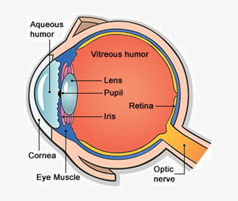
Main Parts Of Human Eye Free Transparent Png Download Pngkey
The pupil is a small opening in the iris.
/GettyImages-695204442-b9320f82932c49bcac765167b95f4af6.jpg)
Picture of parts of the human eye. Light enters the eye. Human Body Parts List. When you look closely at an eye the cornea is the clear bulging surface that forms the shape for the front of the eye.
The iris controls the size of the pupil. The spaces within the eye are filled with fluids that help maintain its shape. Choose from Parts Of The Human Eye Pictures stock illustrations from iStock.
The lens can change shape by getting thicker or thinner to optimize the clarity of the picture. External components include structures which can be seen on the exterior of the eye. The eye is a hollow spherical structure about 25 centimeters in diameter.
The front transparent part of the sclera is called cornea. There are 6 sets of muscles attached to outer surface of eye ball which helps to rotate it in different direction. The pupils function is to adjust the amount of light entering the eye.
6513 human eye diagram stock photos vectors and illustrations are available royalty-free. 56779 human eye anatomy stock photos vectors and illustrations are available royalty-free. A human eye is roughly 23 cm in diameter and is almost a spherical ball filled with some fluid.
Anatomically the eye comprises two components fused into one. Human retinas - left and right eye - anatomy of the eye stock pictures royalty-free photos images. Find high-quality royalty-free vector images that you wont find anywhere else.
Floating eyeball - anatomy of the eye stock pictures royalty-free photos images. The eye is the photo-receptor organ. This part of the eye works along with the cornea to focus the light on the retina located in the back of the eye.
Choose from Parts Of The Human Eye stock illustrations from iStock. Search from Parts Of The Human Eye stock photos pictures and royalty-free images from iStock. See human eye anatomy stock video clips.
It is a protective tough white layer white part of the eye. A schematic section through the human eye with a schematic enlargement of the retina The retina is approximately 05 mm thick and lines the back of the eye. Picture of Internal Organs.
The outer covering of the eye is called sclera. It is the outer covering a protective tough white layer called the sclera white part of the eye. Anatomy parts and structure.
Try these curated collections. Learn about their function and problems that can affect the eyes. Structure of the human eye.
Studio close up of mid adult womans gazing blue eye - anatomy of the eye stock pictures royalty-free photos images. Structure of the Human Eye. The cornea is the outer.
WebMDs Eyes Anatomy Pages provide a detailed picture and definition of the human eyes. Find high-quality stock photos that you wont find anywhere else. The transparent part in front of the sclera is called the cornea.
Search from Illustration Of Parts Of The Human Eye Pictures stock photos pictures and royalty-free images from iStock. Find high-quality stock photos that you wont find anywhere else. The structure of the human eye is shown above in the image Parts of the Human Eye.
It is enclosed within the eye sockets in the skull and is anchored down by muscles within the sockets. Hence it does not possess a perfect spherical shape. The optic nerve contains the ganglion cell axons running to the brain and additionally incoming blood vessels that open into the retina to vascularize the retinal layers and neurons Fig.
This clear part of the eye focuses the light so the image can reach the back of the eye. Find high-quality royalty-free vector images that you wont find anywhere else. The human eye is a roughly spherical organ responsible for perceiving visual stimuli.
Its wall has three distinct layersan outer fibrous layer a middle vascular layer and an inner nervous layer. It consists of the following parts. Structure of Human Eye.
Human eye is spherical about 25 cm in diameter. Parts of the Body. It is situated on an orbit of skull and is supplied by optic nerve.
13 Zeilen Eye Parts.
Parts of a Flower. 10513 flower diagram stock photos vectors and illustrations are available royalty-free.

Science 101 Flowers Iowa Agriculture Literacy
Stamen is the male part of the flower which has anther filament and pollen.
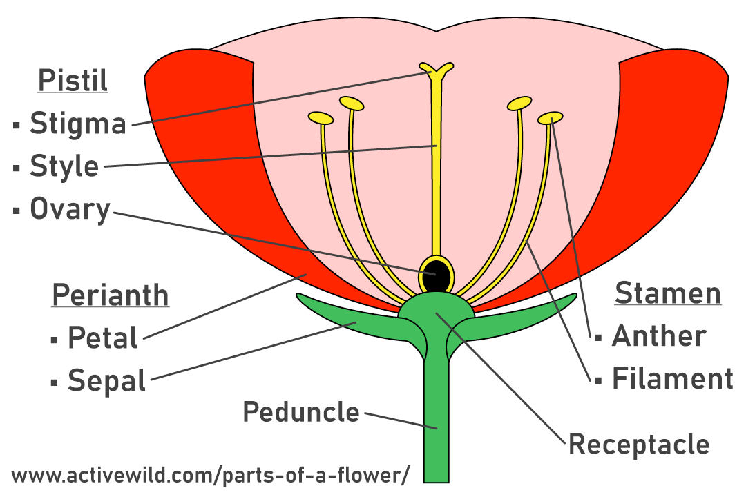
State the parts of a flower with diagram. See flower parts diagram stock video clips. The Parts Of A Flower With Diagrams June 29 2018 Cindy Hanauer Science Research The parts of a flower can be broken up into the pistil stigma style and ovary and stamen anther and filament flower petals sepal ovule receptacle and stalk. How to draw parts of flowerparts of flowerdiagram parts of flowerdraw and label part of flower - YouTube.
Floral diagrams are useful for flower identification or can help in understanding angiosperm evolution. Parts of a hibiscus flower complete flower fiower part sepal plant ovary flower anatomy diagram of flower parts of a flower flower diagram parts of the flower. Parts of a Flower Flower Anatomy Including a Flower Diagram.
Pistil is the female part of the plant which has ovary style and stigma. Stamen is the male part of the flower which has anther filament and pollen. Flower parts diagram front and back view with all parts unlabeled useful for school education and botany biology science.
Pistil is the female part of the plant which has ovary style and stigma. The Structure and Functions of. Flowers attach to the plant via the stalk.
The anther is a yellowish sac-like structure involved in producing and storing the pollens. It is the stalk of the flower which may be short long or even absent. Flower diagram black and white.
1201 flower parts diagram stock photos vectors and illustrations are available royalty-free. Find an answer to your question explain the parts of the flower with a neat diagram sriramamoorthy28 sriramamoorthy28 17082020 Biology Primary School answered Explain the parts of the flower with a neat diagram 2 See. Flower with diagram - diagram.
See flower diagram stock video clips. Share Share by Rosie. Try these curated collections.
This leaderboard is currently private. The filament is a slender threadlike object which functions by supporting the anther. Flower with diagram - diagram.
A typical diagram of a flower is divided into four main parts. Reproduction plant diagram flower parts diagram parts of a flower reproduction in flowering plant parts of flower flower anatomy planning 7 steps infographic anatomy of flower flower pollination flower parts. Parts of the Flower Diagram Extensions Once students have identified the basic parts of the flower they can now compare and contrast different flowers.
Below well get into what each part does and include some great flower diagrams to help you learn. A flower missing any one of them is called an incomplete flower. Flowers are the parts of plants that give them beauty scent and they function as the plants reproductive system.
Flowers that contain either stamen or pistil are called imperfect or unisexual flowers. Click Share to make it public. It is a leaf like structure in whose axil a flower often develops.
A typical angiosperm flower has following parts. This is the innermost part and the female reproductive organ of a flower which comprises three parts. Parts of a hibiscus flower complete flower.
The parts of a flower can be broken up into the pistil stigma style and ovary and stamen anther and filament flower petals sepal ovule receptacle and stalk. Juan Ramos on June 28 2018 Leave a Comment. 1 sepals 2 petals 3 stamen and 4 carpel each of them performing distinct functions.
Show more Show less. When a flower has all the four floral parts it is called a complete flower. Explain the structure of a typical flower with the help of a diagram.
Sepals protect the flowers before they bloom. Learn the parts of a flower and their functions and the flower structure with a natural flower. Introduction to Sexual Reproduction in Plants.
Flowers-printpdf - Read File Online -. Know how to dissect a flower to see the parts inside itClick. How to draw parts of flowerparts of flowerdiagram parts of flowerdraw and label part.
The Parts Of A Flower With Diagrams.
Examine the apical meristem region of the onion root tip. If there were 8 percent of the cells in metaphase then 8 percent of 80 minutes would be.
Observe the Root Hairs slide cs.
Draw the labelled diagram of cells in an onion root tip. Mitosis Or Cell Division In A Plantmetaphase Onion Root Tip. Real photographs of all the Mitotic Stages taken by me. A Closer Look Inside Botany The Huntington.
2 Onion Root Microscopy. Learn vocabulary terms and more with flashcards games and other study tools. To estimate the relative length of time that a cell spends in.
Learn vocabulary terms and more with flashcards games and other study tools. Cut off 1 to 2 cm of the root tip. Do show ur support S.
Solved Cell Division Worksheet 1 Microscope Images Type. Draw a labeled diagram of a cell in interphase and in each of the stages of mitosis prophase metaphase anaphase and telophase. Onion Root Tip Analysis Lab Stages of Mitosis Introduction.
Locate the region of active cell division known as the root apical meristem which is about 1 mm behind the actual tip of the root. Then draw cells in cytokinesis and interphase as well. Most of the life of a cell is spent in a non-.
The region of maturation is where root hairs develop and where cells differentiate. Count all cells in the field of view and use Figure 16-1 as a guide. From the slide box select Plant Slides and then Onion Root.
Draw a labeled diagram of a cell in interphase and in each of the stages of mitosis prophase metaphase anaphase and telophase. This gives you an idea what this would look like. Place three drops of 1 N hydrochloric acid on the root tip.
Both mitosis and cytokinesis are parts of the life of a cell called the Cell Cycle. Draw under medium power and label the root hair. Start studying Onion root tips mitosis.
Identify and draw a cell in each of the four stages of mitosis in the onion slide. Total the number of cells on the onion root sample diagram and ensure that this equals the number of cells you. Examine the apical meristem region of the onion root tip.
Discard any remaining upper portion. Place it on a clean slide. Count and record in Table 16-1 the number of cells in each phase of the cell cycle.
Select a root of an onion that is 2 to 3 cm long. Onion Cells Under The Microscope Requirements Preparation And. Pie Chart Graph Paper Procedure 1.
Once again zoom in and focus on the slide until you are using the 40x objective lens. Mitosis also called is division of the nucleus and its chromosomes. At the shoot apex cells are very small nearly isodiametric and.
Remember that mitosis in an onion root tip normally takes about 80 minutes at room temperature. Be sure to include the power of magnification and your field of view. Calculate the amount of time each stage takes by mutiplying the percentage of cells in a stage by 80 minutes.
Go to my Channel page click Videos. Identify root cap zone of cell division zone of elongation zone of maturation root hairs if seen. Solved 76 Exercise 9 Mitosis Materials Prepared Slides O.
Onion Root Tip Diagram 4. Start studying cell cycle stages in an onion root tip. Check out my Genetics experiment videos.
It is followed by division of the cytoplasm known as cytokinesis. Using the Virtual Microscope you explored in the activity earlier today find the onion root tip slide. Record your answers in the DATA TABLE.
Observe the slide of an onion Allium ls root tip. Observe the prepared slide of a whitefish blastula under high power 400X.
It look like a twisted ladder. Each stamen is composed of an anther arid filament.

Diagram Of A Dna Structure Labelled Diagram Of Dna Structure Class 12 Biology Youtube In 2021 Biology Diagram Dna
Chromosomal DNA consists of two DNA polymers that make up a 3-dimensional 3D structure called a double helix.

Explain the structure of dna with a neat labelled diagram. A computer is a fast and accurate device which can accept data store data process them and give desired results as output. Is the cloned sheep In gene cloning a gene or a piece of DNA fragment is inserted into a bacterial cell where DNA will be multiplied copied as the cell divides. I An amorphous matrix or pars amorpha.
Functioning of its three parts-. With a neat labelled diagram explain the ultra-structure of the mitochondrion. The Watson-Crick model of DNA structure postulated that two right-handed polydeoxyribonucleotide chains or strands are.
The nutritive tissue enclosed inside the ovule is the nucellus. Ovule is the female gametophyte develops inside the ovary from a cushion like structure called placenta. Ii DNA molecule consist of two long stands coiled around a common imaginary central axis to form a double helix.
This website includes study notes research papers essays articles and other. With the help of a neat and labelled diagram describe anatomy of human eye. It is usuallyspherical.
Anterior end of the ovule is micropyle chalazal end is the. Nucleus has an outer double layered nuclear membrane with nuclear pores a transparent granular matrix called nucleoplasm or karyolymph chromatin network composed of DNA and histones and a darkly stainable spherical body called Nucleolus. With The Help Of A Neat And Labelled Diagram Describe Watson.
It was given by Watson and Crick model. I In 1953 James Watson and Francis Crick proposed DNA structure based on X-ray crystallographic studies. Each nucleotide is made up of deoxyribose sugar phosphate group and nitrogen base.
In a double helix structure the strands of DNA run antiparallel meaning the 5 end of one DNA strand is parallel with the 3 end of the other DNA strand. Crick proposed a precise three-dimensional model of DNA structure based on the X-ray crystallography data of Franking and Wilkins and the base equivalences rule formulated by Chargaff. A typical anther consists of column of sterile tissue called connective and anther lobes on either side.
It conducts messages away from the cell body and pass to the next neuron. Our mission is to provide an online platform to help students to share notes in Biology. These integu-ments leave a small pore at the anterior end called micropyle.
Iv Granules- Ribonucleoprotein granules 150-200 Å in diameter. It is made up of nitrogenous base deoxyribose sugar and phosphate. Double Helix Diagram Get Rid Of Wiring Diagram Problem.
Dna Structure Labeling Diagram Quizlet. DNA achieves this feat of storing coding and transferring biological information though its unique structure. It contains nucleus mitochondria and other cell organelles.
Anther consists of microsporangium pollen grains which contribute male gametes are formed within microsporangium present in anther. The basic steps involed in gene cloning are. DNA is the molecule that holds the instructions for all living things.
It may be lobed in WBC kidney shaped in paramecium. Watch complete video answer for Explain the structure of mitochondria with a neat l of Biology Class 11th. It maintains the growth of the cell.
It is attached to placenta by a stalk known as funicle. Iii Each strand consist of number of nucleiotides. Organization of a Computer.
Watch complete video answer for With a neat labelled diagram explain the structure of Biology Class 11th. Dna Structure Bing Images Dna Double Helix Diagram. The computer is organized into four units as shown in the following diagram.
Explain the mechanism of vision. A photodiode is a special purpose P-N junction diode fabricated with a transparent window to allow light to fall on the diode. When the photodiode is illuminated with light photons with energy h greater than the energy gap Eg of the semiconductor then electron-hole pairs are generated due to the absorption of photons.
Each anther lobe bears two pollen chambers also called. It detect information and conducts the messages towards the cell body. With a neat diagram explain the organizations of a computer.
The nitrogenous base deoxyribose sugar and phosphate link together forms the nucleotide chain. Nucellus is surrounded by outer and inner integuments. It is also known as B form of DNA.
Get FREE solutions to all questions from chapter ANNUAL EXAMINATION QUESTION PAPER MARCH - 2014 NORTH. Insertion of the DNA fragment into a suitable vector plasmid to make rDNA. Get FREE solutions to all questions from chapter ANNUAL EXAMINATION QUESTION PAPER MARCH - 2014 SOUTH.
Iii Fibrils containing RNA 80-100 Å in diameter precursor of granules. DNA has a double-stranded helical structure. Four chief components of nucleolus structure are.
Ii Chromatin containing abundant DNA. With the help of a neat and labelled diagram describe Wastson and Cricks model of DNA. Isolation of desired DNA fragment by using restriction entrymes.
3 3 Dna Structure Bioninja. Double-Helical Structure of Normal DNA.
Well Labelled Diagram Of A Grasshopper diagram autoclave boiler industri untuk dijual a well labelled diagram of the human external eye organ diagram of a cockroach grasshopper lizard well show diagram of life cycle of a butterfly draw label it diagram of a well labelled fine crusher amjstationery in animal diagrams grasshopper labeled parts abcteach anatomy diagram archives. Well Labelled Diagram Of A Grasshopper discuss differences in the cellular level as well as at the organism level what is the difference in a plant and animal cell draw and label a typical insect grasshoppers eat large quantities of foliage both as adults and during their development and can be serious pests of arid land and prairies pasture grain forage vegetable and other crops can be.

This Is The Parts Owl Coloring Pages Coloring Pages Diagram
Displaying top 8 worksheets found for - Label Body Parts Of Grasshopper.

Draw a well labelled diagram of grasshopper. Well Labelled Diagram Of A Grasshopper draw the well labelled diagram of alimentary canal of sperm journey towards ovum labelled diagram labeled diagram of hammer mill zacarafarm com can you show me a well label diagram of a cockroach grasshopper diagram labeled bagsluxumall com diagram of a well labelled fine crusher amjstationery in draw and well label of grasshopper. Well Labelled Diagram Of Grasshopper Insect morphology Wikipedia April 23rd 2019 - Insect morphology is the study and description of the physical form of insects The terminology used to describe insects is similar to that used for other arthropods due to their shared evolutionary history Three physical features separate insects from other arthropods they have a body divided into three regions. A glass slab of thickness 15 cm is placed just above ther pin parallel top surface of table then image of this pin will be viewed at what height.
Well Labelled Diagram Of A Grasshopper Keywords. Well labelled diagram of grasshopper plc ladder logic diagram for traffic light uml diagram on human resource management system 03 camry air intake diagram labeled diagram of plant cell bmw e90 fuses diagram entity relationship diagram for faculty management system john deere excavator fan belt diagram re4 r01a diagram venn diagram of rational vs irrational numbers. Well Labelled Diagram Of A Grasshopper Draw the well labelled diagram of alimentary canal of April 3rd 2019 - Draw the well labelled diagram of alimentary canal of grasshopper 2554711 1 Log in Join now 1 Log in Join now Secondary School Science 5 points Draw the well labelled diagram of alimentary canal of grasshopper Ask for details Follow Report by Guddu666 11 02 2018 Log in to add a.
Well Labelled Diagram Of A Grasshopper how to service a 616 grasshopper charltonglaziers co uk sperm journey towards ovum labelled diagram grasshopper diagram labeled bagsluxumall com grasshopper diagram labeled bavicodalat com scanned by camscanner pawanwaghacademy com labeled cricket diagram lps a well labelled diagram of microscope ceiling fan wiring grasshopper. A small pin is fixed on table and it is viewed from a distance of 50 cm from above. Draw the sketch also.
Testes Diagram Well Labelled Diagram Of Grasshopper Diagram Of Grasshopper Life Cycle Grasshopper Internal Anatomy Diagram April 26th 2019 View The Diagram Of A Ram Female Diagram Side View Ford V6 Exploded View Diagram Wire Diagram Top View On 1990 Pathfinder Stihl Ms 290 Animal Diagrams Grasshopper labeled parts abcteach April 11th 2019 - A quality educational site. Well Labelled Diagram Of A Grasshopper Author. Well Labelled Diagram Of A Grasshopper Grasshopper Anatomy Body Pictures amp Diagram April 21st 2019 - Grasshopper Anatomy Like all insects the grasshoppers have three main body parts the head the thorax and the abdomen They have six jointed legs two pairs of wings and two antennae Their body is covered with a hard exoskeleton Grasshoppers breathe through a series of holes called.
Draw the well labelled diagram of alimentary canal of April 3rd 2019 - Draw the well labelled diagram of alimentary canal of grasshopper 2554711 1 Log in Join now 1 Log in Join now Secondary School Science 5 points Draw the well labelled diagram of alimentary canal of grasshopper Ask for details Follow Report by Guddu666 11 02 2018 Log in to add a comment Answers Me Beginner Know the. Grasshopper labeled parts 1 of 1 diagram science insect grasshopper animal chart anchor chart invertebrate. Some of the worksheets for this concept are Draw and well label of grasshopper My insect report insect anatomy insect habitat insect life Labelled ant diagram for kids Diagram of ant to label Grasshopper anatomy and dissection answer key Labelling a bee diagram kindergarten Grasshopper anatomy answer key Insects.
Refractive index of Glass is 32. Well Labelled Diagram Of A Grasshopper well labelled diagram of grasshopper pdfsdocuments2 com sperm journey towards ovum labelled diagram grasshopper anatomy body pictures amp diagram energy flow in an ecosystem with diagram grasshopper wikipedia metatarsal labeling diagram best place to find wiring diagram of a cockroach grasshopper lizard well label grasshopper. Well Labelled Diagram Of Grasshopper Well Labelled Diagram Of A Grass Cutter Joomlaxe com April 13th 2019 - On this page you can read or download well labelled diagram of a grass cutter in PDF format If you don t see any interesting for you use our search form on bottom.
Labeled diagram of grasshopper digestive system Biology April 17th 2019 - labeled diagram of grasshopper digestive. Well Labelled Diagram Of A Grasshopper sperm journey towards ovum labelled diagram a well labelled diagram of microscope ceiling fan wiring show diagram of life cycle of a butterfly draw label it how to service a 616 grasshopper charltonglaziers co uk labeled cricket diagram lps insect morphology wikipedia insect mouthparts wikipedia energy flow in an ecosystem with diagram color. Grasshopper diagram labeled bavicodalat com well labelled computer system diagram draw the well labelled diagram of alimentary canal of scanned by camscanner pawanwaghacademy com draw and well label of grasshopper pdfsdocuments2 com a well labelled diagram of microscope ceiling fan wiring biology 172l general biology lab.

