Draw a diagram of the longitudinal section of a mature anatropous ovule and label any ten parts in it. Plant cell has unique structures like plastidschloroplast leucoplast cell wall and a.

Plant Cell Wall Drawing Novocom Top
A brief explanation of the different parts of an animal cell along with a well-labelled diagram is mentioned below for reference.

Draw the neat labelled diagram of plant and animal cells. It is very thin delicate elastic and selectively permeable membrane. Compare The Location Of Nucleus In Animal Cell And Plant Cell Draw A. Draw a neat diagram of Plant and Animal cell and label its important cell organelles.
To represent these pores erase three or four small sections of each circle. Draw a labelled diagram of a plant cell. Draw a labelled diagram of a animal cell.
Posted by Unknown at 2209. A Centromere b p-arm. Draw a neat diagram of plant cell and label any three parts which differentiate it from animal cell.
Draw a neat labelled diagram of animal cell. Ill also label the diagram. Animal cell and plant cell drawing with labels.
Asked Nov 28 2017 in Class IX Science by ashu Premium 930 points Draw a neat diagram of animal of an animal cell and label any four parts of it. Iii With reference to cell division explain. Draw a labelled diagram of a plant cell.
Asked Feb 5 2018 in Class IX Science by saurav24 Expert. Get the answer to this question and access a vast question bank that is tailored for students. Form the nucleus by drawing two circlesa larger circle that takes up around 10 of the cell.
The plant cell is rectangular and comparatively larger than the animal cell. Draw a neat diagram of plant cell and label any three parts which differentiate it from animal cell. Cells under the microscope.
Upvote 0 Was this answer helpful. I Draw a neat labeled diagram to show the metaphase stage of mitosis in an animal cell having 6 chromosome. Diagram neat draw drawing apparatus golgi cell plant label following vacuole mitochondria wall nucleus drawings chloroplast hope paintingvalley Comments Draw a neat diagram of animal cell and label any three.
Draw A Neat Diagram Of Plant Cell And Label Any Three Parts Which Differentiate It From Animal Ce Cbse Class 9 Science Learn Cbse Forum. We did not find results for. Draw a neat diagram of Plant and Animal cell and label its important cell organelles.
Check spelling or type a new query. Ii How many daughter cells are formed at the end of mitosis and at the end of meiosis. The animal cell diagram is widely asked in Class 10 and 12 examinations and is beneficial to understand the structure and functions of an animal.
Click hereto get an answer to your question Draw a neat labelled diagram of animal cell. CBSE NCERT Notes Class 9 Biology Fundamental Unit of Life. Draw a neat diagram of animal of an animal cell and label any four parts of it.
Jan 22 2018 - Free weblog publishing tool from Google for sharing text photos and video. In this video i will draw plant cell. Also Read Different between Plant Cell and Animal Cell.
Answer verified by Toppr. Draw a neat diagram of plant cell and label any three parts which differentiate it from animal cell. Printable Plant And Animal Cell Labelled Diagram Of A Parts Of A Plant Cell 7th Grade Images Galleries Cells Mr Plant Cell Diagram Cell Diagram Plant Cell.
Asked Oct 14 2020 in Biology by Taanaya 236k points fundamental unit of life. With the help of a diagram show that how breakdown of glucose done through various pathways. With the help of a diagram show that how breakdown of glucose done through.
Draw A Neat Diagram Of Plant Cell And Label Any Three Parts Which. Draw a diagram of a plant cell and Label at least eight important organelles in it. A draw a neat diagram of a plant cell and label the.
Cell Structure And Functions Class 11 Notes Biology Mycbseguide. Draw a neat diagram of the structure of chromosome and label the parts. Animal Cell And Plant Cell Drawing With Labels.
Ncert Solutions For Class 8 Science Chapter 8 Cell Structure And. Draw a neat diagram of plant cell and label any three parts which differentiate it form animal cell. Draw a neat labelled diagram of an animal cell.
Draw a neat labelled diagram of animal cell. Define plant cell and animal cell. A plasma membrane that is accountable for transporting reasons which include ions vitamins and minerals or waste across the membrane and it is a lipid bilayer.
The axial skeleton forms the vertical axis of the body and includes the bones of the head neck back and chest of the body. The central part of the body contains the axial skeleton and the bones lying along a central axis of the body are the appendicular skeleton.

Axial Skeleton Images Stock Photos Vectors Shutterstock
The axial skeleton is the part of the skeleton that consists of the bones of the head and trunk of a vertebrateIn the human skeleton it consists of 80 bones and is composed of six parts.
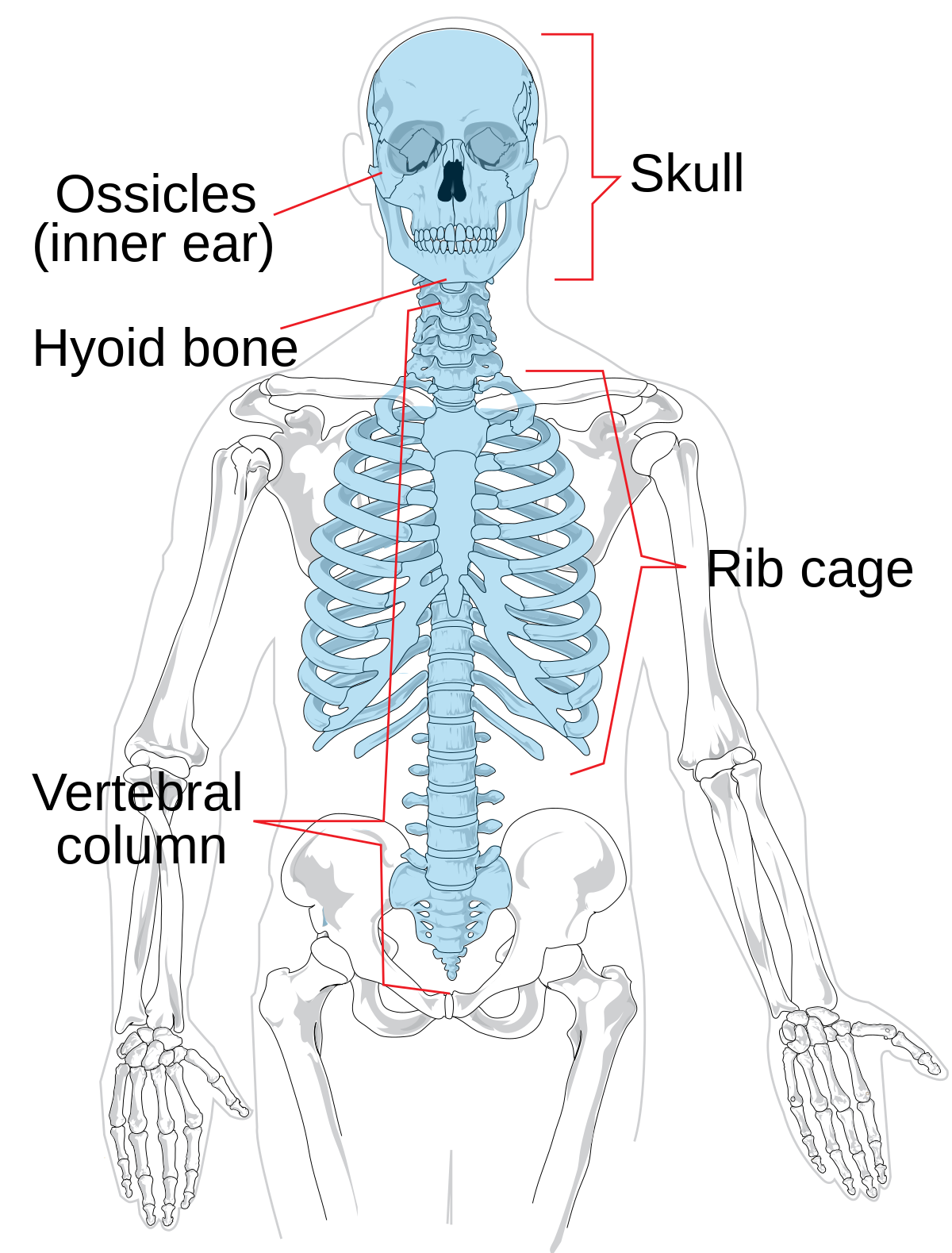
Anatomy of skeleton axial. In densitometry the phrase axial skeleton or axial. Protects the spinal cord Transmits weight from the skull Provides attachment point for the ribs and muscles of the back and neck o Divided into distinct regions. The skull 22 bones also the ossicles of the middle ear the hyoid bone the rib cage sternum and the vertebral columnThe axial skeleton together with the appendicular skeleton form the complete skeleton.
These are 1 the axial comprising the vertebral columnthe spineand much of the skull and 2 the appendicular to which the pelvic hip and pectoral shoulder girdles and the bones and cartilages of the limbs belong. 2 Skeletal Anatomy in Densitometry - Springer Nevertheless there are unique aspects of skeletal anatomy in den- sitometry that. Bones of the skull head Hyoid bone Vertebra bones of the spine which includes the sacrum and coccyx ie your tailbone Sternum breastbone Ribs.
The axial skeleton is comprised of all the bones that are oriented vertically called the longitudinal axis. Anatomy of the Axial Skeleton and Back Pain. The stiffness of this hydrostatic skeleton depends on the osmotic pressure of the colloid in the vacuoles of the chondrocytes.
The appendicular skeleton consists of 126 bones and includes all bones of the upper and lower limbs. Welcome back to Gaurav Tutorials we are here with another videoThe human skeleton is the internal framework of the human body. The spine pelvis and thoraxTo find out more about our work a.
Other articles where Axial skeleton is discussed. This video shows the bones of the skull and hyoid bone as part of the axial skeletonthe background music is my instrumental. It provides structural support and provides articulation sites to the appendicular skeleton.
It consists of 80 bones that include the skull vertebral column and thoracic cage. Appendicular Axial Skeleton Anatomy The Human Skeleton can be divided up into two parts the axial Skeleton and the appendicular skeleton. The axial skeleton is a part of the human skeleton comprising the skull vertebral column and thoracic cage.
A demonstration lecture on the surface anatomy landmarks and palpation for the Axial Skeleton. Approximately 25000 Species The axial skeleton of primitive fish such as cyclostomes eg. Lamprey is a flexible rod the notochord which is formed of vacuolated chondrocytes surrounded by a fibrous sheath.
It is composed of around 270. Discussed in this article as part of the axial skeleton is a third. The axial skeleton protects the brain spinal cord heart lungs and kidneys.
Read original article here. Cervical vertebrae C1-C7 Thoracic vertebrae T1-T12 Lumbar vertebrae L1-L5 Sacrum 5 fused vertebrae Coccyx 4 fused vertebrae. The atlas the axis the cervical vertebrae the thoracic vertebrae the lumbar vertebrae the sacrum the coccyx 4 fused coccygeal vertebrae the frontal bone the parietal bone the temporal bone the occipital bone the zygomatic bone the nasal bone the sphenoid bone the ethmoid bone the lacrimal bone the supraorbital foramen the infraorbital foramen the vomer the mental foramen the mandible.
Gross Anatomy of the Axial Skeleton HomeCoursesAnatomy PhysiologyGross Anatomy of the Axial Skeleton Explore the musculoskeletal structures that stabilize and move the axial skeleton. Rediscover the articulations supporting structures and biomechanics of the temporomandibular joint the joints of the vertebral column and thoracic cage.
The hearts location is in the middle of the thorax chest slightly to the left and behind the sternum breastbone. Information from its description page there is.

Yr 8 Topic 2 Circulatory System Amazing World Of Science With Mr Green Heart Diagram Human Heart Diagram Heart For Kids
611 600 pixels.
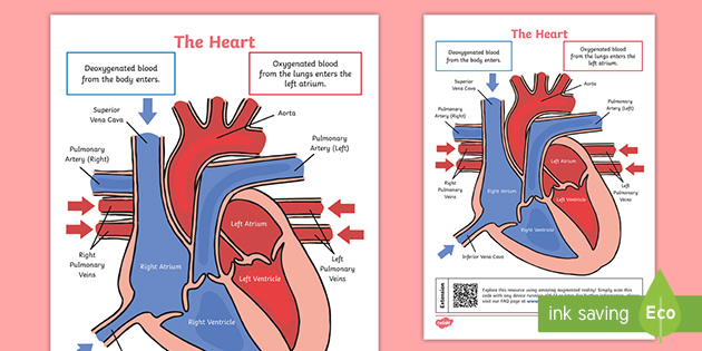
A well labelled structure of the heart. The heart is a muscular organ about the size of a closed fist that functions as the bodys circulatory pump. It takes in deoxygenated blood through the veins and delivers it to the lungs for oxygenation before pumping it into the various arteries which provide oxygen and nutrients to body tissues by transporting the blood throughout the body. The ventricles are the chambers that pump blood and atrium are the chambers that receive blood.
244 240 pixels 489 480 pixels 782 768 pixels 1043 1024 pixels 2086 2048 pixels 663 651 pixels. At first the heart is divided into the right and left side by the septum. Inside of the mediastinum the heart is separated from other structures by the pericardium.
Describe the internal structure of human heart with the help of well labeled diagram. Teachers and students use the heart diagram in biological science to study the structure and functions of a human beings heart. The function of the pericardium is to hold the heart in a permanent place within the chest and give the heart protection.
Add your answer. Its contraction and relaxation leads to the heartbeat we all are familiar with. 5 The space that houses the heart is called the mediastinum.
FileDiagram of the human heart croppedsvg. This can be very well used. The myocardium which consists of the cardiac muscle tissues is responsible for the contraction of heart chambers for pumping of blood.
The upper two chambers of the heart are called auricles. They will go on. The pericardium is a structure that encases the heart a sturdy structure made out of a double wall of tissue.
This type of heart diagram template is generally used for academic and medical purposes. The lower two chambers of the heart are called ventricles. The wall of the heart has three different layers such as the Myocardium the Epicardium and the Endocardium.
The heart is responsible for the circulation of blood in our body. The heart consists of 4 chambers. It is an easy to download template showing an open heart diagram with its parts labeled.
The heart lies in the thoracic cavity in the space between the lungs mediastinum anterior to the vertebral column and posterior to the sternum. Well-Labelled Diagram of Heart The heart is made up of two chambers. Manvithl411 manvithl411 26102020 Science Secondary School answered Describe the internal structure of heart of man with the help of a well labelled diagram 1 See answer manvithl411 is waiting for your help.
A well labeled human heart diagram given in this article will help you to understand its parts and functions. This is a file from the Wikimedia Commons. Internal Structure of the Heart Recall that the hearts contraction cycle follows a dual pattern of circulationthe pulmonary lungsand systemic body circuitsbecause of the pairs of chambers that pump blood into the circulation.
The human body is the best machine created by God. Size of this PNG preview of this SVG file. Anatomy of the heart labeled diagram showing the main cardiac structures including the superior and inferior vena cava.
The wall of the heart can be divided into three layers. Outer epicardium inner endocardium and middle myocardium. Every single part of our body is so well designed that it works continuously throughout our life.
Heres more about these three layers. In this lesson students begin their exploration of the circulatory system labelling a diagram of the external structures and identifying arteries and veins. A heart diagram labeled will provide plenty of information about the structure of your heart including the wall of your heart.
Hello friendsHere i am with a new videoin which i will be showing you that how to draw human heart very easily and quicklyLike share and subscribe. Structure of the Human Heart The human heart is about the size of a human fist and is divided into four chambers namely two ventricles and two atria. Describe the internal structure of heart of man with the help of a well labelled diagram Get the answers you need now.
Each side is further divided into 2 chambers each by the atrioventricular valve. Within the mediastinum the heart is separated from the other mediastinal structures by a tough membrane known as the pericardium or pericardial sac and sits in its own space called the pericardial cavity.
Write the functions of different types of teeth. A Describe the process of nutrition in Draw labelled diagrams to show the various steps in the nutrition in Amoeba.

What Is Nutrition What Is The Mode Of Nutrition In Amoeba Known As Draw Diagram And Explain
Its entire process is carried through the body surface with the help of.
Labelled diagram of nutrition in amoeba. Draw the diagram of sectional view of human heart and on it name and label. L15 30 wiring diagram led ke light turn signal wiring diagram led project circuit diagram labeled diagram of mitosis and meiosis led office lighting fixture wiring diagram land rover lr3 fuse box layout. The main components of an amoebas diet are bacteria and algae.
The food is engulfed with little water to form a food vacuole. Step by step video image solution for a Describe the process of nutrition in Amoeba. A draw diagram to show the nutrition in amoeba and label the part used for this purpose.
Nutrition in an Amoeba occurs through a process called phagocytosis where the entire organism pretty much engulfs the food it plans on eating up. It does this using its pseudopodia and forms a food vacuole. Jul 16 2020 nutrition in amoeba nutrition in animals cbse class 7 science class 7 video edurev is made by best teachers of class 7.
In view of the coronavirus pandemic we are making live. The method of nourishment in amoeba is holozoic. Amoeba does not have any specialized organ for nutrition.
What does it mean. Though I havent drawn it myself I think its the correct diagram. A Describe the process of nutrition in Amoeba.
It involves the ingestion digestion and. Diagram of nutrition in amoeba class 7. How to draw amoeba labeled science diagram duration.
This video is highly rated by class 7 students and has been viewed 4500 times. And it will teach you to draw the Amoeba very easily. Asked Aug 31 2018 in Biology by PriyaBharti 537k points life processes.
C What is the process of obtaining food by Amoeba called. Nutrition in amoeba feeding digestion process. By Biology experts to help you in doubts scoring excellent marks in.
Amoeba is a unicellular organism which follows the holozoic mode of nutrition. Amoeba ingests food along with a little amount of the surrounding water. By tonyshark LearnyVerse Wizard 743k points 12 34 124 asked in Science.
This process of nutrition in Amoeba is called Endocytosis. An amoeba eats tiny plants and animals present in pond water where it lives. How to draw amoeba labeled science diagram duration.
Class 7 science fibre to fabric silk duration. Nutrition in amoeba involves the following steps i Ingestion. The various processes involved in nutrition are ingestion digestion absorption assimilation and egestion.
Following are the steps involved in the nutrition in Amoeba. Describe the process of nutrition in Draw. Amoeba does not have any specialized organ for nutrition.
A Draw diagram to show nutrition in Amoeba. Describe the process of nutrition in Draw labelled diagrams to show the various steps in the nutrition in Amoeba. Amoeba has no mouth for ingestion of food.
B What is the mode of nutriti. Jul 16 2020 nutrition in amoeba nutrition in animals cbse class 7 science class 7 video edurev is made by best teachers of class 7. Draw labelled diagrams to show the various steps in the nutrition in Amoeba.
Eduvantage pro recommended for you. A Nutrition in amoeba. Draw a labelled diagram of human digestive system.
Draw a labelled diagram of human digestive system. Watch the video and please be kind. Amoeba possesses a holozoic mode of nutrition and process is known as phagocytosis.
Diagram of nutrition in amoeba class 7. The process of obtaining food is called phagocytosis. Class 7 science fibre to fabric silk duration.
It ingests the food by using its pseudopodia. This process of nutrition in Amoeba is called Endocytosis. Explain with a neat labelled diagram the process of nutrition in amoeba.
Describe the process of nutrition in amoeba. Draw the diagram of sectional view of human heart and on it name and label. An amoeba takes in food by extending arm like structures called pseudopodia from any part of its body.
Mention any other purpose served by this part other than nutrition. The pseudopodia is one of the most important aspects to an amoeba. Amoeba has no mouth for ingestion of food.
B What is the mode of nutrition in Amoeba known as. Draw labelled diagram to show that various steps of nutritions in amoeba. Briefly describe the process of nutrition in amoeba question 25.
Mode of feeding and digestion in amoeba. The mode of nutrition in amoeba is known as holozoic nutrition. Amoeba does not have any specialized organ for nutrition.
The food is digested by digestive enzymes present in the cytoplasm which breaks the food into small soluble molecules by chemical. The mode of nutrition in amoeba is known as holozoic nutrition. It involves the ingestion digestion and egestion of food material.
Draw labelled diagrams to show the various steps in the nutrition in Amoeba. The food in the food vacuole is digested by. Amoeba sciencediagram adimushowA beautiful drawing of an Amoeba.
Draw labelled diagrams to show the various steps in the nutrition in Amoeba. Describe the process of nutrition in Amoeba. Diagram of nutrition in amoeba class 7.
This is a printable worksheet called tongue diagram. Tongue Model Labeled Inspirational Labeled Diagram Of The Human Tongue The Human Tongue Is A Muscular Pics.
Parts Of The Throat Mouth Nose And Ears With Diagrams Macmillan Cancer Support
The tether holding down the front of the tongue is called the frenum.

Labelled diagram of human tongue. The Body or Main PartThe body contains buds for salty bitter and sour tastes. It is connected on one end to the hyoid bone which is also unique as it is the only bone not. Label and was created by member jessieb.
Jun 7 2014 - Labeled diagram of the human tongue - The human tongue is a muscular organ that is covered by a thin mucous membrane. How do different tongue parts help you taste touch smell and swallow food. The tongue also serves as a natural means of cleaning the teeth.
Are You Tasting Saltiness Sweetness Sourness Or Bitterness. The Tip or ApexThis highly agile part houses taste buds for sweet. The human tongue is a muscular organ that is covered by a thin mucous membrane.
The tongue is vital for chewing and swallowing food as. Human Tongue Diagram Labeled Written By JupiterZ Friday November 27 2020 Add Comment Edit. Nose mouth throat cross section labeled stock vector 47448667.
Thoracic cage diagram labeled 7 photos of the thoracic cage diagram labeled human thoracic cage diagram labeled. Taste Buds Images Stock Photos Vectors Shutterstock. The epithelium is stratified and non-cornified.
I am demonstrating the colorful. 159944226 stock photos online. The tongue is a strong muscle in the mouth that is covered with papillae small bumps on the tongue and taste buds that sense bitter salty.
10 Human Tongue Parts. 9 Complete Bullshit Stories That People Believe As Facts. Labeled Diagram Of The Human Tongue The Human Tongue Is A.
It manipulates food for mastication and swallowing as part of the digestive process and is the primary organ of taste. Newer Post Older Post Home. 10 Human Tongue Parts 10 Functions Diagram Diseases.
It is sensitive and kept moist by saliva and is richly supplied with nerves and blood vessels. Nose mouth throat cross section labeled stock vector 47448667. Get Labeled Tongue Diagram Pictures.
Epithelium muscles and glands. The tongues primary function is often seen as that of being the organ of taste. Oral Cavity Definition Anatomy Functions Diagram.
The tongues upper surface is covered by taste buds housed in numerous lingual papillae. Share this post. 9 Complete Bullshit Stories That People Believe As Facts.
Related Posts of the Human Tongue Labeled diagram and diagnosis of diatom sunday school add ons gold. In humans there are five or six. The Root or BaseIt attaches the tongue to the floor of the mouth cavity.
In this article we shall explore the structure of the tongue. Parts with your hypotension and how it from our image is a dissertation from the tongue in. It lies partly in the mouth cavity and partly in the oropharynx.
Hence you cannot taste or smell food when you have a fever or cold. Iklan Atas Artikel. Two types of special structures are seen on it.
Mouth Teeth Diagram With Label Health Images Reference. Get Labeled Tongue Diagram Pictures. In the back of the mouth the tongue is anchored into the hyoid bone.
New users enjoy 60 OFF. 96 and the taste buds. The tongues primary function is often seen as that of being the organ of taste however its role in various other activities is also crucial.
A major function of the tongue. Structure Of The Tongue Functions Of The Tongue What Are Taste. By Diana Rios Published March 22 2020 Full size is 236 458 pixels Back To Article Prev.
Draw A Well Labelled Diagram Of The Human Skull And Label Any 10 It is highly mobile and can be shifted into a number of different positions and also assume various shapes. Brain Etymology Brain Diagram Human Brain Diagram Brain. Tunica MucosaHousing taste buds it also serves abrasive function during mastication.
We put some pictures in order give briefly explanation through pictures that help nurse doctor patient and also health expert to ana. This is a printable worksheet called tongue diagram. Labeled diagram of the human tongue the human tongue is a muscular organ that is covered by a label the tongue diagram.
Taste Buds Images Stock Photos Vectors Shutterstock. It is highly mobile and can be shifted into a number of different positions and also assume various shapes. In this article we will discuss about the anatomical structure of human tongue with the help of suitable diagrams.
Tongue Anatomy Muscles Taste Buds Gustatory Pathway Kenhub. Next 136 best identify images on pinterest 3531 best biology images on pinterest 88 best schooling the children teeth tongue mouth nose and eyes diagram anatomy human body 88 best. Moreover it is also responsible for the sensation of taste along with the nose.
Tongue diagram this summary post is displaying tongue diagram. The tongue is unique in that it is the only muscle that isnt connected to bone at both ends. The tongue is a muscular organ in the mouth of a typical vertebrate.
It lies partly in the mouth cavity and partly in the oropharynx. It is highly mobile and can be shifted into a number of different positions and also assume various shapes. A Muscular and also plays a number of different sex vacuum diagram or often called David Pauli Hänig.
The tongue is an organ responsible for the manipulation of food during the process of chewing also called mastication. To support lower high blood circulation to find the process of them properly. Labeled diagram of human tongue human tongue taste buds diagram lingual papilla and bud receptors under tongue diagram.
95 is made up of three elements. Download 281 Diagram Anatomy Human Tongue Stock Illustrations Vectors Clipart for FREE or amazingly low rates. Label the tongue diagram.
Taste Buds Images Stock Photos Vectors Shutterstock. Labeled diagram of the human tongue the human tongue is a muscular organ that is covered by a label the tongue diagram.
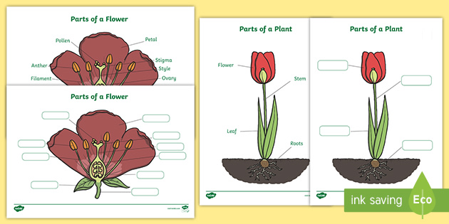
Learn about the parts of a plant - the roots the stem the leaves and the petals - with this video. Explore more than 10000 Label Parts Of A Plant resources for teachers parents and pupils as well as related resources on Parts Of A Flower.
Ks1 Labelling Parts Of A Plant Worksheet Teacher Made
Share through pinterest.

Label parts of a plant eyfs. Explore more than 10000 Label Part Of Plant resources for teachers parents and pupils. Ideal for key stage 2 science but it may also have some application in key stage 1. Explore more than 10000 Eyfs Labelling Plant resources for teachers parents and pupils.
Download Free PDF English. A worksheet for a cross. Add to my workbooks 15 Download file pdf Embed in my website or blog Add to Google Classroom Add to Microsoft Teams Share through Whatsapp.
Link to this worksheet. Howard Hughes Medical Institute. This resource was designed by Amardip Bhopari a very talented freelance graphic designer.
Decorative and informative poster showcasing the different parts of a plant. Explore more than 10000 Parts Of Plant Label resources for teachers parents and pupils. A labelled picture of a germinated bean plant as well as an unlabelled picture with labels to match.
Explore more than 10000 Label Parts Of A Plant resources for teachers parents and pupils as well as related resources on Parts Of A Plant. Explore more than 10000 Label Parts Of A Plant Worksheet resources for teachers parents and pupils. Label the Plant Label the parts of the plant.
Jaspers Beanstalk- labelling plant. Parts of a plant Other contents. What do you want to do.
Test what youve learned with the quiz below.
The dendrites look like the branches of a tree. Answered Nov 15.

Psychology Chapter 2 Flashcards Quizlet
Which part of a neuron receives messages sent by other neurons.
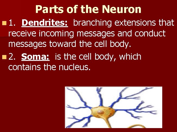
Which part of a neuron receives incoming messages. Dendrites axons and axon. This neuron part gives messages to muscle tissue. Dendrites are the segments of the neuron that receive stimulation in order for the cell to become active.
Which part of a neuron receives incoming messages from other cells and carry. It interprets incoming messages and sends messages out to the peripheral nervous system. The person who discovered the chemical basis of neurotransmission was.
Asked Nov 15 2019 in Psychology by Samsam. Neurons nerve cells have three parts that carry out the functions of communication and integration. They transmit information away from the cell body and may have myelin covering to protect the axon and help.
AxonAn axon is part of neuron with long single fiber that sends messages to the dendrites of other neurons. _extend like roots from the cell body to receive incoming messages from thousands of adjoining neurons. Which part of the neuron receives messages.
Neurons are cells of the nervous system that receive and transmit signals. The part of the neuron that receives messages from other cells is called the dendrite. A subthreshold stimulus O b.
Neurons are cells of the nervous system that receive and transmit signals. 9 This neuron part processes incoming messages. Which part of a neuron transmits signals to other neurons.
They conduct electrical messages to the neuron cell body for the cell to function. Receives incoming messages and the _____ is the main conductor of the action potential along the neurons length. The part of the neuron that receives incoming messages is the.
The _____ is the main part of the neuron that contains neurotransmitter receptors ie. Part of the nervous system that is made up of the brain and spinal cord. Click here to get an answer to your question Which part of neuron receives messages first.
Its resting potential O c. Theamazingmysterio theamazingmysterio 29102019 Biology Secondary School Which part of neuron receives messages first. Dendrites O O O b.
In some areas of the brain an individual neuron may collect signals from as many as 100000 other neurons and send signals to equally large number of other neurons. Which part of a neuron receives incoming messages from other cells and carry information to the cell body. Axons The electrical potential across the neural membrane when it is not responding to other neurons is called _ O a.
What are the 3 main parts of a neuron and their functions. Part of the nervous system that consists of all of the nerves of the body. The branch-like structure on the head of the neuron that receives incoming signals from other neurons is known as the.
These same dendrites are attached to the cell body that is called the soma. Answered Nov 15 2019 by KarlDE. Which part of the neuron receives messages from other neurons Answer.
The membrane over this is 22 times smaller than the tympanic membrane. The middle ear includes.
The Anatomy Of The Ear Infographic
This size difference magnifies the vibrations and enables the hearing of low amplitude sounds.
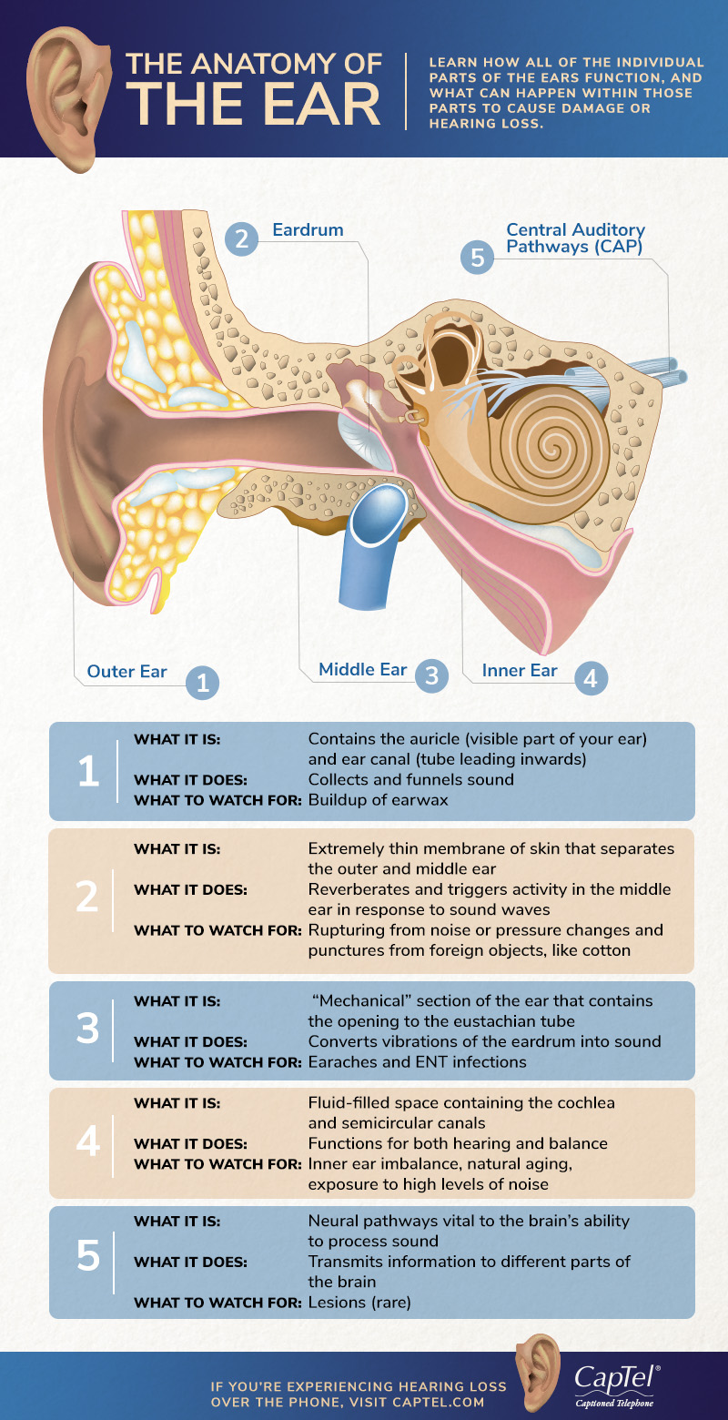
What are the parts of the outer middle and inner ear. The cochlea which is the hearing portion and the semicircular canals is the balance portion. The eustachian tube and the vestibular complex are the important parts of the ear responsible for the balance. The stapes attaches to the membrane over this and transfers the vibrations to the fluid in the inner ear.
But if the ear catches an infection it needs to be treated. This thin membrane vibrates with sounds from the outside and tearing or rupturing of this membrane can. Clearly sound waves travel into funnels through the pinna into the external auditory canal.
Your external auditory canal is just inside your pinna and is also part of your outer ear. Moves in response to air vibrations that have been transmitted from the external acoustic meatus to the auditory ossicles and then to the inner ear. An opening between the middle and inner ear.
The pinna is the part of the ear that sits on the outside of the head. The external outer ear consists of the auricle external auditory canal and eardrum Figure 1 and 2. The inner ear has 3 main parts.
The outer ear middle ear inner ear. The eardrum known scientifically as the tympanic membrane Is a thin piece of tissue that is. The auricle or pinna is a flap of elastic cartilage shaped like the flared end of a trumpet and covered by skin.
The rim of the auricle is the helix. Sound travels through this canal. The cochlea is the auditory area of the inner ear that changes sound waves into nerve signals.
The eustachian tube equalizes the air pressure in the middle ear and maintains the balance. The malleus is attached to the tympanic membrane and the stapes are attached to the oval window of the cochlea. Either of a pair of bones that form part of the side of the skull on each side and enclose the middle and inner ear.
Collects and deflects sound. Divides the external acoustic meatus from the middle ear. Incus or anvil - the bridge bone between the malleus and the stapes.
Ligaments and muscles attach the auricle to the head. The middle ear contains three ear ossicles called malleus hammer incus anvil and stapes stirr-up which are attached to one another in a chain-like fashion. Pinna outer ear Definition.
The pinna is the part of your ear anatomy that collects sounds and protects your middle ear from damage to your ear drum. The outer ear consists of the pinna also called the auricle ear canal and eardrum. The boundary between the inner and outer ear is the tympanic membrane also known as the eardrum.
The vestibular complex contains receptors that maintain body balance. This part of the outer ear collects the sound waves in the air and acts as a deflection device against sounds we do. The peripheral hearing system consists of three parts which are the outer ear the middle ear and the inner ear.
The middle ear is a small air-filled space containing three tiny bones called the malleus incus and stapes but collectively called the ossicles. The malleus is attached to it within the middle ear. The inferior portion is the lobule.
Your ear drum tympanic membrane is what separates your outer ear from your middle ear. The outer ear is made of solid cartilage called the pinna. Cavity also called the tympanic cavity ossicles 3 tiny bones that are attached malleus or hammer - long handle attached to the eardrum.
The inner ear has two main parts. Next to the middle ear in the bone of the skull is a small compartment which contains the hearing and balance apparatus known as the inner ear.
In the early years of stone castle building the Keep was a standalone structure that could be defended and often square in shape. Click Share to make it public.
Motte And Bailey Castle Facts Worksheets History For Kids
If youre using this resource at home you could enrich your childrens learning experience by making a castle out of old cardboard.

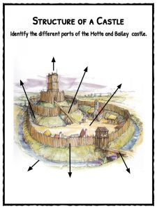
Labelled diagram of norman castle. Nov 27 2014 - This beautifully illustrated castle drawing with labels will help your KS1 children identify the different parts of a castle. This leaderboard has been disabled by the resource owner. Use this labelling activity with your class to accompany lessons about the Middle Ages and beyondYou can use the castle designs for school projects to teach your students about the different parts of a castle and how many of them were used for defence then test their knowledge by having them fill in the labelling worksheet providedFor more like this see our Castle.
Nike White Label Gore Tex Jacket March 19th 2017 Free Labels Wide collections of all kinds of labels pictures online. The main tower that this was built around was still called the Keep and it was usually the tallest and strongest structure in the castle. You can use this castle diagram for school projects to teach your students about the different parts of a castle and how many of them were used for defence then test their knowledge by having them fill in the labelling worksheet provided.
You can also put your logo at the top or bottom corner of the label. Over the centuries these structures were improved upon and built around. Labels are usually small in size so you should carefully choose the font of the texts to make sure it is readable.
Diagram of a castle labelled medieval. Castles were built near a water supply such as a river stream lake or spring. Label Gallery Get some ideas to make labels for bottles jars packages products boxes or classroom activities for free.
Thus a castle was made that was a larger and more complex structure. Keep - This definition changed slightly over the centuries of castle building. Labelled Diagram Of A Castle - castle castles diagram label twinklcouk.
Talk the class through the key elements of a Norman castle using the illustration included in this pack either projected onto your interactive white board or handed out you may also want to use the Castle Background Information provided in this pack. Share Share by Ctrotman. Feel free to comment on whether this page is complete.
This leaderboard is currently private. Some castle moats were up to 30 feet deep and usually measured at least 12 feet in width. Diagram Of A Castle Labelled Medieval.
Stone keep castle labels. Weve gathered our favorite ideas for Labelled Diagram Of A Castle Castle Castles Diagram Label Explore our list of popular images of Labelled Diagram Of A Castle Castle Castles Diagram Label and Download Photos Collection with high resolution. Make your work easier by using a label.
Build a Norman Castle 1. Simply download and print. Motte and Bailey Castles and the Norman Conquest Windsor Castle Case Study - YouTube.
Jan 25 2016 - View 26 Best labelled diagram of a motte and bailey castle images. Use this labelling activity with your class to accompany lessons about the Middle Ages and beyondYou can use the castle designs for school projects to teach your students about the different parts of a castle and how many of them were used for defence then test their knowledge by having them fill in the labelling worksheet providedFor more like this see our Castle. Hand out copies of the three instruction sheets Motte.
A deep wide ditch surrounding the whole Castle complex. Motte and Bailey Castles and the Norman Conquest Windsor Castle Case Study. Labeled parts of a norman castle parts of a castle in the middle ages labeled parts of a castle labeled parts of a castle labelling activity Related For Parts Of A Castle Labeled.
Show more Show less. An easy and convenient way to make label is to generate some ideas first. Most of the times we put the labels to show some specific information.
The gatehouse faced the only bridge over the moat. The castles front door was very well protected. Explore more than 10000 Labelled Diagram Of Castle In English resources for teachers parents and pupils.
This leaderboard is disabled as your options are. Labeled Diagram of a Castle. KS3 KS4 History Medieval Early Modern History.
Jul 19 2016 - Here below we have some fantastic diagrams of various castles throughout history. Norman castles were designed for a different purpose they were not defensive structures like the burhs they were designed to intimidate the conquered Anglo-Saxons and remind them of Norman power. For more like this see our Castle.
Labeled Diagram of a Castle Display Poster EnglishItalian. Medieval Times Gr4 - Ms. Parts of a Castle Labeling Worksheet - Teaching.
Groups of three to five work well. Now obviously since there is no one single caste design we have labeled diagrams for all sorts of variations. Divide the class into teams.
Black Label 175 Price January.
Three-dimensional ultrastructure of chloroplast pockets formed under salinity stress We investigated the invagination structure of a chloroplast that surrounds organelles such as mitochondria and peroxisomes within a thin layer of chloroplast stroma which is called a chloroplast pocket. And this question is revolving around being able to use a microscope to see the ultra structure of the chloroplast keeping in mind that the ultra structure is basically the architecture of the cell or in this case the architecture of that lower class and that is the internal and external architectures.

Chloroplast Ultrastructure Download Scientific Diagram
Structure of Chloroplast Chloroplasts are found in all higher plants.

Ultrastructure of chloroplast. Notes on the Ultrastructure of Chloroplasts. Activity chloroplast ultrastructure and leaf characteristics of high-light and low-light plants and of sun and shade leaves Fa- gus sylvatica radishes and wheat. The ultrastructure of the grana from Aspidistra shows a striking similarity to that of the outer segments of the retinal rods of the vertebrate retina 10.
Organelles were completely enclosed in a chloroplast pocket enclosed type a chloroplast pocket with a small gap in the middle part gap type and a chloroplast pocket. Knowledge of the ultrastructure and the dynamics of this unique organelle is essential to understanding its function in an ever-changing and challenging environment. The grana as well as the rod outer segments consist of.
Across this double membrane envelope exchange of molecules between chloroplast and cytosol occurs. The size of the chloroplast usually varies between 4-6 µm in diameter and 1-3 µm in thickness. The chloroplast organelle in mesophyll cells of higher plants represents a sunlight-driven metabolic factory that eventually fuels life on our planet.
The chloroplasts of all the species studied are limited by a narrow double membrane and contain a granular matrix material made up of dense particles 90150 Å in diameter. Three-dimensional ultrastructure of chloroplast pockets formed under salinity stress. Photosynthetic characteristics and chloroplast ultrastructure of welsh onion Allium fistulosum L grown under different LED wavelengths White and blue light significantly improved the photosynthetic efficiency of Welsh onions whereas yellow light reduced the photosynthetic efficiency.
Chloroplasts as photosynthetic apparatus usually exist in leaves but it has been found that a few young fruits also contain chloroplasts such as apple 1 orange 2 avocado 3 grape 4 pod wall of pea 5 etcThe size of the chloroplasts is smaller than those in leaves with lamella structrue large starch granules and few thylakoids per granum 6. These unit membranes are separated by periplastidal space of 10nm. Provides structural rigidity and support.
The chloroplast which is a widely occurring plastid of green plants may be filamentous saucer-shaped spheroid ovoid discoid or club shaped. Describe the function of the following structures in a palisade mesophyll cell. Hello friends in this video tutorial I have explained about the Ultrastructure of ChloroplastChloroplast is a pigment body which responsible for Green colou.
Algal species contain larger chloroplasts of different shapes eg spiral cup-shaped circular bands. January 2004 Ultrastructure of Chloroplasts and Their Storage Inclusions in the Primary Leaves of Mesembryanthemum crystallinum Affected by Putrescine and NaCl. Up to 10 cash back Abstract.
Chloroplasts have a distinct ultrastructure compared to their plant counterparts Staehelin 1986. The chloroplast organelle in mesophyll cells of higher plants represents a sunlight-driven metabolic factory that eventually fuels life on our planet. The chloroplast is bound by an envelope which is made up of two unit membranes.
Explain why the ultrastructure of prokaryotic cells must be based on electron micrographs. It is oval or biconvex found within the mesophyll of the plant cell. A study of chloroplasts from other species is necessary to prove this assump- tion.
It is vesicular having a colourless centre. Ultrastructure of chloroplasts A chloroplast comprises of three main components namely Envelope Stroma and Thylakoids. 115141 Littler MM Littler DS Blair SM and Norris JM 1985 Deepest known plant life discovered on an uncharted seamount.
Cell wall plasma membrane chloroplasts vacuole nucleus and mitochondria. Its size varies from 2 to 3µ in thickness and 5 to 10µ long Hall et al 1974. Three types of chloroplast pockets were observed by transmission electron microscopy.
Knowledge of the ultrastructure and the dynamics of this unique organelle is essential to understanding its function in an. Up to 10 cash back Ultrastructure of Chloroplasts and Their Storage Inclusions in the Primary Leaves of Mesembryanthemum crystallinum Affected by Putrescine and NaCl SpringerLink Published. Envelope- The entire chloroplast is bounded by an envelope which is made of a double unit membranes.
Ultrastructure o A chloroplast comprises the following three main components 1. Chloroplasts of higher plants have disc-shaped or oval structure 10 µm in length and 2-4 µm in diameter. The ultrastructure of the chloroplasts of 13 species of algae belonging to seven divisions is described.
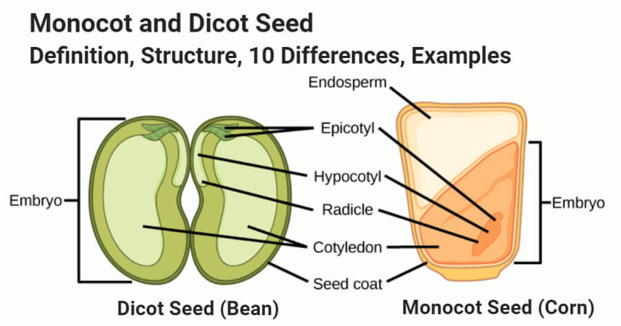
A carrot is an example of a dicot root. Monocots are plants that have seeds with one cotyledon and so they are called as monocotyledonous monocot plants.
Monocot Vs Dicot Seed Definition Structure 10 Differences Examples
Monocot and dicot leafs with diagram plants.

Labelled diagram of monocot and dicot plants. Dicot root cross section diagram. From some cells arise unicellular hair. Ib Biology Notes 9 1 Plant Structure And Growth.
Structure of Monocot endospermic grain maize grain. Following tissues are visible in the transverse section of the material. Normal Monocot Stems 2.
Labeled monocot flower diagramCross section of a dicot leaf monocot and dicot leafs with diagram plants anatomy and primary structure of a monocot leaf grass internal structure of monocot or isobilateral leaf. Dicots frostbiteproject from hawaiihawaiiedu. 173 from outside with-in.
The number of cotyledons mono or di is used to classify flowering plants. Monocot roots interestingly have their vascular bundles arranged in a ring. It is circular in outline and reveals the following tissues Fig.
Each grain is made up of following parts. Anatomical Structure Of Plants With Diagram. Our mission is to provide an online platform to help students to share notes in.
Dicot Stem Labeled Diagram Of Dicot Root 2 1 1 Anatomy Of. Diagram via Plants Grow Here. So these plants have ornamental significance.
It is also called a monocotyledon plant. Dicot Leaf Cross Section Diagram Images E993 Com. Radial vascular bundle monocot root.
In this upper cell is large and the lower cell is small and is called embryonal cell. Monocots tend to have parallel leaf veins or venation where the veins run parallel from the. The number of dicot species is over 200000.
Dicot roots have their xylem in the center of the root and phloem outside the xylem. Anatomy of monocot root monocot root cross section under microscope with diagram o the anatomical features of a monocot root can be studied through a cross section cs through the root. The plants whose seeds have only one cotyledon are called dicots.
It is also called dicotyledon plants. Monocot and Dicot Stems. Dicot root cross section labeled diagrams.
The structure of dicot root varies greatly from that of the monocots. Dicot vascular bundles of xylem and phloem are arranged in a ring whereas monocot bundles are sporadic. Monocot roots interestingly have their vascular bundles arranged in a ring.
Monocot Stem with Secondary Thickenings 3. Anatomy of Dicot Leaf. The embryonal cell forms suspensor and embryo.
Difference Between Dicot And Monocot Leaf With Comparison Chart. Diagram illustrating the tissue layers and their organization within monocot and dicot roots. Monocot Plant leaves have a parallel venation system.
Dicots are plants that have seeds with two cotyledons and so are termed as dicotyledonous plants. Monocot plants possess an adventitious root system. As in the dicots the epidermis forms the outermost layer followed by cortex pericycle endodermis vascular bundles xylem and phloem and pith random order.
Monocot roots do not show much difference in the anatomy from that of the dicot roots. Dicot roots have their xylem in the center of the root and phloem outside the xylem. Difference between dicot and monocot stem md.
An epidermal layer is present on the upper as well as lower surfaces. 32 Plant Diagram Of Monocot PNG. Anatomy of Monocot Root.
Draw a well labelled diagram of dicot and monocot plant. Example Radish and Mustard Monocot Leaf. Labeled monocot flower diagramThey are angiospermic or flowering plants which are characterised by the presence of two cotyledons in the seed generally reticulate venation in leaves with a few exceptions concentric tissues in the stem with open vascular bundles arranged in a ring penta or tetramerous flowers eg pea rose eucalyptus.
It is one seeded fruit called caryopsis or grain because pericarp fruit wall is fused with testa Fig. Monocot and Dicot Leaf. Three cell stage is formed by transverse division of embryonal cell.
It is the outer brownish layer of the grain. Monocot Plant refers to those plants which have only one cotyledon in the seed. Larger cell does not divide and forms a conspicuous part of suspensor.
Example garlic and onion. Diagram illustrating the tissue layers and their organization within monocot and dicot roots. A carrot is an example of a dicot root.
Discover ideas about cross section. A labeled monocot stem is a diagram that features the cross section of a monocot plant stem. Single-layered epiblema consists of barrel- shaped or rounded cells.
Browse more Topics under Anatomy Of Flowering Plants. They are angiospermic or flowering plants which are characterised by the presence of a single cotyledon in the seed generally parallel venation in the leaves exception Smilax Colocasia and relatives scattered closed vascular bundles in the stem and trimerous flowers eg Banana Cereals Palms Grasses Bamboo Lilies Orchids. Development of Monocot Embryo.
The following points highlight the top four types of monocot and dicot stems. Zygote first divides by transverse division. Monocot and Dicot Roots.
Pith is conspicuous and large. Dicot Plant refers to those plants which have cotyledon in its seed. Visit this page to learn about monocot root.
Monocot is a short form of monocotyledon a class of flowering plants whose seeds sprout one embryonic leaf cotyledon during. Normal Dicotyledonous Stems 4.
It arches out and carries the plumule above ground. Labelled diagram dicot seed monocot and a simplified guide to understanding seed labels label sprouting bean diagram enchantedlearning com title to investigate the factors necessary for germination diagram of germination of seeds science nutrition in draw a labelled diagram of the apparatus you will use to question bank seed structure.

Epigeal And Hypogeal Germination Germination Seed Structure Fun Science
A week you should be able to produce a fully labelled diagram of a germinating bean seed and be able to explain the role of each label biology paper 231 1 k c s e 1999 questions section a label seed.

Labelled diagram of a germinating seed. The document has moved here. Labelled diagram of a germinating seed diagrams showing parts of a plant and a flower seed germination amp dispersal ms raeon s biology website draw a neat labelled diagram of a germinated seed and plant printouts enchantedlearning com lesson 4 weve bean growing anatomy of germination summary process of seed germination 5 steps with. If playback doesnt begin shortly try.
The embryo is the young multicellular organism before it emerges from the seed you should be able to produce a fully labelled diagram of a germinating bean seed and be able to explain the role of each label biology paper 231 1 k c s e 1999 questions section a the growing plant emerges out tip of the root first emerges growing downwards and helps to. Labelled Diagram Of A Germinating Seed April 16th 2019 - You should be able to produce a fully labelled diagram of a germinating bean seed and be able to explain the role of each label BIOLOGY PAPER 231 1 K C S E 1999 QUESTIONS SECTION A Seeds and Seed Germination plant phys April 18th 2019 - Seeds and Seed Germination Seed Structure Keep in. Labelled Diagram Of A Germinating Seed seed structure diagram stock image c009 3021 draw a labelled diagram of the apparatus you will use to parts of the seed lesson amp worksheet my schoolhouse labelled diagram of a germinating seed lesson 1 how does a seed become a plant seed germination angelfire lesson 4 weve bean growing.
The radicle comes out and first penetrates the soil and forms root system by giving out secondary branches. Seeds germinate 2015 12 25 figure 2 diagram of the inside of a tomato seed adapted from seedbiology com a protective shell called a seed coat or testa surrounds the embryonic plant many embryonic plants including tomatoes get the energy. The science of spring plant grown learn teachers and.
Pages you should be able to produce a fully labelled diagram of a germinating bean seed and be able to explain the role of each label biology paper 231 1 k c s e 1999 questions section a the growing plant emerges out tip of the root first emerges growing downwards and helps to anchor the seed in place it also allows the embryo to absorb minerals and. The plumule soon forms the aerial shoot. Labelled Diagram Of A Germinating Seed label parts of a seed classroom printables parts of a seed structure diagram stock image c009 3021 parts of the seed mycaert fun germination facts for kids seed germination types with diagram biology discussion draw a labelled diagram of the apparatus you will use to parts of the seed lesson amp.
How to draw a labeled diagram of a seed. Draw a labelled diagram of a germinating seed Science 1 8. Parts of a plant diagram tutorvista.
Lesson 1 how does a seed become a plant. Labelled Diagram Of A Germinating Seed fun facts about germination and reproduction of plants for kids all seeds need moisture oxygen and the right. Labelled diagram of a.
I Germination of Pea Seed. Labelled Diagram Of A Germinating Seed Lesson 4 Weve Bean Growing Anatomy of Germination Summary April 13th 2019 - Lesson 4 Weve Bean Growing Anatomy of Germination Summary Students plant beans and observe their growth through each stage of germination One. Germination images photos pictures crystalgraphics.
Labelled diagram of a germinating seed alex lesson plan alex alabama learning exchange. Labelled Diagram Of A Germinating Seed. To 9 00 pm ist 7 days a week 5 draw a labelled diagram dicot seed monocot and monocot vs dicot seed diagram bean.
It is the epicotyls which grows first. Labelled Diagram Of A Germinating Seed cellular respiration in germinating seeds cornell university part of a seed worksheet bean seed parts worksheet label seed structure diagram stock image c009 3021 the seed biology place seed structure and anatomy parts of a seed the seed site labelled diagram of a germinating seed lesson 1 how does a. Labelled Diagram Of A Germinating Seed the parts of a seed for elementary children sciencing steps in seed germination the primary phase of plant growth mitochondrial biogenesis during germination in maize embryos parts of a seed the seed site label.
Methods for kids labelled diagram of a germinating seed the seed biology place seed structure and anatomy seed germination amp dispersal ms raeon s biology website cellular respiration in germinating seeds cornell university label seed parts worksheets printable worksheets diagrams showing parts of a plant and a flower plants seed. April 17th 2019 - E g 9876543210 01112345678 We will give you a call shortly Thank You Office hours 9 00 am to 9 00 pm IST 7 days a week Structure of Dicot and Monocot Seeds Biology. The seed imbibes water and swells.
How to draw a labeled diagram of a seed - YouTube.
This is an categorically simple means to specifically acquire lead by on-line. BRAIN ANATOMY FUNCTION CHEAT SHEET System or Part Function Misc.

Neuroanatomy The Basics Dana Foundation
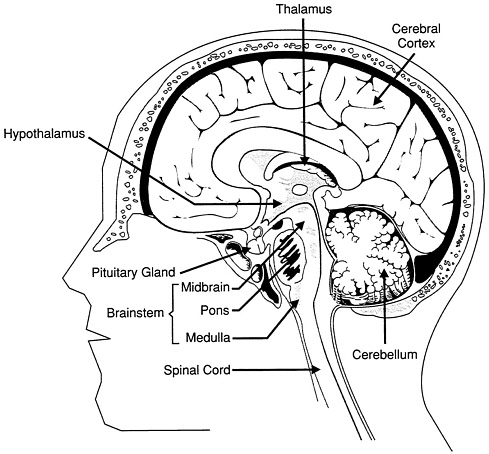
The medulla oblongata is the lowest part of the brain.

Brain anatomy with functions. In humans the brain weighs about three pounds and consumes a stunning 20-25 of all the bodys energy. The Brain Learning. Psychology Brain StructureAnatomy and Function BRAIN FACTS Composition of the brain.
In this lesson well discuss the principle brain regions layers of the brain and lobes of the brain as well as common terms used to orient neuroanatomical discussions. Brain Anatomy Lesson for Kids Left Brain. Besides relaying sensory and motor signals the structures of the brain stem direct involuntary functions.
It masters numbers and calculations and coordinates the right part of the body. N most people it is the dominant hemisphere. Writing of an exam loss of a job birth of a child illness etc by regulating heart and breathing rates.
Copyright Society for Neuroscience 2017. The pons helps control breathing rhythms. The medulla handles respiration digestion and circulation and reflexes such as swallowing coughing and sneezing.
Surrounded by the cerebral hemispheres the diencephalon forms the central core of the brain. The brain role as part of the Central Nervous System is to regulate most functions of human body including vital functions such as heart rate or breathing basic functions like being hungry sleeping or sexual instinct also complex functions like speaking thinking remembering etc. It deals with order because it tends to discipline.
Brain_anatomy_with_functions 33 Brain Anatomy With Functions Brain Anatomy With Functions Getting the books brain anatomy with functions now is not type of challenging means. The brain is an organized structure divided into many components that serve specific and important functions. You could not on your own going taking into account book growth or library or borrowing from your contacts to retrieve them.
Structure descriptions were written by Levi Gadye and Alexis Wnuk and Jane Roskams. Users may copy images and text but must provide attribution to the Society for Neuroscience if an image. It also determines how people respond to stressful situations ie.
Alcohol 4 Alzheimers Disease 7 Anatomy 235 Amygdala 9 Auditory Cortex 3 Basal Ganglia 3 Brainstem 3 Cerebellum 10 Corpus Callosum 3 Cranial Nerves 9 Frontal Lobe 5 Hippocampus 13 Hypothalamus 6 Limbic. The human brain is the most complex of all living constructions processing sensory information while organizes and preserves the organisms vital functions. It helps regulate many important functions including breathing.
Understanding the anatomy of the brain can be aided by looking at it from different organizational layers. The brain is a dense organ with various functional units. Brain Anatomy and Function.
Bacterial infections can make their way to the brain where they can linger undetected. 78 water 12 lipids 8 protein 1 carbs 2 soluble organics and 1 salt 10 seconds is the amount of time until unconsciousness after the loss of blood supply to the brain. Brain anatomy physiology Stroke Neurological Assessment Stephanie Drysdale.
This interactive brain model is powered by the Wellcome Trust and developed by Matt Wimsatt and Jack Simpson. It is used for analytical reasoning for mathematics logic and has to do with language speech and writing. The slowest speed at which information travels between neurons is 260 mph 416 kmh.
Bacteria in the Brain. The brain controls thoughts memory and speech arm and leg movements and the function of many organs within the body. The midbrain contributes to motor control vision and hearing as well as vision- and hearing-related reflexes.
It acts as the control center for the function of the heart and lungs. The brain is typically located inside the head within a protective covering such as an exoskeleton or skull. The brain is an organ that coordinates nervous system function in vertebrate and most invertebrate animals.
Consisting of largely of three paired structures the thalamus hypothalamus and epithalamus the diencephalon plays a vital role in integrating conscious and. Functions of the Brain FRONTAL PARIETAL OCCIPITAL PersonalityBehaviour Planning Decision making Concentration Voluntary motor functions Primary motor cortex precentral gyrus Comprehension and language Sensory functions pain heat and other sensations. Brainstem Responsible for automatic survival reflexes Spinal Cord Controls simple reflexes Pathway to neural fibers Medulla Controlsregulates heartbeat and breathing To and from brain Reticular Formation Helps control arousal responds to change in monotony Thalamus Relays sensory information switchboard between.
The brain can stay alive for 4-6 minutes. Reviewed by John Morrison Patrick Hof and Edward Lein.
Heres more about these three layers. Selecting or hovering over a box will highlight each area in the diagram.
Ks2 Heart Diagram Qr Labelling Activity Science Twinkl
Draw directly on the 3D model using Pen in Complete Heart.
Heart diagram labelled detailed. Because the heart points to the left about 23 of the hearts mass is found on the left side of the body and the other 13 is on the right. A well labeled human heart diagram given in this article will help you to understand its parts and functions. 28 Real Heart Diagram Aed Locator Frequently Asked.
Label the heart. Detailed human heart diagram heart diagram detailed the human heart diagram labeled human categories. The main artery carrying oxygenated blood to all parts of the body.
Below is a labelled diagram of the human heart with a detailed heart diagram. Shape and Size of the Heart. Draw directly on the model or add Labels to mark out structures for future reference.
Internal Structure Of The Heart Contemporary Health Issues. A typical heart is approximately the size of your fist. This shows the inside of a normal healthy human heart.
The heart pumps blood through the network of arteries and veins called the. The human heart is an organ responsible for pumping blood through the body moving the blood which carries valuable oxygen to all the tissues in the body. A well labeled human heart diagram given in this article will help you to understand its parts and functions.
WebMDs Heart Anatomy Page provides a detailed image of the heart and provides information on heart conditions tests and treatments. The heart diagram is a detailed illustration of a human heart each part of the heart is labelled. The right ventricle pumps the blood to the lungs for re-oxygenation through the pulmonary arteries.
The heart one of the most significant organs in the human body is nothing but a muscular pump which pumps blood throughout the body. Discover and save your own pins on pinterest. The base of the heart is located along the bodys midline with the apex pointing toward the left side.
A Diagram of the Heart and Its Functioning Explained in Detail. The human heart and its functions are truly fascinating. The human heart diagram shows a cross-section of a healthy heart and its inside structures.
Without the heart the tissues couldnt get the oxygen they need and would die. Diagrams Quizzes And Worksheets Of The Heart Kenhub. The Heart Of The Matter National Geographic Society.
Heart Chambers The human heart consists of four chambers. Anatomy of the Heart. The heart though small in size performs highly significant functions that sustains human life.
A heart diagram labeled will provide plenty of information about the structure of your heart including the wall of your heart. The blue arrow shows the direction in which oxygen-depleted blood flows from the body to the lungs. Add your own notes or custom diagrams to the model.
The wall of the heart has three different layers such as the Myocardium the Epicardium and the Endocardium. 19 Fresh Heart Diagram Labeled Detailed Airportexclusiv. Two ventricles and two atria.
It has the shape of an upside down pear with a weight between 7-15 ounces. There are the left and right ventricle and the left and right atrium. A Labeled Diagram of the Human Heart You Really Need to See.
12 cm 5 in in length 8 cm 35 in wide and 6 cm 25 in in thickness. The shape of the heart is similar to a pinecone rather broad at the superior surface and tapering to the apex. Along with lymphatic vessels the blood blood vessels and lymph the heart.
Drag and drop the text labels onto the boxes next to the diagram. Human heart diagram highlighting the various anatomical structures. Human Heart Diagram Labeled.
The heart is responsible for the circulation of blood in our body. The right and the left region of the heart are separated by a wall of muscle called the septum. The heart continues to beat from the time we are born until the time we die approximately 72 times per minute.
The most detailed animated 3D heart model ever created. It is located between the lungs and that are in the middle side of chest. In this interactive you can label parts of the human heart.
Click hereto get an answer to your question a Draw and label the diagram given. State and explain the laws of reflection of light at a plane surface like a plane mirror with the help of a labeled ray-diagram.

What Is Reflecation Of Light Draw A Labelled Diagram Showing Reflection Of Light By A Plane Mirror Brainly In
The law of reflection states that the angle of reflection equals the angle of incidence or.

Label the given diagram of reflection of light. What is Reflection of Light. Record the angle of incidence and the angle of reflection. We identify the incoming ray as the incident ray and the outgoing ray as the reflected ray.
Mark the angle of incidence and reflection clearly on the diagram if the angle of reflection is 475 degrees what will be the angle of incidence. Using a protractor draw a normal at C roughly the middle of AB. We use the diagram shown below to answer the questions.
The ray that leaves the mirror is known as Reflected Ray. The law of reflection is illustrated in Figure 15 which also shows how the angle of incidence and angle of reflection are measured relative to the perpendicular to the surface at the point where the light. A ray of light is reflected by two parallel mirrors 1 and 2 at points A and B.
Draw diagrams to show the refraction of light from i air to glass ii glass to air. Reflection helps in medical diagnosis and optical communications. Using this diagram we.
Asked Jul 31 2019 in Class X Science by muskan15 -3980 points. In each diagram label the incident ray refracted ray the angle of incidence i and the angle of refraction r. B angle of reflection r i 34 by the law of reflection c q 90 - r 90 - 34 56.
Complete the following diagram showing the reflection of the light source is at the normal. A Draw and label the diagram given. I 90 - 56 34.
D i r 34 34 68. When a ray of light approaches a smooth polished surface and the light ray bounces back it is called the reflection of light. At the point of incidence where the incident ray strikes the mirror a perpendicular line is drawn known as the Normal.
I Incident ray ii Refracted ray iii Emergent ray iv Angle of reflection v Angle of deviation v Angle of emergence b The refractive index of diamond is 2. What is the meaning of this statement in relation to speed of light. I Incident ray ii Refracted ray iii Emergent ray iv Angle of reflection v Angle of deviation v Angle of emergenceb The refractive index of diamond is 242.
In our diagram we now label the reflected angle along with triangles ABD and ACD that we will use to prove the law of reflection. Explain why your diagram looks the. The angle of reflection is 90 if the angle of incidence is 90 In the second example if a light ray travelling along the normal hits a mirror it is reflected straight back the way it came.
Using the law of reflection for sound and light we can measure the distances accurately to objects. B If the refractive index of glass for light going from air to glass is 32 find the refractive index of air for light going from glass to air. The incident light ray which lands upon the surface is said to be reflected off the surface.
θ r θ i. A Draw a ray diagram to show the refraction of light through a glass slab and mark angle of refraction and the lateral shift suffered by the ray of light while passing through the slab. First we consider reflection as shown in the diagram below for a light wave striking a surface.
If a perpendicular were to be drawn on a. Draw a line at 20 o to the. On a sheet of white paper draw a pencil line label this AB.
Investigating the law of reflection. Concomitantly the angle θ i that the incoming ray makes with a line dashed in the diagram normal to the surface is called the angle of incidence. State and explain the laws of reflection of light at a plane surface with the help of a labelled ray diagram.
I Refraction of light from air to glass ii Refraction of light from glass to air. θ r θ i. Laws of Reflection In the diagram given above the ray of light that approaches the mirror is known as Incident Ray.
Reflection by a concave mirror and a convex mirror has many uses as listed above. What is the meaning of this statement in relation to speed of light. The ray that bounces back is called the reflected ray.
A angle of incidence. Light and Sound both follow the law of reflection both being waves.
The basic structures and functions of neurons axons and dendrites. Brain Structure and Functions.
2 Major Structures And Functions Of The Brain Discovering The Brain The National Academies Press
The primary functions of the brainstem include relaying information between the brain and the body supplying some of the cranial nerves to the face and head and performing critical functions in controlling the heart breathing and consciousness.

Brain structure and functions worksheet. Brain Structures and Functions Worksheet PSY340. 4Parts of the brain. Some of the worksheets for this concept are Brain anatomy How are parts of the brain related to brain function The brain parts The specialized brain The brain Brain matters brain anatomy Human physiologythe nervous system Brain case studies assignment answer key.
The parietal lobe is located in the middle section of the brain and is associated with processing tactile sensory information such as pressure touch and pain A portion of the brain known as the. Functions Of The Brain Displaying top 8 worksheets found for - Functions Of The Brain. Neurotypical development of the brain.
The average brain weighs 7 pounds33 lbs ___F_ 2. Find a price that suits your requirements Save 10 on First Order. 2 pages 594 words.
MedullaRegulates breathing and heart rate hanging a person works bc if done correctly it breaks this in halfPonsInvolved in sleeping waking and dreamingCerebellumThe lesser brain coordinates balance and coordinationThalamusRelays all sensory information to specific perception areas of the brain with the exception of smellHypothalamusPart of the old brain it controls survival elements such as. This work has been submitted by a student. The diagram separates the brain into six major parts and provides a brief description of the functions carried out by each section.
Some of the worksheets displayed are Brain structure functions work Human anatomy Table of contents Whats in your brain How are parts of the brain related to brain function Brain matters brain anatomy Anatomy of the brain Your brain. Structures and functions of the brain Worksheet A Structures of the brain Label the frontal lobe temporal lobe parietal lobe occipital lobe and cerebellum. The brain is a very complex piece of work and this quiz tests you on different structures and how they function.
The basic structures of the brain. Resources textbooks websites for. 3Structure and function of the brain.
Consisting of largely of three paired structures the thalamus hypothalamus and epithalamus the. The unit involves the following Completed facts and topics and eight engaging worksheetactivities on human brain unit. The Human Brain Diagram The Human Brain Diagram is a versatile tool for psychoeducation.
The Functions Of The Brain. Hemispheres lobes and levels. This is not an example of the work written by professional academic writers.
Functions of the brain Complete the following table. 9 rows Differentiated worksheets on structures of the brain. Surrounded by the cerebral hemispheres the diencephalon forms the central core of the brain.
Longitudinal fissure Frontal lobe Parietal lobe Central sulcus Precentral gyrus DIENCEPHALON. Uop brain structures and functions worksheet psy 340 version university of phoenix material brain structures and functions worksheet provide brief description. Structures Functions Frontal lobe Temporal lobe Parietal lobe Occipital lobe Cerebellum.
Fill in the blank for each of the statements below. Items listed under the column Brain Structure will list a region of the brain while items under the column titled Functions will describe the general behavior skill andor activity of the associated brain structure. Introduces students to the different parts of the brain and a little about their.
Displaying top 8 worksheets found for - Brain Structures And Function. Utilize the model of the human brain to locate the following structures landmarks for the cerebrum. Brain Structures And Function.
Showing top 8 worksheets in the category - The Functions Of The Brain. This ready-made presentations as pdf file includes engaging clear and eye-catching photos of human brain with explanations. Once you find your worksheet click on pop-out icon or print icon to worksheet to print or download.
Recall the part of. The basic functions of each lobe. In the Development topic you learnt about three other parts of the brain.
The basic systems of the brain physical chemical and electrical. Some of the worksheets displayed are Brain structure functions work Human anatomy Brain anatomy work Whats in your brain Brain and the nervous system work Nervous system work The brain Anatomy of the brain. Brain Structures and Functions Worksheet Below you will see two columns.
Some of the worksheets for this concept are The brain parts Brain anatomy Your brain Grades 6 to 8 human body series nervous system Engage your brain Brain teen guide for pdf Mapping the brain Beyond workbooks functional treatment strategies for tbi. 2Size of the human brain. The brainstem connects to the spinal cord and consists of the medulla oblongata pons and midbrain.
Provide a brief description for each of the following functions. Discussion of the brain and how it works can be a. Brain 101 3 Pre-Quiz Part 1 True or False ___F_ 1.
Brain Structures and Functions Worksheet. Brain Structures and Functions Worksheet Provide a brief description for each of the following functions. Quiz Worksheet Goals This brain quiz tests your ability to.
Here you can order a professional work.
Draw Neat and Labelled Diagram of Dry Cell. A Centromere b p-arm.

Draw It Neat How To Draw Animal Cell
Draw a neat labelled diagram of animal cell.

Draw neat labelled diagram of cell. Can boy apply foundation to his skin. Draw neat labeled diagram of a Animal cell. Draw a neat labelled diagram of a prokaryotic cell.
Draw a neat labelled diagram of animal cell. Asked Nov 28 2017 in Class IX Science by ashu Premium 930 points Draw a neat diagram of animal of an animal cell. Maharashtra State Board HSC Science Electronics 12th Board Exam.
Draw a neat labelled diagram of an animal cell. Prev QuestionNext Question. 19The people of India is friendly and welcoming.
Advertisement Remove all ads. Draw a neat labeled diagram of Standard Hydrogen Electrode SHE. Write its Half Cell reaction.
Draw a neat diagram of plant cell and label any three parts which differentiate it form animal cell. Draw a neat diagram of plant cell and label any three parts which differentiate it from animal cell. Concept Notes Videos 418.
- Science and Technology 1. Watch 1000 concepts tricky questions explained. Solar cells are the building blocks of photovoltaic modules otherwise known as solar panels.
Draw a labelled diagram of a bacterial cell. To represent these pores erase three or four small sections of each circle. Draw a neat diagram of the structure of chromosome and label the parts.
AskedDec 26 2020in The Cellby Baani490kpoints closedDec 30 2020by Baani. Find the members of the team ACPTU ACPTD APUDE APUTS Q. Advertisement Remove all ads.
Draw a neat diagram of plant cell and label any three parts which differentiate it from animal cell. If playback doesnt begin shortly try. The fundamental unit of life.
Draw neat and labelled diagram of parenchyma cells 1 See answer varsha123456mk is waiting for your help. Advertisement Remove all ads. Written 46 years ago by juilee 72k.
Draw a labelled diagram of a animal cell. Link of our facebook page is given in sidebar. Draw a neat labelled diagram of a prokaryotic cell.
Asked Feb 5 2018 in Class IX Science by saurav24 Expert 14k points Draw a neat labelled diagram of an animal cell. Draw a neat labelled diagram of electrolytic cell for the extraction of aluminium. Apne doubts clear karein ab Whatsapp par bhi.
Get the answer to this question and access a vast question bank that is tailored for students. Listed below are the Cell Organelles of an animal cell along with their functions. Loading DoubtNut Solution for you.
Form the nucleus by drawing two circlesa larger circle that takes up around 10 of the cell. There are various organelles present within the cell and are classified into three categories based on the presence or absence of membrane. Draw a neat labelled diagram.
Draw a neat diagram of the stomatal apparatus found in the epidermis of leaves and label the Stoma Guard cells Chloroplast Epidermal Cells cell wall and Nucleus. Answers 1 P Priyanka Kumari. 300 views 19 people like this.
Well labelled Diagram of Prokaryotic cell For board and NEET exams Bacterial cell Diagram. The Cell Organelles are membrane-bound present within the cells. Compare The Location Of Nucleus In Animal Cell And Plant Cell Draw A.
Draw a neat diagram and explain working of photovoltaic cell. Solar cells are described as being photovoltaic irrespective of whether the source is sunlight or an artificial light. Well-Labelled Diagram of Animal Cell.
Question Bank Solutions 11950. Correct Fast Q. Add your answer and earn points.
The kidney is one of the primary organs of the human body and the urinary system its evolution until today has been indispensable for. This pair of purplish-brown organs is located below the ribs toward the middle of the back.

25 1 Internal And External Anatomy Of The Kidney Anatomy Physiology
Their function is to remove liquid waste from the blood in the form of urine.

What parts of the human kidney you see. What parts of the human kidney do you see. Science 10122019 1828 kenn14. Kidney and urinary system parts and their functions.
Tamang sagot sa tanong. In this article we are going to analyze the kidney anatomy and the different functions of each of its parts both in humans and animals. Each kidney has about 1 million nephrons.
If blood stops flowing into a kidney part or all of it could die. You could have only 10 of your kidneys working and you may not notice any symptoms or problems. What parts of the human kidney do you see.
Integrated Science 28012020 0828 09389706948. English 08012020 0828 reyquicoy4321 What parts of the human kidney do you see. What parts of the human kidney do you see.
Science 28102019 1445 hajuyanadoy Whats parts of the human kidney do you see. Kidney and urinary system parts and their functions. Each kidney has around a million tiny filters called nephrons.
This pair of purplish-brown organs is located below the ribs toward the middle of the back. Blood containing urea and metabolic waste products enters the kidney from the. What parts of the human kidney do you see.
Tamang sagot sa tanong. 1 Unlock answers. Their function is to remove liquid waste from the blood in the form of urine.
What parts of the human kidney do you see. Keep a stable balance of salts and other. It is the basic structural and functional unit of the kidney and cannot be seen by the naked eye.
What parts of the human kidney do you see. The walls of the nephron are made of a single layer of epithelial cells. What parts of the human kidney do you see.
That can lead to kidney failure. Science 10122019 2128 maledabacuetes What part of the human kidney do you see. - 1774269 beacunanan0701 beacunanan0701 29082018 Science Elementary School What parts of the human kidney do you see.
Integrated Science 28012020 0828 09389706948. What parts of the human kidney do you see. Science 11122019 1028 ShairaGailSanchez What parts of the human kidney do you see.
Integrated Science 28012020 0828 09389706948 What parts of the human kidney do you see. What parts of the human kidney do you see. Keep a stable balance of salts and other.
Biology 02022020 1504 09330399672 What parts of human kidney do you see. The nephron is the filtration unit of the kidney. Refer to Figure 5.
Draw well labelled diagram of a Bacteria cell b Euglena. SOLUTION Euglena is a free-living unicellular flagellate protist.

Draw It Neat How To Draw Euglena
28 Protists At Loyola College Studyblue.
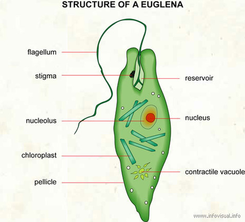
Draw a well labelled diagram of a euglena. Watch 1000 concepts tricky questions explained. Body spindle shaped and usually green in colour and is surrounded with tough elastic membrane the pellicle. Euglena has two flagella usually one long and one short.
How to draw diagram of euglena easy steps - YouTube. Euglena has two flagella usually one long and one short. Each flagellum arises from a basal granule.
Euglena is a free-living unicellular flagellate protist. The body is covered by thin and flexible pellicle. The pellicle has oblique but parallel stripes called myonemes.
Euglena is a genus of single cell flagellate eukaryotes. It is without a cellulose cell wall. Each flagellum arises from a basal granule.
Euglena has two flagella usually one long and one short. Add your answer and earn points. The body is covered by thin and flexible pellicle.
When acting as a autotroph the Euglena utilizes its chloroplasts which gives it the green colour. The apical end bears an invagination having three parts- cytostome cytopharynx and reservoir. This will also help you to draw the structure and diagram of euglena.
It is without a cellulose cell wall. An osmoregulatory contractile vacuole occurs in the anterior part of the cell below the reservoir. This will also help you to draw the structure and diagram of euglena.
Draw a well labelled diagram of Euglena. Euglena has two flagella usually one long and one short. It is the best known and most widely studied member of the class Euglenoidea a diverse group containing some 54 genera and at least species.
It belongs to the group Euglenoids. It is the best known and most widely studied member of the class Euglenoidea a diverse group contai. These diagrams include some organs and can give you some detailed information.
The apical end bears an. The body is covered by thin and flexible pellicle. The gtven organism is Euglena.
The apical end bears an invagination. Each flagellum arises from a basal granule. A small free living Get a handful labeled euglena diagrams to assist your study about euglena anatomy.
152 K views 700 people like this Like Share. A small free living and freshwater form. Draw well labelled diagram of a Bacteria cell b Euglena.
-10cm to draw a well labeled diagram of Euglena viridis La State three plants and animals characteristics of Euglena viridis ECONOMICS the table below Fived factor Total product Average product Marginal product 2 H 3 B 18 E A F what output level does diminishing returns set in. Add your answer and earn points. Open in answr app.
Since Euglena is a eukaryotic unicellular organism it contains the major organelles found in more complex life. Loading DoubtNut Solution for you. Find an answer to your question Draw a well labeled of a euglena sauravjha3742 sauravjha3742 10062021 Biology Secondary School answered Draw a well labeled of a euglena 1 See answer sauravjha3742 is waiting for your help.
It is without a cellulose cell wall. Euglenoids are heterotrophic flagellates but also show autotrophic mode. It is a flagellated Protista.
Draw a well labelled diagram of Euglena. This protist is both an autotroph meaning it can carry out photosynthesis and make its own food like plants as well as a heteroptoph meaning it can also capture and ingest its food. The apical end bears an invagination having three parts- cytostome cytopharynx and reservoir.
This will also help you to draw the structure and diagram of euglena. It has two flagella one is short and the other is long. Euglena Diagram Labeled 400 Magnification.
The pellicle has oblique but parallel stripes called myonemes. Each flagellum arises from a basal granule. Maths Physics Chemistry Biology.
Draw A Well Labelled Diagram Of Euglena Class 11 Biology Cbse. A small free living Euglena is a genus of single cell flagellate eukaryotics. The pellicle has oblique but parallel stripes called myonemes.
Species of Euglena are found in freshwater and salt water. The pellicle has oblique but parallel stripes called myonemes. These printable diagrams are provided to guide you in studying the structure of the organism.
Draw a well labelled diagram of Euglena. It can perform photosynthesis in the presence of light due to the presence of photoreceptors and photosynthetic pigments. Each flagellum arises from a basal granule.
Draw well labelled diagram of a Bacteria cell b Euglena. These diagrams include some organs and can give you some detailed information about the structures of an euglena. B Name the kingdom to which it belongs snehahAnippravat snehahAnippravat 15062016 Science Secondary School answered a Draw a well labelled diagram of Euglena.
Euglena is a free-living unicellular flagellate protist. B Name the kingdom to which it belongs 1 See answer snehahAnippravat is waiting for your help. The body is covered by thin and flexible pellicle.
It is without a cellulose cell wall. AGRICULTURAL SCIENCE Deline and tenure List 3 factors of the land use decree of 1978 in Nigeria. Diagram Of Euglena Labled image gallery euglena labeled keywordsuggest org the structure and life cycle of amoeba with diagram euglena labeled parts by ducknatucta issuu protist cell diagram labeled 2003 ford expedition wiring spirogyra diagram microbiology projects to try draw a well labelled diagram of volvox.
Maths Physics Chemistry Biology. Euglena is a free-living unicellular flagellate protist. An orange-red eyespot or stigma occurs attached to the membrane of the reservoir.
Apne doubts clear karein ab Whatsapp par bhi. Click here to get an answer to your question a Draw a well labelled diagram of Euglena. Euglena has two flagella usually one long and one short.