5 out of 5 stars. 12oz Beehive Honey Jar Kitchen Decor Kitchen Storage Glass Canister Glass Kitchen Canister Home Decor Glass Storage Jar Glass Honey ModernFarmhouseStore.

Love Sweet Love How To Make Mini Honey Jars Wedding Favors The Pretty Life Girls
Favorite Add to Honey Bee Mason Jar Mason Jar Honey Bee Decor KNBDesign.

How to decorate honey jar. Cut the fabric in stars or just leave it as it is. Spray the glue on the glass jar and glue as much fabric as you wish to it. Artistically designed to coordinate beautifully with any tabletop decor.
Take a pinch of the herb you chose and sprinkle it on top of the paper. If you are only putting the image on one section of the jar just paint that section. Most honey I have seen sold in stores come in narrow mouth containers.
Thats how we bottle honey. Invest in a uniquely shaped clear glass honey bottle to showcase the natural look of your honey. Poke a hole in the top of the lid to pull the string through as you use it.
Take a spoonful of honey and add it to the jar. Place the lid onto your jar and close it. To decorate a glass jar with fabric for Christmas you will need some pray glue and some red embroidered fabric.
Bay leaf Lavender and Cinnamon all have purifying and protective qualities. This matter is made worse when most of the content is used and the remaining honey sits on the bottom making it impossible to scoop out. Dip your sponge brush in the decoupage glue and carefully paint a thin layer on your glass jar.
Take your candle and place it on top of the sealed jar. Touch your honey covered finger to your lips and think of the person as you lick the honey from your lips. This Honey Jar is a part of the Tabletop Collection that has been an all-time favorite.
Rest assured this Honey Jar will make a statement of elegance thereby changing the ambiance from the simplest affair to an upscale event. Paint a thin layer of decoupage glue on the jar. A creatively-shaped bottle can instantly set your product apart from the rest.
Make your own by adding a few tablespoons each of olive oil and red wine vinegar to your honey jar and vigorously shaking it. After a period of time of non-use the honey tends to get gummy and doesnt flow out of the container easily. The jars didnt need to be washed they came sterilized from the supplier and we just put them on a large dinner plate to make it easier to clean up an drips and spills.
5 out of 5 stars 28 300. Honey adds a touch of sweetness to balance out salad dressings. Perfect to give as a gift or for personal use.
We tare a sanitized cooled jar on a scale and then fill it to the weight indicated on our labels. Mar 11 2017 - We chose glass jars for our honey for several reasons but one was to limit plastic and for customers to be able to reuse their glass jars. Heres some great ways of recycling your old Sweetree Honey Jars.
Express your own personality and style bright and eye-catching or quaint and comfortable to represent your honey products. Another option you have is to cover the glass jar with a old red sock and decorate it with a ribbon. If you are putting images around the entire jar.
Finally we slap on a new clean canning-jar lid for each jar and its done. When all is ready we place the 5-gallon bucket of honey onto an elevated surface. See more ideas about glass jars mason jars jar.
Decorate a glass jar with glass paint or crafting materials to match your décor to create a decorative flower vase or bud jar. Honey pot with dipper honey jar Northern Light color honey keeper with a honey stick sugar pot sugar jar. Season with salt pepper and any dry or fresh herbs you desire.
Use glass jars with a lid to keep your string tangle-free. 5 out of 5 stars 48 2000. When a jar was almost full hed use a knife to cut off the.
Fold the paper once towards you and put it inside the jar. Id setup a plate with jars and my dad would start to carefully fill the jars with honey from the pitcher.
First the purpose of a microscope is to magnify a small object or to magnify the fine details of a. The parts of a compound microscope are of two categories as given below the eyepiece is a drum which fits loosely into the draw tube.

Parts Of A Microscope With Functions And Labeled Diagram
It is the topmost part of the microscope.
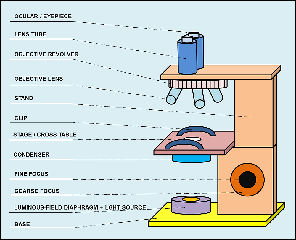
Draw and label the parts of a light microscope. 872018 2 Simple. 1 2 3 and 4 Let us take a look at the different parts of microscopes and their respective functions. Draw and label a light microscope.
Draw and label the different parts of a light microscopeIN THE PARTSpe post navel hirte in the wine The RealistaAmState Cupan Am KasTaphragmEyepieceFine Adjustment nobHigh Power CrveStat201aFigure T. Similar to a magnifying glass and has only one lens. Microscopy from www2nauedu.
It is a u shaped structure and supports the entire weight of the compound microscope. This forms the illuminator or light source of the microscope. Before exploring microscope parts and functions you should probably understand that the compound light microscope is more complicated than just a microscope with more than one lens.
Easy and simple step by step tutorial for beginnersThanks for Watching and Subscribing Minutes DrawM. Observe the skin cells under both low and high power of your microscope. Base As the name suggests the base is the lowest portion on which the whole structure of the microscope rests.
How to use a microscope. How to draw microscope slides. Animal cells under a light microscope.
How to draw a microscope diagram. BASE Supports the MICROSCOPE D. Light Microscope With Labeled Parts Written By MacPride Thursday October 22 2020 Add Comment Edit.
STAGE CLIPS HOLD the slide in place C. Microscope drawing and label at. Use the diagram to show how the microscope works ie.
Viewing Cells Using The Compound Light Microscope Stains Ppt. Draw and label the parts of the microscope. Light Microscope Drawing At Getdrawings Free Download.
Optical parts a mechanical parts of a compound microscope. COARSE ADJUSTMENT KNOB Moves the stage up and down for FOCUSING I. Remember to subscribe to my channel and turn on notification.
1 14 13 2 12 3 11 4 10 5 an 8 6 7. Draw it in a paper and label each part numbered 1-14. A light is needed to shine on the object and then reflected by the mirror into the lenses hence causing greater magnification.
Arm The arm connects the body tube to the base. Stereoscopic Microscope Gives a three dimensional. Draw your own light microscope and label the parts.
Parts of the Light Microscope T. FINE ADJUSTMENT KNOB Moves the stage slightly to SHARPEN the image G. Microscope Parts and Functions With Labeled Diagram and Functions How does a Compound Microscope Work.
Draw a cylinder at the base of the arm. Draw and label the parts of microscope. Body Tube It is the part of the microscope that holds the eyepiece.
Through the eyepiece you can visualize the object being studied. It also carriers the microscopic illuminators. Use straight lines to enclose a rectangular shape around the bottom of the microscope.
Microscope drawing and label at getdrawings com free for. Add notes to the diagram. Use straight parallel vertical lines for its sides and curved lines for its top and bottom.
Parts Of A Microscope With Functions And Labeled Diagram. Https Ntrs Nasa Gov Archive Nasa Casi Ntrs Nasa Gov 20170000349 Pdf. Microscope Parts and Functions Microscope One or more lenses that makes an enlarged image of an object.
872018 3 Compound Microscope Lets light pass through an object and then through two or more lenses. Microscope drawing - step 9. Label the parts of the compound light microscope in the figure below and then draw the path of light from the illuminator to your eye.
All the best Microscope Drawing And Label 33 collected on this page. EYEPIECE Contains the OCULAR lens J. The user must hold this part in order to move the microscope from one place to another.
Parts Of Violin Diagram. In some of the diagrams youll see we mention low 1 and 1.
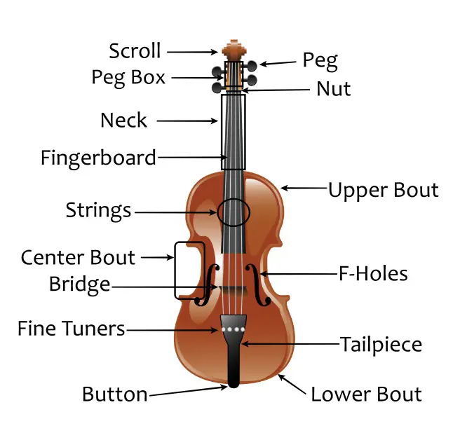
Violin Parts Complete Piece By Piece Guide And Bow
The bridge supports the strings at the lower end of the violin.

Labelled diagram of violin. Discover and save your own Pins on Pinterest. The endpin is the attachment point of the tailpiece and it is often assorted to pegs and tailpiece. The high E string is sometimes colloquially called the top string and the low G string might colloquially be called the bottom string Strings were originally made from sheep gut confusingly called catgut but.
Nelson Ray D. The top of each instrument is made of spruce and the rest from maple. The position of the bridge is essential as it directly relates to the quality of sound produced by the violin.
Posted on April 5 2019 April 4 2019. Modern violin peg fitting is always done with a 130 taper but if you work with old instruments you often will be faced with 120 holes as well as varying steep tapers from. Its main function is decoration but right below it is the pegbox where the strings thread through the tuning pegs.
When the string vibrates the bridge also vibrates. The document also includes a labeled image of a violin bow. Violin And Bow Diagram Violin Parts Violin Learn Violin.
Purfling is found on the edges of the top and. Dijelovi violine i gudala Share Share by Pepica76. Oct 30 2019 - This Pin was discovered by Danna Acevedo.
Violin Fingering for notes played in the 1st position are to the right of the fingerboard. While most violins have two-piece backs that are joined together with a seam down the middle one-piece backs are preferred due to their increased resonance. A simplified drawing showing the appearance structure or workings of something.
A smaller angle makes it easier to play two or three strings at the same. Newer Posts Older Posts Popular Post. The interior of the instrument also contains a sound post bass bar corner blocks end block and neck block as well as the linings throughout.
This leaderboard is currently private. A Box Plot-Density Trace Synergism. Image Info File Name.
The bridge is held in place by the strings tension. Your email address will not be published. These notes require the violinist to shift the position of their hand to a higher position on the keyboard in order to play these notes.
The sound post is the round post inside the violin that runs from the front-piece to the back-piece under the bridge of the violin. How To Make A Venn Diagram On Word 2007. Glava violine puž ključevi vrat violine hvataljka tijelo violine F otvori konjić žičnjak podbradak štap gudala strune žabica vijak kožica vrh gudala čep.
Fingering for notes played in 3rd position are to the left of the fingerboard. G3 D4 A4 E5. The sounding post plays a key role in how the violin produces sound and it also helps to support the structure of the violin.
Bobcat 610 Parts Diagram. Diagram For 220v Wiring 3 Prong Plugs Club Car Wiring Diagrams 48 Volts Cub Cadet 54 Inch Deck Belt Diagram Circuit Diagram Using Nand Gate Craftsman 1 2 Hp Garage Door Wiring Diagram Double. Look below for a complete diagram and description the parts of a violin.
The upper nut is meant to elevate the strings and when well adjusted it increases the playing ease and comfort. Honda Fourtrax 300 Rear Differential Diagram. A figure composed of lines that is used to illustrate a definition or statement or to aid in the proof of a proposition.
The American Statistician 522 181184. For an explanation of shifting visit. Show more Show less.
Check out a helpful worksheet of a violin that has its parts labeled. Violins is reached near the large end of the reamer. Tuning Pegs - Used to tune the violin.
Labelled Diagram Of A Violin. Four strings tuned in 5ths. For violas a full-size reamer is required however and thats also fine for 44 violins that have had larger pilot holes drilled if they are properly positioned.
10108000031305199810480559 Besten Dank an Holger für den Hinweis. The upper and lower nuts. There are two violin nuts.
These parts are the tuning pegs the fingerboard the F-hole the bridge the chin rest and the fine tuners. Violinen-Diagramm ist eine Weiterentwicklung des Boxplots. What youll achieve will still be a chord-based rhythmic accompaniment.
This simply refers to. Most violin music for beginners uses 1st position. The bridge of the violin comes in varying angles of curvature.
Und soll die Visualisierung der Verteilungen verbessern. Scroll - located at the top of the violin. Das Boxplot-Skript kann man durch.
Play Violin Viola Parts Of The Violin Viola Violin Violin Parts Viola Music. The violin end pin or end button is a piece of hardwood placed into the violin enfoncée sous pression dans le violon in the lower block. An image of a violin found here identifies six parts of the violin.
There are many parts to a violin viola and cello and these diagrams show the basic parts and names of areas on the stringed instrument shown. Er wurde 1998 vorgestellt 1 Hintze Jerry L. Leave a Reply Cancel reply.
As you can see each finger is given a number weve labeled the thumb but it isnt used in violin playing. 1722 File Size.
Compare The Location Of Nucleus In Animal Cell And Plant Cell Draw A. Labelled diagram of animal cell class 8.
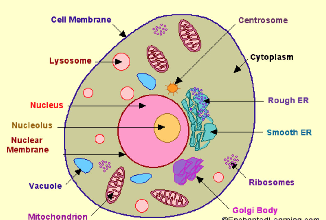
Structure Of Cell Cell Structure And Functions Class 8
The cell is the structural and functional unit of life.

Labelled diagram of animal cell class 8. Onion Cell Diagram Labeled Vaculoe Wiring Diagram Database. Labelled diagram of animal cell class 8. Plant cell read biology ck 12 foundation.
These solutions for cell structure and functions are extremely popular among class 8 students for science cell structure and functions solutions come handy for quickly completing your in an animal. Here It Is The Labeled Diagram Of Plant And Animal Cell Class 9. Plant Cell Labelled Diagram Class 8.
Lets go over the individual components of plant cells listed on a diagram such as the one above and explore the roles that each of the organelles has. Draw a labelled diagram of a animal cell. So they do not contain chloroplast.
In this video im going to draw labelled diagram of animal cellin this video you will see the diagram of animal cell and its labellingthis diagram of. To draw a well labelled diagram of plant and animal cells the golgi apparatus is placed near the endoplasmic reticulum. However they differ as animals need to adapt to a more active and non-sedentary lifestyle.
Plant cells vs animal cells with diagrams a plant cell is a eukaryotic cell enclosed by a cell wall containing a membrane bound nucleus and other description simple diagram of animal cell ensvg english. Below the basic structure is shown in the same animal cell on the left viewed with the light microscope and. Join the discussion forum to ask questions or reply to 2.
Ncert Solutions For Class 8 Science Chapter 8 Cell Structure Functions. Chapter 8 of class 8 science provides a detailed study about various parts of plant and animal ncert books for class 8 science is designed beautifully for kids. Check spelling or type a new query.
Animal Cell Class 8 Diagram. Maybe you would like to learn more about one of these. Class 8 Mensuration Factorisation Linear Equations in One Variable Understanding Quadrilaterals The Making of the National Movement.
Nucleus Cell Structure And Functions Class 8. Draw Diagram Of Animal Cell And Plant Cell Animal Cells And Plant Cells Cell Structure And Functions Class 8 Ncert Notes Cbse Class Notes Online Classnotes123 Draw a out line of animal cell put lot of bends as shown to represent flexible plasma membrane. Animal Cell Diagram For Class 8 Cbse Telangana Scert Class 8 Biology Chapter 2 Cell The Basic Unit Of Life Solution The most important structures of plant and animal cells are shown in the diagrams below which provide a clear illustration of how much these cells have in common.
Diagram of plant and animal cell for class 8 brief sketch. Label the structures a b and c. Label The Parts Of The Diagrams Given Belowidentify Class 11 Biology Cbse.
The animal cell diagram is widely asked in Class 10 and 12 examinations and is beneficial to understand the structure and functions of an animal. A comparison of plant and animal cells using labelled diagrams and descriptive. Answer verified by Toppr.
Labelled diagram of animal cell class 8. 8The cell membrane is a thin sheet of skin all around the cellThe main function of cell membrane is to control the passage of materials which go into the cell or go out from the cellIt also protects the cell and gives shape to the cell. Plant Cell Simple English Wikipedia The Free Encyclopedia - Diagram of plant cell and animal cell.
Labelled and annotated colour animal cell diagram for ks5 biology updated. Lets begin with the components of the animal cells a cell is always surrounded by a thin membrane called plasma membrane. Animal Cell Diagram For Class 8 Ncert - A Well Labelled Diagram Of Animal Cell With Explanation - The most important structures of plant and animal cells are shown in the diagrams below which provide a clear illustration of how much these cells have in common.
Labelled and annotated colour animal cell diagram for ks5 biology updated. Draw a neat labelled diagram of animal cell. A comparison of plant and animal cells using labelled diagrams and descriptive explanations.
Question 3 draw a labelled diagram of plant cell. Asked nov 28 2017 in class ix science by ashu premium 930 points. Animal Cell Class 8 Diagram bookfanatic89.
Ncert Solutions For Class 8 Science Chapter 8 Cell Structure And. Ncert Exemplar Class 8 Science Chapter 8 Cell Structure And Functions Learn Cbse. Make sketches of animal and plant cells state three differences.
We can say that the size of the cell depends on the function it performs. These cells differ in their shapes sizes and their structure as they have to fulfil specific functions. Draw a labelled diagram of a animal cell and plant cell.
I spelt it wrong in the diagram sorry. Click hereto get an answer to your question Draw a neat labelled diagram of animal cell. 27 Jul 2021 Post a Comment The part of the cell that contains dna and aids in several body functions including reproduction metabolism and growth.
A brief explanation of the different parts of an animal cell along with a well-labelled diagram is mentioned below for reference. Draw a labelled diagram of a animal cell and plant cell. Ncert solutions for class 8 science.
Animal Cell Diagram Class 8 Ncert. General Science subject concept of Plant cell and Animal cell for 8th std students explanation of labeling the diagram Thumbs up if you like the video and. Plant cells and animal cells share some common features as both are eukaryotic cells.
Draw a labelled diagram of a animal cell and plant cell. Cell Structure And Functions Class 11 Notes Biology Mycbseguide. The function of a vacuole in an animal cell is to hold air water and particles of food.
Draw A Neat Diagram Of Plant Cell And Label Any Three Parts Which. Animal Cell Diagram Class 8 CBSE Class 9 Science chapter 5. This green pigment is important for photosynthesis in.
Cell Structure And Functions Study Material For Neet Aipmt. Plant Cell Diagram For Class 8 Ncert Label parts and thousands of other science skills.
Rd and 4th grade activity for the water cycle hi-lo activities for learning about the water cycle. Stages of Water Cycle.

Correctly Label The Parts Of The Water Cycle Make Sure To Indicate In Your Answer Which Part You Brainly Com
This will show evaporation.
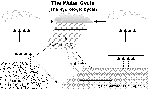
Draw and label the parts of the water cycle. Complete the picture of the water cycle and label each component. Complete the picture of the water cycle and label each component. I used PRISMACOLOR pencil in this drawing.
Evaporation and transpiration condensation. Other Parts of the Water Cycle Evaporation condensation and precipitation are the three main parts of the water cycle but there are some other stages that water can cycle through. Draw a landscape showing plenty of atmosphere ocean and land both above and below.
Visit to my channel. Use erasable pencil at first then color it in with crayons colored pencils andor pens. Water cycle also called hydrologic cycle cycle that involves the continuous circulation of water in the Earth - atmosphere system.
Find an appropriate scene from the Outdoor or Country Rustic categories. Label the main parts of the water cycle with text and arrows. Presenting and defending results of activity.
Of the many processes involved in the water cycle the most important are evaporation transpiration condensation precipitation and runoff. Ecological Cycles Part I - Draw an illustrated Diagram of The Hydrologic Water Cycle Take an 11 x 17 sheet of paper and draw an illustrated diagram of the hydrologic cycle according to the following directions. Describe whats happening at each stage.
Draw and label the water cycle. Write down the change of state of the water in each part of the cycle in the above diagram. Label the parts of the water cycle.
The complete water cycle is carried into four stages which are as follows. The process by which water from its liquid state changes to vapour a gaseous state is termed as evaporation. The cycling of water in and out of the atmosphere is a significant aspect of the weather patterns on earth.
The water vapor or steam leaves the river lake or. This cycle is made up of a few main parts. Label the main parts of the water cycle with text and arrows.
Draw arrows in the diagram below to indicate where water moves through the water cycle. The water cycle describes how water evaporates from the surface of the earth rises into the atmosphere cools and condenses into rain or snow in clouds and falls again to the surface as precipitation. Water Cycle Diagram Create Biology Diagram examples like this template called Water Cycle Diagram that you can easily edit and customize in minutes.
Role of plants in water filtration activity. The Water Cycle Condensation Precipitation Transpiration Evaporation Percolation. Subscribe to my channel to get more drawing videos.
The water falling on land collects in rivers and lakes soil and porous layers of rock and much of it flows back into the oceans where it will once more evaporate. Evaporation Condensation Precipitation and Collection. Natural forces such as the sun air land trees river seas and mountains play an important part in completing the water cycle.
Label the Parts of the Water Cycle. Label the Parts of the Water Cycle. Evaporation is when the sun heats up water in rivers or lakes or the ocean and turns it into vapor or steam.
Label the Parts of the Water Cycle For Google Apps. Explain that in the experiment to follow we will be creat-ing a mini water cycle. This is the initial stage of the water cycle.
During the water cycle water in the water bodies. Create your own model of the Water Cycle. Diagram Of Water Cycle.
Evaporation condensation and precipitation and what hap-pens during each part. Saved by Marni Bickford. Add extra information about the water cycle with text boxes.
How do you draw and label a diagram of the water cycle and explain how water moves through it. Draw diagram of activity and label parts of the water cycle. Materials Markers Warm Water Plastic wrap.
Click to edit this example. Use arrows to show the movement of water in the water cycle. Demonstrate the cyclical movement of water either by drawing the water cycle at the board or sharing a poster of the water cycle.
Draw a diagram of the water cycle. These teaching activities for the water cycle are in google apps and in printables. Draw diagram of inversion layers and label parts.
Precipitation condensation evaporation transpiration and surface runoff. Be sure to label the following steps. Visit to my channel.
Lets learn how to draw water cycleFollow my step by step water cycle drawing and I am sure you will be able to draw the same quite easilyThis water cycle.
Have the students compare this new drawing to their original one and to note what they Filename. Label The Parts Of A Volcano.
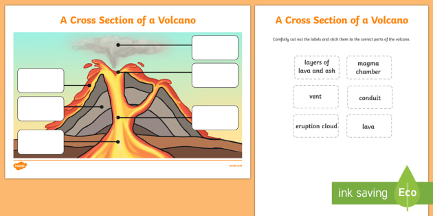
Volcano Cross Section Labelling Activity Ks1 Geography
9 Parts of a volcano from the inside.
Label of parts of volcano. Clearly put the name of your volcano type or types at the top of your diorama either on a background or with a label. When it erupts from being at the surface lava flows and ash deposits over the surrounding areas. Magma is lighter than the solid rock around it so it rises.
Lava - Molten rock that erupts from a volcano that solidifies as it cools. Portrait volcano diagram google search superior specifically for you from label the parts of a volcano worksheet label the source. Tell the students to draw another volcano and label as many parts as possible.
Ash Steam and Gas - cloud which is pushed out of the volcano. What are the parts of a volcano. Gip99_chapter4pdf - Read File Online - Report Abuse.
To prepare you should color this one in nicely. The Anatomy of Volcanoes 1 Magma. Crust the crust is earths outermost rocky layer.
Conduit - An underground passage magma travels through. From the depths of the lithosphere to the Earths crust these are the parts of a volcano according to science. It also looks at different types of plate boundary and where volcanoes are located.
Parts of a volcano labeling worksheet students label the parts of a volcano diagram using words provided in the word bank. Parts Of A Volcano Diagram. Label the parts of a volcano.
The composition of a volcano introduces a few complicated concepts to children so it can be quite. Label the important parts of your volcano. Secondary Vent - place where magma reaches the surface without going through the main vent.
If a volcano continues to erupt it will grow and grow into a larger volcano. When rocks become so hot they can become a substance called magma. Secondary Cone - a cone that builds up around secondary vents.
Summit - Highest point. Label our Volcano Diagram to understand the different parts. Vent an opening in earths surface through which volcanic materials escape.
Flank - The side of a volcano. Crater - Mouth of a volcano - surrounds a volcanic vent. Lava flow and more.
This is the part of the volcano located deepest underground even beneath the Earths crust. This fantastic parts of a volcano interactive labelling activity is a fun and engaging way of introducing your class to the different parts of a volcanoLabelling activities like this one are brilliant for introducing more visual topics to your class. It then goes on to look at the different features of volcanoes by looking at a cross-section of a volcano.
Throat - Entrance of a volcano. Main Parts of a Volcano. The part of the conduit that ejects lava and volcanic ash.
Label the parts of a volcano. In this activity students will label a model of a volcano. It usually comes out of erupting volcanoes.
LP_Volcanopdf - Read File Online - Report Abuse. This Parts of a Volcano Interactive Labelling Activity is a great way for KS2 pupils to develop their understanding of the different parts of a volcano. Place it on cardstock or laminate it so that it will last longer.
It collects in magma chambers on average 1. This parts of a volcano interactive labelling activity is a great way for ks2 pupils to develop their understanding of the different parts of a volcano. Sill a flat piece of rock formed when magma hardens in a crack in a volcano.
Students label the parts of a volcano diagram using words provided in the word bank. Label other environmental features for a very detailed or specific representation of a volcano. The parts of a volcano that are included in this activity are.
Crater - circular depression at the top of the volcano. Pupils can drag and drop the vocabulary to label the diagram of a volcano. Volcanoes Differentiated Labelling Diagram Teacher Made The part of the conduit that ejects lava and volcanic ash.
The resource is self-checking making this perfect for independent learning. For example if identifying features on one of the large stratovolcanoes such as Mount Hood or Mount Fuji you might also label drainages which for such cones are often radial in form--ie streams spilling from all directions from the central height. Volcanoes form when magma that sits within the earths mantle magma reservoir and works its way up to the surface.
Crater Vents Conduits Slopes Magma Chamber. Parts of a volcano labeling worksheet teachervision students label the parts of a volcano diagram using words provided in the word bank this printable can be used as post unit assessment for earth science or volcanoes and earthquakes or can be assigned as a take home or independent pletion activity an answer key is provided volcano diagram printout enchantedlearning. 13 Parts of a Volcano.
In this topic we look at how movements in the earths crust can cause both volcanic eruptions and earthquakes. Eventually some of the magma pushes through vents.
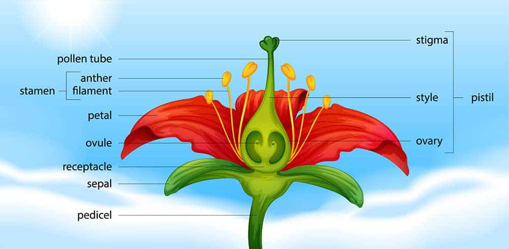
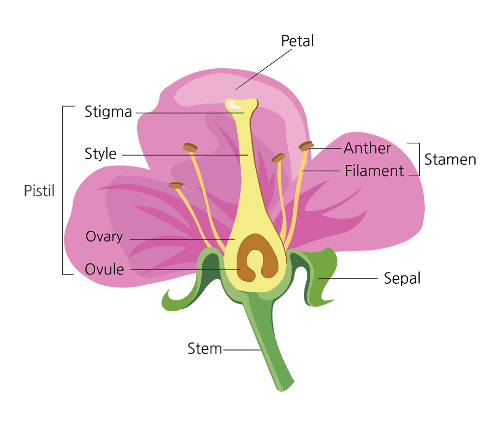
The pistil has three parts. Flowers Flowers are the most obvious part of many plants.

Parts Of Plants Online Pdf Activity
Vascular Anatomy of Floral Parts of Flower.
Parts of flower and their functions pdf. Responses should include roots stems leaves flowers fruits and seeds. In some cases they are extremely showy. Important Parts of a Flower 3.
The stamen has two parts. Anthers Pollen producing part. A flower missing any one of them is called an incomplete flower.
Parts of a Flower and their Functions. Parts of a microscope notes pdf a basic lesson with an overhead key and student worksheet related to the parts of a microscope. 1 sepals 2 petals 3 stamen and 4 carpel each of them performing distinct functions.
Sexual reproduction takes place in flowers. Classroom with tables for children to work in small groups Objectives. Female part Pistil.
In other cases they are not easily seen. March 16 2019 by admin. A flower on the front half as shown at right.
Sepals are the outermost layer and protect the flower in bud Petals are the next row in and are colorful to attract pollinators Stamens come next. The parts of a plant and their functions Reproduction and life cycles Part 1. Introduction to a Flower 2.
After fertilization the ovary of the flower develops into a fruit containing a seed. March 16 2019 by admin. Sexual reproduction takes place in flowers.
The first flap includes the flower the next flap includes only the stem the next includes the leaves and the bottom flap includes the roots. Blank flower parts worksheet plant parts worksheet and cell organelles worksheet. The attraction parts are the petals sepal and receptacle.
Next learners take apart dissect their flower and. Lets now see the parts of a flower and their functions. Parts of a flower and their functions worksheet pdf.
Many flowers have male parts and female parts. When a flower has all the four floral parts it is called a complete flower. A participle is a verbal that functions as an adjective.
Parts Of Gumamela Flower And Their Functions Pdf. Cut the top fold into four flaps. Parts of a flower and their functions pdf.
The important functions of flowers are mentioned below. Parts of a Flower and Their Functions. Gametophytes develop in the flowers.
Functions Of Flower. Leave a Comment Uncategorized Uncategorized. Stamen is the male reproductive part of a flower.
Life cycle of a plant. Describe the parts of flowers and their functions the types of flowers and inflo-rescences. Whether they are showy or not the purpose of a flower.
The female parts of a flower consist of an ovary which contains one or more ovules a style and the stigma. Let us learn about Flower. The most important function of flowers is reproduction.
Stamen contains 2 parts. After reading this article you will learn about. Flower Parts And Functions Pdf.
Flower parts and functions worksheet pdf. Morphology Of Flowering Plants Class 11 Notes In Pdf Plant Parts And Their Functions Pmf Ias Lesson 01b Parts Of A Flower Lp. Stigma Sticky surface at the pistils top where the pollen germinates.
Have learners choose a flower and sketch it on the Parts of a Flower. Identify the basic parts that make up a seed and explain the function. Learners will 1 identify the different parts of a flower and understand their function.
Flower Parts Pollination Worksheet Fill in the boxes with the name of the flower part from the words in the box below. Flower Parts And Functions Pdf. The typical flower consists of four parts as follows.
Parts of a flower with their structure and functions. What Are The Four Main Parts Of A Flower Quora Parts Of A Flower And Its Functions Parts Of A Flower And Plant Do You Know Them All. Parts of a flower and their functions pdf.
Male part Stamen. Data collection observation asking questions analysis communication. It has three main parts called stigma style and ovary.
The flowers can produce diaspores without fertilization. Parts of a flower and Part 2. Identify the basic parts that make up a seed and explain the function.
In a flower the female reproductive part is called the Pistil. Stamen contains 2 parts. 2 understand the importance of pollen for plant reproduction.
Most flowers have four main parts. Anther and the filament. The pistil has three parts.
Filaments They hold up the anthers. Open each flap and write the function of each plant part. A typical diagram of a flower is divided into four main parts.
Male part Stamen The stamen has two parts Anthers Pollen producing part Filaments They hold up the anthers Female part Pistil The pistil has three parts Stigma Sticky surface at the pistils top where the pollen germinates Style Holds up the stigma Ovary Contains the ovules o Ovules Become the seed after fertilization by pollen Other Parts of the Flower Petals Usually bright to attract pollinators Sepals Protect the flower. To learn more visit The Four Whorls of a Flower. Learners observe and dissect a flower to discover its anatomy and the how each part contributes to its.
Parts Of Gumamela Flower And Their Functions Pdf. Pollination fertilisation fruits and seed dispersal Living processes and what plants need to grow Grouping and classification Plants in their natural environment. Flowers are composed of the same Location.
Plant parts and their functions structural organization in plants. They are the male part of the flower and produce pollen for pollination The pistil or female part is in the center of the flower. Extensions wash and cut enough celery so that each student will be able to taste two stalks of cut celery.
Even mamono mana which has the power to erode other types of mana. Each woodcut was counted individually totaling 16494 Many woodcuts depict more than one object or image although all still remain part of an individual woodcut.

Water Cycle Wikipedia
9781420000948 1420000942 Percutaneous Absorption - Drugs Cosmetics Mechanisms Methods Robert L.

Explain the mechanism of water cycle with the help of well labelled diagram. The first attempt to present Capt. 37 Full PDFs related to this paper. Unlike gasoline- or diesel-powered generators natural gas generators must be able to burn a gaseous fuel rather than a liquid one.
To Which are Added Observations on the Dangerous Effects of Altering the Accustomed Diet of Patients and the Ill Effects of Salt Water Hemlock c. Добірки джерел і теми досліджень. For instance the two sites Wizernes and Evoisson which possess similar general indices poor water.
Indeed I think a good case might be made for thirty. The Memorial Art Gallery Rochester New York January nineteen hundred nineteen. Also a gun fitted with this type of lock.
The land use along the Gombak River has not changed very much over the years. The makers of our large dictionaries have been exceedingly crotchety in their choice of what. The computation of simple proportions revealed a great deal about the publishers priorities as well as those of the English religious authorities which were to encourage people to live well as Protestants in order to die well.
An Abstract of the Methods of Curing Diseases of the Eyes Legs and Breasts. As he explains it then if starless spheres as well as partial epicycles and eccentric spheres are not physically real they would be regarded as useful by astronomers for determining the positions and trajectories of the stars and planets given that the human mind was not endowed by God with the ability to apprehend the true quality of the celestial order though it was made able to access. The Anthropology of Globalization provides an ethnographic introduction to the world of flows and interconnections.
That would be to say that one-fifth of the miracles of the great cycles were artistic units in themselves and. A figure characterized by her deviant desires and sexual secrets depraved acts and dangerous agenda. Thus apoptosis and necrosis decrease could only be explained by total destruction of damaged cells without renewal.
The mana that all life forms discharge blends with the mana that fills the world and the mana that fills the world nurtures plants and animals while at the same time dwelling within them. Download Full PDF Package. With this storage time we showed that the selected immune biomarkers were able to discriminate between different sites which were similarly classified by general indexes such as water and ecological qualities and FBI.
The 1918 rotary exhibition of the American Water Color Society and a group of oils by Andrew T. It is concerned with tracking the paths taken by the various cultural flows that crisscross the globe as well as with exploring the. This could explain why the rate of ionic.
Список тез доповідей конференцій на тему Italian language Etymology Early works to 1800. Before that and since the days of their original printing only scattered bits had been republished for one or another reason -- on occasion even merely to disparage or glorify the man or what he wrote depending on the publishers bent. Schwartz University of Rochester Memorial Art Gallery and American Watercolor Society page images at HathiTrust 1960 joint economic report.
This chapter analyzes the mechanism that produces a witchthe output so to speak of the witch trial scene. The slow but resolute pace of English as the language of science. In this manner the mana that fills the world continuously cycles.
Maibach 9782287003899 2287003894 Le Labyrinthe Du Continu - Colloque De Cerisy Springer Publishing. Firearms now historical an early type of flintlock usually of English manufacture having an external safety in the form of a pivoted hook which engages a notch in the rear underside or breast of the cock. A short summary of this paper.
This requires a carburetor -- a device that blends a precisely metered amount of fuel and air and injects the mix into the engines cylinders --. The affectively supercharged cycle by which this occurs is projective and attributive. I have gone carefully through the four English cycles with Professor ten Brinks censures in mind and I conclude that at least twenty of the individual plays have central motive consistent action and well-rounded dramatic plot.
In short the production-through-demonization of the witch has a particularly queer shape. CD4 T cell functions include activating other immune cells releasing cytokines and helping B cells to produce antibodies. Наукові публікації для бібліографії з повним текстом pdf.
A major portion of the land along the Penchala River was considered as squatter areas which have been replaced by housing estates and office buildings in the last few years. Preying on plants and animals aids the predators in producing new mana. From knowledge to power.
Bronaugh Nina Dragicevic Howard I. If Mr Bond would share the grounds for explaining the diminution instead of injuring himself we could perhaps help him somewhat63. And on the Cure of Cancerous Scrophulous and other Chronic Diseases by Mild Internal Remedies.
John Smiths works objectively and with sympathetic understanding of their character was made by Edward Arber in 1884. The Gallery 1919 by Andrew T. The slow but resolute pace of English as the language of science.
This may explain why the levels of salt pollution are generally higher for the Penchala River. They help to shape activate and. He has to explain also that a few words will probably be noticed in the Slang and Cant Dictionary that are questionable as coming under either of those designations.
MHC Class II molecules interact with CD4 on the T helper cells which helps identify this cell type. Newbery 1774 by William Rowley page images at. These have been admitted because they were originally either vulgar terms or the compiler had something novel to say concerning them.
Experiments on lodestones such as immersing them in water or cutting pieces off them were reported62 and offers of help made. From knowledge to power.
Tes classic free licence. 1801 Top Labelling A River Teaching Resources.
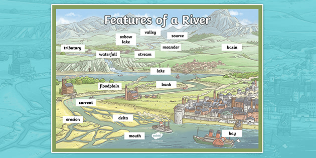
Features Of A River Ks2 Labelled Display Poster Ks2 Rivers
Or this brilliant UK Rivers and Seas MapGet creative with our wonderful River.
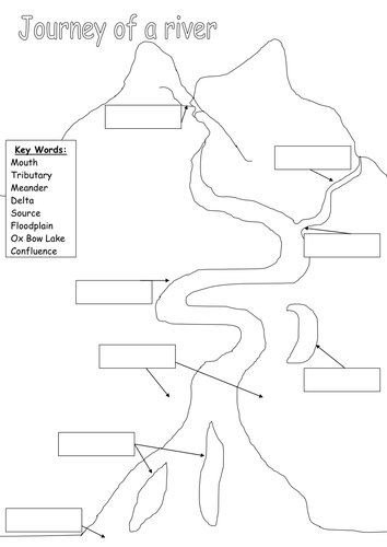
Labelling parts of a river ks2. Parts of a River labelling sheet. Some of the worksheets displayed are Name date components of a watershed States of water label the diagram Use the words in the box to fill in the Watersheds 4 congruence and triangles Egyptian project 1 Grammar for high school Name canoeing merit badge final test. If youve found this river labelling worksheet useful then you might also like this Diagram of a River KS2 Labelling Activity and beautifully illustrated matching posterTake a look at this fantastic Rivers Lesson Plan next.
If youve found this river labelling worksheet useful then you might also like this Diagram of a River KS2 Labelling Activity and beautifully illustrated matching posterTake a look at this fantastic Rivers Lesson Plan next. Created to be used with my year 5 class though suitable for other KS2 classes. A lesson plan explanation text and worksheet activity on the features of a river during its different stages.
Or this brilliant UK Rivers and Seas MapGet creative with our wonderful River. Children have to label the physical features of a river from source to mouth. Geography- Stages of a River.
Labelling Parts of a River Worksheet. Major Rivers of the World Labelling Worksheet -. The parts of a river.
Showing top 8 worksheets in the category - Parts Of A River Labeling. Children have to match the description to the correct area of the river. Pupils can cut out and position the labels to identify key features - great for class discussion and group collaboration.
This practical resource provides pupils with the diagrams and labels needed for them to identify parts of a river. Rivers lead to the sea or a lake and the place where it enters the sea or lake is called the river mouth. A river is a moving body of water that flows from its source on high ground across land and then into another body of water which could be a lake the sea an ocean or even another river.
Labelling Parts of a River Worksheet -. Label the features of a river. Labelling a River tributary confluence delta estuary floodplain levee meander mouth oxbow lake source waterfall main channel Use the labels at the bottom of the page to identify each of the parts of the river system.
Rivers that flow into the sea form an estuary where fresh water from the river mixes with. Major Rivers of the World Labelling Worksheet. Rivers comprehension A-Z rivers Rivers glossary Identifying different river features Locating river features in the lower middle or upper course of a river.
Children can practise identifying the parts of a river with labelsPupils can cut out and position the labels to identify key features - great for class discussion and group collaborationThis resource complements the Go Teach Label Parts of a River Interactive Activity. Through looking at these diagrams it is easier to understand the nature of V-shaped valleys the river ordering system the water cycle and other aspects related to rivers. What is the river like at its source.
Parts Of A River Labeling. This practical resource provides pupils with the diagrams and labels needed for them to identify parts of a river. These colorful and child friendly activities help you to teach all about the topic of rivers.
Labelling Parts of a River Worksheet. Geography- Stages of a River - YouTube. Aimed at 10-13 year olds it has activities which look at.
Give children labels with the parts of a river and ropes and or chalk and ask them to make a large-scale version of their diagram in teams of 8 When finished ask them about their part eg. I have created this worksheet to accompany the PowerPoint I created of the same name. Children are given statements about the width speed etc of a river and asked to identify which stage of a rivers journey they belong to.
These river diagrams help to explain the geography topic of rivers. Arrows point to different areas of a river. If playback doesnt begin shortly try restarting your device.
Typical flower diagram with labels. Flower Pieces Parts A.
Parts Of A Flower And Their Functions With Diagram Trees Com
Diagram Of A Typical Hibiscus Flower Images 134.

Label the parts of typical flower. Parts of a Flower - Science Quiz. Collectively the male parts of the flower are called the stamen. 1 Label a diagram of the external parts of a typical flowering plant Shoot root stem leaves flower fruit seed.
Filament Receptacle Ovule Stigma Carpel Anther Style Stamen. Plants A Basic Overview. Label the parts of a typical flower.
Flower Structure Diagram Untpikapps. It is the stalk of the flower which may be short long or even absent. Science.
Corolla red arcs consists of five fused petals antepetalous stamens are joined to petals by hairy filaments. Most roots grow underground. Students know and understand the characteristics and structure of living things the processes of life and how living things interact with each other and their environment.
The main parts of a flower that are important for its function are its male and female parts the carpel and the stamen. Bracts are specialized leaves from the axil of which bracteate flowers arise. Diagram Label the Flower anther - the anther is the tip of a flowers stamen the male reproductive organs of the plant - it contains the pollen.
Flowers contain vital parts including petals which form flowers. Sepals Protect the flower bud when it is developing. 4th Grade Science 15 teachers like this.
C C. A typical flower consists four whorls of floral. The pistil contains the stigma style and ovary.
Not all species produce flowers with all four parts. Label The Parts Of A Flower Answers - Fun for my own blog on this occasion I will explain to you in connection with Label The Parts Of A Flower AnswersSo if you want to get great shots related to Label The Parts Of A Flower Answers just click on the save icon to save the photo to your computer. Flowers Label the parts of a typical flower.
Make a model of a typical flower u2013 label all the parts p642 Filename. Although all flowers are different they have several. Label the parts of a typical flower.
Terms in this set 22 stigma. The anther produces pollen which is held in the small round pouches that sit on top of the filament. ROW_3_model_ideaspdf - Read File Online - Report Abuse.
2 State the function of the root and shoot 3 Identify tap and fibrous root systems 4 Explain the term Meristem and give its location in the stem and root 5 Name and give the function of four zones in a longitudinal section of a root. Ovary - the ovary is a female reproductive organ in plants that produces ovules. Chromebooks computers iPads iPhones Android tablets Android phones Kindle Fire.
Filament - the filament is the part of the flower that holds the anther and part of the stamen the male reproductive organs of the plant. What are the major Functions of Flo. Students will edit this template.
Fruit label the orange lemon apple strawberry watermelon avocado banana pear cherry and grapes in english. Label Parts of a Flower. Main Parts Of A Plant Their Functions Structure Diagram.
5_16_58_622pdf - Read File Online - Report Abuse. Diagram Of A Typical Plant Cell Structure Print Zazzle. About Press Copyright Contact us Creators Advertise Developers Terms Privacy Policy Safety How YouTube works Test new features 2020 Google LLC.
Label the parts of a typical flower 1 - peduncle 2 - petal 3 - sepal 4 - style 5 - stigma 6 - ovary 7 - pollen sac 8 - stamen 1 - receptacle 2 - petal 3 - s Grade Label the parts of a typical flower 1 - peduncle 2 - pe. Life Under The Microscope Naturphilosophie. Sepals protect the flowers before they bloom.
The female part of the flower the pistil is located at the center of the bloom. Plants G L. 4th 5th grade O.
Each student will label and list the parts and functions of a flower. Label the Parts of a Flower Click Use the tool to correctly label each flower part. .
Ria went to a plant nursery with her mother. Dissect and label each part of a flower. It is a leaf like structure in whose axil a flower often develops.
A typical angiosperm flower has following parts. Iii - Life Science. 12 11 6 13 10 1.
It is at the base of. Youll recognize the pistil in a plant diagram because it looks like a small knob that protrudes from the flower. Draw The Diagram Of A Typical Flower And Label The Parts.
Label the two lower nodes the first and second nodes on the plant diagram. Label the parts 1 to 10 in the figure shown below. Filament I 13 7.
The Life Cycle Of A Flower Discover How Flowering Plants.
It is composed out of the ossicles three tiny bones that link the eardrum to the inner ear. Lets Learn about Human Ears with this video.

Structure And Function Usherkids Uk
This causes the eardrum to vibrate and sound is produced.
Diagram and function of ear. It runs from the pinna to the ear drum or tymapanic membrane. This outside structure called the pinna acts like a satellite dish or. The outer ear which includes the complex shell that is the visible ear we see on the outside of our heads.
The ear ossicles present in the middle ear help in the amplification of the sound waves. The resonance of the outer. Just as the eyes turn certain wavelengths of light into images so the ear turns certain wavelengths of vibration into sounds.
The intensity of the sound waves is increased ten times by the ossicles. It transmits sound waves from the external ear to the internal ear through the chain of ear ossicles and thus transforms the air-borne vibrations from the tympanic membrane to liquid-bone vibrations in the internal ear. The ear performs the functions of hearing and balancing equilibrium.
The outer part of the ear ie pinna helps in the collection of sound vibrations and sends them to the eardrum. The diagram of ear is important from Class 10 and 12 perspective and is usually asked in the examinations. The perilymph of the internal ear.
Function of Ear. This creates an electrochemical signalwhich is sent. The sound waves pass through the auditory canal and reach the eardrum.
The Structure of Human Ear. In this part of the outer ear modified sweat glands that produce ear wax are located. The sound waves entering the ear get converted into electric impulses for the brain to understand and interpret.
The outer ear includes an ear canal that is is lined with hairs and glands that secrete wax. Inner ear and Central auditory nervous system. The middle ear extends from the ear canal.
Image will be Uploaded Soon Human Ear Structure and Function. It is divided into three fluid-filled chambers called scalae that spiral around a bony core. The ear catches sound waves and converts it into impulses that the brain interprets making it understandable and helps the human body differentiate between different sounds.
When the stapes moves in and out of the oval window of the cochlea it creates a fluid motion hydrodynamic energyIt causes membranes in the Organ of Corti to shear against the hair cells. Different structures of the human ear help in different functions. The vibrations produced pass through the tympanic membrane to the tympanic cavity.
Three major parts of a human ear are the outer middle and inner respectively each having its own functions anatomy and ear diseases as well. The middle ear is a narrow air-filled cavity in the temporal bone. It may be noted that the frequency sound does not change.
The mechanism of hearing involves the following steps. The vibrations are transmitted across the middle ear by the malleus incus and to the stapes bones. The outer ear receives the sound waves and transmits them down the ear canal to the eardrum.
They pass through the external auditory meatus to the tympanic membrane which is caused to vibrate. As you can see in the human ear diagram it is designed in such a way that it captures the maximum auditory stimuli from the atmosphere and transfers them to the brain for translating into hearing response. The pinna of the outer ear protects the eardrum from intense sound and channels the sound to the eardrum through the auditory canal.
The cochlea is the most critical component of the inner ear. It is spanned by a chain of three tiny bones the malleus hammer incus anvil and stapes stirrup collectively called the auditory ossicles. The cochlea present in the inner ear is responsible for the hearing.
Function of Ear Hearing. Again the results of such experiments suggest that the two primary structures contributing to the resonance of the outer ear are the concha and the ear canal. The sound waves are collected by the external ear up to some extent.
Human ear is a sense organ responsible for hearing and body balance. It does this through a system of many parts including. One such organ is the ear that helps us in the process of hearing and balancing.
Ear Diagram And Function. Functions of Ear. Functions of the Middle Ear.
This part of the ear provides protection and channels sound. The scala media or cochlear duct. The latter fits into the fenestra ovalis.
Following are the important function of the ear. The external auditory meatus or an ear canal is approximately ¼ inch in diameter. The function shown is for the entire outer ear.
Experiments similar to the one just described can be conducted to isolate the contribution of various cavities to the resonance of the total outer ear system. In the human ear diagram we can distinguish between the inner ear middle ear and the outer ear. Structure and Functions of the Ear Explicated With Diagrams The ear is another extraordinary organ of the house of wonders that is the human body.
Let us take a look at the human ear structure with the help of a diagram and understand its functions a little more closely. Its function is to recognize the pathway of the sound. The function of the outer ear is to collect sound waves and guide them to the tympanic membrane.
It transports collected sound of Outer ear to Ear drum Eardrum It is a thin membrane skin It collects compressions and rarefactions of sound Compressions make eardrum move inward Rarefactions make eardrum move outward Ear bones There are 3 ear bones - Hammer Anvil and Stirrup They amplify sound vibrations increase its volume They also transmit sound to inner ear Inner ear Cochlea The cochlea is part of inner ear. 1988 rx7 alternator wiring diagram fiat 415 wiring diagram draw a logic diagram online tip ring sleeve diagram 2006 audi a6 ignition amp located 2000 lexu rx300 engine part diagram wiring schematic redarc wiring diagram earbud wiring diagram new beetle engine diagram 3976 fuel filter nissan 200sx wiring diagram 1966.
Brassica rapa can be purchased under the trade name Wisconsin Fast Plants and used in this activity. The pistil is considered the female part of a flower because it produces seeds.

Nature Cultural And Travel Photography Blog Brassica Flower Parts
Thursday December 14 2017.

Labeled diagram of brassica flower. Rapa Turnips - Plantinfo. The vegetative shoot shows unlimited growth whereas the flower shows the limited growth. Plants primary teaching resources and printables sparklebox.
Make 10 MCQs draw a labeled diagram of brassica flower also collect flower and dry it. Find flower anatomy diagram lesson plans and teaching resources. 12 When a terminal flower is depicted the axis is not present and therefore cannot be shown.
One especially fast-bolting bolt means to set flowersusually making the leaves bitter variety of brassica was developed by the University of Washington for research and education and has a life-cycle of just one month. They are not only involved in reproduction but are also a source of food for other living. Brassica Campestris Flower Diagram.
Plant Flower Anatomy - Anatomy Drawing Diagram. Diagrams are usually depicted with the subtending bract below and the axis above the flower itself both in the median line. Parts of a plant.
Flower develops on the mother axis stem in the form of floral bud. Napus from other brassicas. The anatomy and morphology of stamens and carpels of cruciferous flower bears testimony to a papaverous ancestory.
Opal Diagram Brassica Flower Youtube. The leaves of turnip Brassica campestris left grasp the stalk completely. For this plant observation lesson students examine a diagram of a Brassica plant and identify its anatomy before planting their own seeds and viewing the changes.
Spring is almost here and flowers are blooming its the perfect time to make a flower anatomy 3D project. Wild Turnip Brassica rapa - Flowers - NatureGate. Diagram of a flowering plant with label.
The reproductive parts of the flower that are necessary for seed production are the stamen the male organ and carpel the female organ. Flower diagram not labeledEach female and male organs are discovered on the identical flower. Shown on the plant cell diagram labeled above that plant cells are surrounded by a thick rigid cell wall.
Labeled hibiscus labeled parts of flower. Flower diagram not labeled. The extraordinary diversity of Brassica oleracea Plant.
A flower is a attribute characteristic of flowering vegetation and is definitely an extension of the shoot meant for copyflowers are enticing and seem in numerous colors and shapes to draw pollinators who assist in pollen switch. Bracteoles if they are present are usually drawn on the sides of the diagram. Education Chart Of Biology For Anatomy Of Hibiscus Flower Diagram Grass Features And Structure Draw A Well Labeled Diagram Of Flower To Show Its Parts Gbzhcrb00.
Diagram Flower Mature Free Vector Graphic On Pixabay Parts Of A Flower Lovetoknow Flower Diagram To Label Wiring Diagram Blog. They are not only involved in reproduction but are also a source of food for other living organisms. The axis corresponds to the position of the main stem relative to a lateral flower.
Quickly find that inspire student learning. Parts and function of the ovary in the flower. Flower diagram not labeled.
Paste it Write definition of set also types of sets i Empty set ii Finite set iii infinite sets iv equal set v disjoint set vi over lapping set Do Ex 11 2nd Learn WriteVerb. U 1935 Office of the Gene Technology Regulator 2008 Figure iv. Flower diagram labeled flower diagram labeled and functions flower diagram labeled parts labeled diagram of brassica flower.
Draw neat and labelled diagram of l s of hibiscus flower. Parts of a flower. The leaves of canola Brassica napus centre only half-grasp the stalk.
But in Brassicaceae the stamens are tetradynamous and not in Papaveraceae. Comparison of floral diagram indicates that Brassicaceae is closely allied to Capparidaceae. Brassica Flower Parts Labeled images similar and related articles aggregated throughout the Internet.
Typical floral diagram of a Brassicaceae Erysimum Bowles Mauve Flowers may be arranged in racemes panicles or corymbs with pedicels sometimes in the axil of a bract and few species have flowers that sit individually on flower stems that spring from the axils of rosette leaves. Life cycle of a plant content classconnect the structure of a flowering plant brassica rapa parts of a flower diagram ks2 beautiful. Flower anatomy flowers consist of reproductive parts and modified leaves.
Its made up of the following parts. By constructing a three-dimensional flower and labeling all the parts children will learn a lot about flower anatomy. Showing posts with label labeled diagram of brassica flower.
Getting an A for outstanding achievement in science by following this step by step 3-D flower project is within your reach. How to draw diagrams on Biology Practical copy Punjab Board Lahore by Naveed Akhtar Uppal calligrapher artist Jhelum Pakistan. Get Free Access See Review.
A bird rape plant Brassica campestris with an associated.
Process of Photosynthesis Diagram Diagram of Photosynthesis How To Draw Photosynthesis - YouTube. In other words we can say that photosynthesis is transformation of solar energyradiant energylight energy ultimate source of.
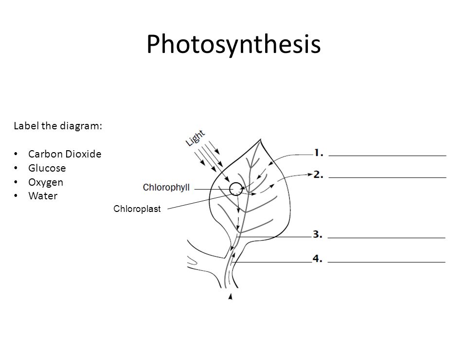
Photosynthesis Label The Diagram Carbon Dioxide Glucose Oxygen Water Ppt Video Online Download
Photosynthesis tree tree oxygen photosynthesis process photosynthesis process of photosynthesis leaf photosynthesis minerals plant plant body photosynthesis vector earth science formulas.

Diagram of photosynthesis without labels. Photosynthesis diagrams worksheet name date period. These are my choices. Blank flower parts worksheet stages of mitosis worksheet answers and plant photosynthesis diagram.
Posted on March 26 2019 by admin. This combination forms glucose C 6 H 12 O 6 Step 3. List of Photosynthesis Diagram Worksheet.
With the venn diagram for the photosynthesis and the respiration it says the terms may be use more than once. In the Photosynthesis circle it shows. Life on earth ultimately depends on energy derived from sun.
The second page breaks down photosynthesis and has students come up with examples of the different sources that plants get their necessities from. Hello there At page below we bring you some cool pictures we have collected in case you need more references for today we are more concern about Photosynthesis Labeling Worksheet. Hydrogen which split from H 2 O combines with CO 2 using energy stored in ATP Step 2.
Plants begin making their food which basically includes large quantities of sugars and carbohydrate when sunlight falls on their leaves. Plants begin making their food which basically includes large quantities of sugars and carbohydrate when sunlight falls on their leaves. 817 photosynthesis diagram stock photos vectors and illustrations are available royalty-free.
Mar 14 2018 - Label the Photosynthesis diagram. Process of Photosynthesis Diagram Diagram of Photosynthesis How To Draw Photosynthesis. Just like humans and animals plants also come with their own set of organs.
Label the diagram structure of chloroplast light vs dark reactions and more. 32 How Does Photosynthesis Work Label The Diagram Answer Key Labels Information List. Diagram Of Photosynthesis 2 stages of photosynthesis a Light phase.
Part of photosynthesis that doesnt require light Step 1. Among the most crucial organs a plant can have are their leaves thin and flattened these structures are adapted to absorb energy from our own Sun to power the processes of photosynthesis. Leaves are primarily composed of three.
A Simple Diagram Of Photosynthesis Hubpages. Broadly plants have two organ systems. Absorbs Calvin cycles Chlorophyll CO 2 H-2-O Krebs cycle Mitochondria Releases.
Get Unlabeled Photosynthesis Diagram ImagesPhotosynthesis explained with a diagram. Water is formed byproduct. This is what I needd help with.
Label Photosynthesis Diagram photosynthesis the process of photosynthesis explained analytical questions 1 molecular structure the process of photosynthesis in plants with. When algae are undergoing photosynthesis the concentration of various molecules changes. Labeled chloroplast diagram engine diagram and wiring.
Water soil and carbon dioxide. By the way about Photosynthesis Labeling Worksheet weve collected various similar pictures to add more info. Photosynthesis is a process used by plants and other organisms to convert light energy into chemical energy that through cellular respiration can later be.
31 Label Photosynthesis Diagram Labels For Your Ideas. Chloroplast Diagram Without Labels. Photosynthesis Diagram According to the diagram of photosynthesis the process begins with three most important non-living elements.
Chloroplast 2 image chloroplast diagram labeled and biology basic units of life with labels thylakoid membrane in photosynthesis definition function structure thylakoid membrane in photosynthesis. Feb 18 2019 - Grab our photosynthesis worksheets containing charts and activities. See photosynthesis diagram stock video clips.
Cannot be formed without the input of if you re looking for photosynthesis diagrams you ve come to. Diagram of a leaf labelling game. Could you please help me.
What does photosynthesis produce what is needed for photosynthesis photosynthesis is also responsible for balancing oxygen and carbon dioxide levels in the atmosphere. Label photosynthesis diagram. A leaf consists of three main parts.
Learn about photosynthesis with interactive diagrams and science games for kids. With the digrams that are circles. Photosynthesis Photon Light Synthesis Putting together is an anabolic endergonic process by which green plant synthesize carbohydrates initially glucose requiring carbon dioxide water pigments and sunlight.
Photosynthesis is a process used by plants and other organisms to convert light energy into chemical energy that through cellular respiration can later be released to fuel the organisms metabolic activitiesThis chemical energy is stored in carbohydrate molecules such as sugars which are synthesized from carbon dioxide and. Labeled Chloroplast Photosynthesis Diagram Hd Png Download Vhv. A the root system and B the shoot system.
This worksheet first has students label a diagram with all of the important parts of photosynthesis. I the petiole ii leaf base and iii lamina or leaf.
Complete the four missing labels and the descriptions of each stage at the sides of the diagram. Photosynthesis occurs in most plant life but may differ in some.

Chapter 8 Flashcards Quizlet
Photosynthesis is a two-stage process in which sunlight photo- powers the production -synthesis of organic molecules.

Which diagram correctly labels the reactants and products of photosynthesis. Label the reactants and products. The products of cellular respiration are glucose oxygen and water molecules. The cellular-respiration diagram would show glucose as the main source of energy.
Photosynthesis and Respiration Review Sheet. Be sure to label the reactants and the products. Photosynthesis is a redox process.
An electron carrier involved in photosynthesis. The diagram below shows the relationship between photosynthesis and cellular respiration and the organelles in which they occur. 6O 2 C 6 H 12 O 6 6CO 2 6H 2 O.
Write the equation for photosynthesis. Cellular respiration occurs in direct synchronicity with this process using the products of photosynthesis as its reactants and producing its reactants. Glucose and Carbohydrates What are the inputs reactants and Outputs.
Li Ming is correct because chlorophyll is a substance that is present before the chemical reaction of photosynthesis takes place. Glucose and Carbon Dioxide. C6H12O6 O2 CO2 H2O 36 ATP 3.
Water and Carbon Dioxide Input. 6CO 2 6H 2 O C 6 H 12 O 6 6O 2. This side represents the substances we have before the reaction takes place.
CO2 H2O Solar energy C6H12O6 O2. Carbon Dioxide and Water. This activity is intended to be given after going over photosynthesis and doing the chapter reading.
REACTANTS before the reaction PRODUCTS after the reaction. It also contains the photosynthesis equation and a label activity to describe the equation. It is extremely important to know the meaning and process of photosynthesis irrespective of the fact that whether it the part of ones curriculum or not.
Cellular respiration uses the products of photosynthesis as reactants. Chloroplasts convert light energy into chemical energy that can be used by mitochondria. What is the relationship between reactants and products of photosynthesis and cellular respiration.
The reactants of photosynthesis are light energy carbon dioxide and water. They are called the products. Which statement describes how photosynthesis and cellular respiration are interrelated.
Water and Oxygen Input. The reactants of photosynthesis are water light and carbon dioxide while the products are oxygen and sugars. Chloroplasts split water into hydrogen and oxygen incorporating the electrons of hydrogen into sugar molecules.
The cellular-respiration diagram would show energy stored in large protein molecules. The products of photosynthesis are carbon dioxide water and energy. The main role of plastid in Chloroplasts is to transform light energy into chemical energy.
The reactants of cellular respiration are glucose and oxygen. Photosynthesis is summarized as 6 CO 2 12 H 2 O Light energy C 6 H 12 O 6 6 O 2 6 H 2 O. They are called the reactants.
Cellulose is a carbohydrate and a polymer of glucose which is constructed by connecting smaller monomer subunits. Be sure to label the reactants and the products. Li Ming is correct because chlorophyll is a substance that is formed from the chemical reaction of photosynthesis.
I encourage my students to use outside sources if they are really stuck on the labeling like their. Light drives electrons from chlorophyll to NADP forming NADPH which provides the high-energy electrons for the reduction of carbon dioxide to sugar in the Calvin cycle. Students label the reactants carbon dioxide and water then the products glucose and oxygen.
This document contains a labeled diagram and a blank diagram of the two stages of photosynthesis. Write the overall reaction of photosynthesis. Photosynthesis Reactants The photosynthetic process requires several simple reactants.
This side represents the substances that we have after the reaction has taken place. The cellular-respiration diagram would show electromagnetic waves as the final product. Draw a picture of a chloroplast.
H 2 O is oxidized and CO 2 is reduced. To the right of the arrow we have the after situation. Ch 4 Photosynthesis Worksheet 1.
Light dependent and light independent reactions. The diagram given in this BiologyWise article is a small pictorial elaboration of the process of photosynthesis that will prove helpful for kids and teenagers to understand this vital process of the plant kingdom. Thylakoid Membrane inside chloroplast NADPH.
The cellular-respiration diagram would show water as the main source of chemical energy. The following diagram describes the reactants and products of each stage. Label the thylakoid grana and stroma.
What are the inputs reactants and Outputs products of Photosynthesis. Write the equation for aerobic respiration. Play this game to review Biology.
Another slide goes into the details of the light dependent reaction and the Calvin cycle. Julian is correct because. The reactants for photosynthesis are light energy water carbon dioxide and chlorophyll while the products are glucose sugar oxygen and water.
Place the words into the diagram.
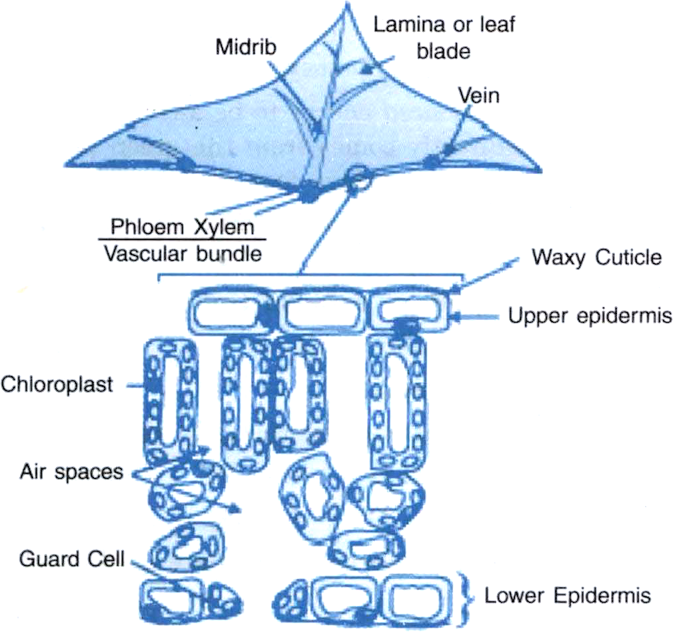
A leaf has two main parts. Leaf base has two small leaf-like structure called stipules.
Draw A Labelled Diagram Of Cross Section Of A Leaf Lamina To Show Chloroplasts From Science Life Processes Class 10 Cbse
Draw the diagram of cross section of a leaf and label the chloroplast and cuticle.

Labelled diagram of a section of a leaf. Cross section of dicot leaf Jul 27 2018 Anatomy of a Typical Dicot Dorsiventral Leaf Cross Section CS Under Microscope with Labelled Diagram Description and PPT Organs. Cross-section of a leaf To get rid of excretory products plants use the following ways. Schematic transverse section through a cross section of a leaf lamina cross section of leaf draw the labelled diagram of cross.
This is the part where a leaf attaches to the stem. Schematic Transverse Section Through A Dicotyledon Leaf Indicating The Scientific Diagram Draw A Labelled Diagram Of Cross Section Leaf Lamina To Show Chloroplasts From Science Life Processes Class 10 Cbse. Draw A Labelled Diagram Of Cross Section Leaf Lamina To Show Chloroplasts From Science Life Processes Class 10 Cbse.
Hi guys today in this video I am going to show you how to draw cross section of a leaf and along with this I am going to tell you some important function of. 2 Lamina- the green flat part of a leaf that is specialized for photosynthesis. C In certain group of plants stomata remains closed during day.
Click here to get an answer to your question Draw a well labelled diagram of a Cross section of a leaf gurnoor550 gurnoor550 29112020 Biology Secondary School answered Draw a well labelled diagram of a Cross section of a leaf 2. Light absorption happens in the palisade mesophyll tissue of the leaf. A protective layer of cells that produces the cuticle.
How is food synthesized by such plants. 2d Labelled Diagram Leaf Epidermis. Leaf anatomy Both Internal and External with Labelled Diagram.
Ii Part of a Leaf. The leaves make food for the plant. File Gold Leaf Electroscope Diagram.
1 Petiole- the stalk that supports a leaf in a plant and attaches the leaf blade to the stem. The epidermis is is also transparent and very thin to. A Draw the diagram of cross section of a leaf and label the following parts i chloroplast ii cuticle b A gas is released during photosynthesis.
1 petiole 2 leaf base and 3 leaf blade or lamina each performing specific functions. The Leaf PDF 檔案how they differ from the rest of the cells forming the epidermal tissue of the leaf. Parts of a Leaf Diagram.
The leaves get rid of excess water from the plant through transpiration. A section through the leaf of tuberose Polianthes tuberosa of family Amaryllidaceae would show the following anatomical structure Fig. Anatomy of the leaf is the detailed study of the internal structure of a leaf usually revealed by its dissectionLeaves are responsible for converting sunlight and carbon dioxide.
Name the gas and also state the way in which the gas is evolved. They are arranged closely together so that a lot of light. Iii Functions of leaves.
Enjoy This Parts Of A Leaf Diagram Freebie The File Includes 4. Both the epidermal layers are uniseriate composed of compactly- arranged rectangular cells with rounded cuticularised outer walls. Labelled diagram - Drag and drop the pins to their correct place on the image.
Gcse Plant Biology Diagrams To Label Teaching Resources. Palisade cells are column-shaped and packed with many chloroplasts. A waxy layer that prevent water loss by evaporation.
A typical leaf shows three main parts. I Excess water is lost by Transpiration. In plants like paddy wheat and other monocotyledons this leaf.
The cuticle is transparent and very thin to allow maximum light penetration. Education Chart Of Biology For Cross Section Of Leaf Diagram. CROSS SECTION OF A LEAF.
Generally leaf base petiole and lamina together form the main parts of a leaf. Leaves are the main photosynthetic organs of the plant. I The leaf is a thin broad flat and green part of a plant which is attached to the stem.
It is the stalk that connects a leaf to the stem of the plant it is made of complex conducting tissues called vascular tissues. November 1 2019 by Ranganr.
47 What Are The 4 Cardiac Valves Suzuki Pictures. This valve is located between the right atrium and the right ventricle.

Roles Of Your Four Heart Valves American Heart Association
Gross anatomy the heart valves are located in the cardiac fibrous skeleton.

What are the 4 cardiac valves. The heart has four valves - one for each chamber of the heart. The cardiac valves function to prevent backflow of blood when a specific part of the heart contracts. The pulmonary valve is located between the right ventricle and the pulmonary artery.
The heart has 4 valves. Structure of the chambers and valves of the heart. View What Are The 4 Cardiac Valves Pictures.
Normal valves have 3 flaps leaflets except the mitral valve. Four valves prevent the backflow of blood during the cardiac cycle. It only has 2 flaps.
The 2 lower chambers are the ventricles. The pulmonary valve is located between the right ventricle and the pulmonary artery. Separates the top right chamber right atrium from the bottom right chamber right ventricle.
Heart Valve Problem Causes Care of Heart Valves Health Continue reading 47 What Are The 4 Cardiac Valves Suzuki Pictures. This valve is located between the left atrium and the left ventricle. The mitral valve and tricuspid valve which control blood flow from the atria to the ventricles.
The atrioventricular valves bicuspid or mitral on the left tricuspid on the right prevent the backflow of blood from the. Open during diastole to direct blood flow from the atria to the ventricles. Separates the top right chamber right atrium from the bottom right.
The 4 heart valves are. The pulmonary valve is located between the right ventricle and the pulmonary artery. The 4 valves are the aortic pulmonary mitral and tricuspid valves.
Tricuspid valve located between the right atrium and right ventricle Pulmonary or pulmonic valve between the right ventricle and the pulmonary artery. The four cardiac valves direct the flow of blood through the heart during the cardiac cycle. The right-sided tricuspid valve TV and left-sided mitral bicuspid valve MV.
The four heart valves are. They receive and collect blood. The cardiac valves function to prevent backflow of blood when a specific part of the heart contracts.
The 2 upper chambers are the atria. Keep reading to know more about these four valves and their functioning. The 4 heart valves are.
What are the 4 types of valves. This valve is located between the right atrium and the right ventricle. Has three leaflets or cusps.
4 heart valves decide the pathway for efficient blood flow through the heart. The heart valves are located in the cardiac fibrous skeleton. This valve is located between the right atrium and the right ventricle.
And are what has always been in my motor. The left side of the heart can be thought of as the hearts powerful engine. Two are atrioventricular AV valves.
What is the cost to diagnose the p0446 toyota code. The 4 heart valves are. The 4 heart valves are.
That is a valve will open to allow fluids to pass in one direction but will close to prevent fluids from passing in the other direction. The valves keep blood moving through the heart in the right direction. Tricuspid Valve Has three leaflets or cusps.
The aortic valve and pulmonary valve. This artery carries blood from the heart to the lungs. This valve is located between the right atrium and the right ventricle.
The four valves in order of circulation are. The mitral valve and tricuspid valve are located between the atria upper heart chambers and the ventricles lower heart chambers. The pulmonary valve is located between the right ventricle and the pulmonary artery.
This valve is located between the left atrium and the left ventricle. They let blood flow forward and prevent the backward flow. Damaged valves can cause loss of power from the engine.
How the heart works 4 chambers. Each side of the heart has a collecting chamber on top atrium and a pumping chamber on the bottom ventricle. Cardiac cath is performed to find out if you have disease of the heartmuscle valves or coronary heart arteries.
This valve is located between the left atrium and the left ventricle. There are 4 valves of the heart 2 on the left side and 2 on the right side.
It have protective function and the outer pinna directs the ear towards the sound wavewithout pinna. The ear is an advanced and very sensitive organ of the human body.

Sense Of Hearing And Equilibrium 3 Parts Sense
Which Ones Have You Heard Of.
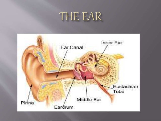
What are the 3 parts of the ear. The ear is divided into three parts. The anatomy of the human ear comprises three major parts namely the outer ear middle ear and inner ear. The three parts of the outer ear are the auricle the ear canal and the tympanic membrane ear drum The auricle is the part that most people see when looking at the ear.
Malleus Incus and. Inner ear consisting of. Different parts of the ear and their functions are very important for hearing.
They are called the malleus the incus and the stapes. The space inside the ear drum is called the middle ear. The external outer ear consists of the auricle external auditory canal and eardrum Figure 1 and 2.
By sensing the movements of these stones the ear can tell our brain where we are relative to the directions up and down and how our body is moving or accelerating. The medical term for. 3 Internal ear.
Three of the smallest bones of the body are found in the middle ear. External ear has-- Pinna - a cartilagenus protective flap External Auditory Meatus -- ear canal surrounded by bone associated with hair and wax glands and Tympanum -- membranar ear drum. The ear canal starts at the outer ear and ends at the ear drum.
The outer ear includes an ear canal that is is lined with hairs and glands that secrete waxThis part of the ear provides protection and channels. What are the 3 major parts of the ear. Middle ear has-- Ear Ossicles -- three in number.
The human ear has three main sections which consist of the outer ear the middle ear and the inner ear. The human ear consists of three parts. Sound waves enter your outer ear and travel through your ear canal to the middle ear.
There are three different parts to the outer ear. The medical term for the outer ear is the auricle or pinna. Eardrum cavity also called the tympanic cavity ossicles 3 tiny bones that are attached malleus or hammer - long handle attached to the eardrum incus or anvil -.
In humans the inner ear contains parts called the semicircular canals where otoliths tiny stone-like structures shift in response to gravity and the movement of our body. The outer ear is made up of cartilage and skin. Malleus or hammer - long handle attached to the eardrum incus or anvil - the bridge bone between the.
Sound waves that reach the tympanic membrane cause it to vibrate. The auditory canal concentrates sons waves and. The ears function is to transmit and transduce sound to the brain through the parts of the ear.
The middle ear is an air-filled space that contains 3 tiny bones known as ossicles called the malleus hammer incus and stapes stirrup. Also asked what are the 7 parts of the ear. Different Parts of the Human Ear.
7 rijen There are three major parts of the ear the outer middle and inner ear. It have two. The ear has three major parts.
External ear Middle ear Internal ear. Click to read full answer. Each part of the ear is again made up of different components.
Lets try to understand the different parts of human ear in detail. Each part has several important components in it. The outer ear the middle ear and the inner earThe major task of the ear is to detect transmit and transduce sound.
Ear is divided in three anatomical parts. External ear middle ear and inner ear. The tragus helix and the lobule.
The middle ear includes. The inner ear is also termed as cochlea which is a dual membrane delicate structure with fluids filled between the membranes and lined with hair cells. It consists of auditory canal and ear drum or tympanum.
The vestibular window separates the middle ear from the inner ear. The auricle or pinna is a flap of elastic cartilage shaped like the flared end of a trumpet and covered by skin. Parts of the ear External Outer Ear.
Inner ear- contains the eardrum 3 small bones namely hammer anvil stirrup its not incus the auditory nerve cochlea 2. The ear canal is a tube. These bones are also known as the hammer anvil and the stirrup.
Each contains several. Three main parts of ear are.