General cargo ship ABDhatches No 1 No 2 No3 C cofferdam 1cargo gear masts and derricks 2. A ship comprises of both visible as well as invisible parts.

Ship For Transport Of Spent Nuclear Fuel Structure Diagram
The house is broken up into the berthing areas the galley and the lounges.
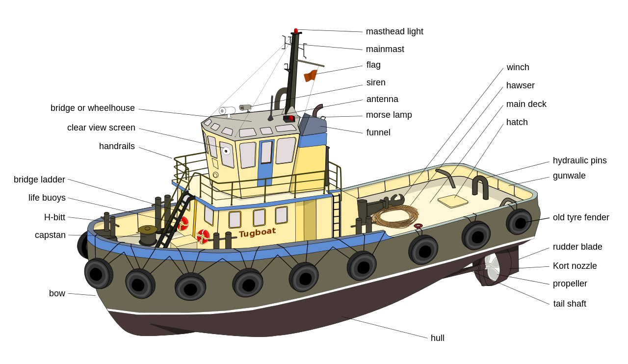
Labelled parts of a cargo ship. C Accommodation or superstructure This where the crew live and operate the ship. Stowage in holds 12. Cargo holds are where we carry the cargo no way right machinary spaces are distributed all around the vessel to make the ship operate and superstructure consists of.
Fore peak tank 3. Bow thruster 5. While common visible parts of a ship are.
SHIP CONSTRUCTION STRUCTURES Image Credit. 3 hatchcover free-fall lifeboat forecastle poop deck poop deck No. Lower hold centreline bulkhead 9.
There may also be several deck areas topside including the poop deck the deck in the rear of the ship and the afterdeck located directly behind the bridge. What are the parts of a cargo ship. Consequently this cargo ship part minimizes the liquid dynamic effect sloshing forces on surrounding structure.
Theres also an area containing washing mach. The forecastle or Focsle Deck. Bulbous bow 2.
Tweendeck centreline bulkhead 8. Rudder anchor bow keel accommodation propeller mast bridge hatch covers and bow thrusters. Cargo holds machinary spaces superstructure and tanks.
Are the invisible parts of a ship. Bow anchor 4. Under deck passage 7.
6 Indicate the parts of the ship below by drawing an arrow to the relevant position. The port may handle one particular type of cargo or it may handle numerous cargoes such as grains liquid. The Captains Cabin is at the rear or stern of the ship.
We always called the area where the crew stays the house. On another hand invisible but structural part of the ship consists of. 3 bridge wheelhouse forepeak afterpeak.
The Hold provides cargo stowage and sleeping space. Container refrigeration ducts 7. Ship Construction Structure Part 1.
A Jibboom is a metal rod that supports the head sail of a ship which makes it sail more easily. Insulated containers in holds 6. Theres also the cargo bays and the engine room machinery spaces.
Not Container hold 8. 6 metres or 10 metres which means that 6 metres of the hull or 10 metres of the hull is above the water. The Forepeak is the part of the hold the space below the lowermost deck of a ship which is nearest the front of the ship at the ships narrowest section.
A Hatch is a door on a ship. Various Other Parts of a Modern Ship. No2 Container hold 9.
Bulkheads frames cargo holds hopper tank double bottom girders cofferdams side. Escuela de Tripulantes y Portuaria de Valparaiso Ship. Bosun store 6.
A swash plate describes the plate employed for this purpose though not stretching to the tank bottom. On larger ships the top deck may have several levels designed to isolate various parts. Rudder anchor bow keel accommodation propeller mast bridge hatch coves and bow thrusters are some common visible parts whereas bulkheads frames cargo holds hopper tank double bottom girders cofferdams side shell etc.
The Hull or skin of the craft keeps the water out. The Fore Castle Area Or The Bow Closed Chalk The Chief Officer stands here when mooring the ship Anchor Chain Mooring Bitts or Bollards This is the Forecastle or fore or forward part of the Ship. D Hull The part of the ship that is partly in the water.
Bridge castle front 2. Cargo ports on the other hand are quite different from cruise ports because each handles very different cargo which has to be loaded and unloaded by very different mechanical means. Bulkheads separate the ship into compartments.
It can lead down into the hold or through bulkheads between compartments. E Freeboard The part of the hull that is above the water. KEEL - At the centre line of the bottom structure is located the keel which is often said to form the backbone of the ship.
A cargo ship generally comprises of 4 areas. Waterline Refers to the line where the hull of a ship meets the water surface. The top deck is broken up by the bridge a covered room which serves as the command center.
An education on the different parts of an Oil Tanker. Freeboard is usually given in metres eg. A cargo ship or freighter is a merchant ship that carries cargo goods and materials from one port to anotherThousands of cargo carriers ply the worlds seas and oceans each year handling the bulk of international tradeCargo ships are usually specially designed for the task often being equipped with cranes and other mechanisms to load and unload and come in all sizes.
Deck cargo A bow. Apart from the major parts of the ships that we read about in the above section there are a few equipment itmes present in almost all modern day ships. Wheel and is used to steer the ship.
Holds Part of the vessel that carries the cargo Escuela de Tripulantes y Portuaria de Valparaiso Ship 18. This describes the hollow or solid longitudinal part of a ship applied in construction of base of the cargo vessel. The cargo is kept in holds or in tanks in the hull.
I hope this has helped you on your basic understanding of ships. Parts of a ship1. The cargo port is also further categorized into a bulk or break bulk port or as a container port.
Theseare the safety systems anchors anchors were present in olden day ships too electrical equipment cranes etc. Foremast and mast top 4. Bow stern funnel mainmast stem sternpost shipboard crane double bottom engine room rudder bulbous bow bow thruster hold No.
Draw a neat diagram of plant cell and label any three parts which differentiate it from animal cell. Get Draw A Neat Labelled Diagram Of Plant Cell And Label Its Parts BackgroundThe nitrates absorbed by the plant roots are converted into various types of amino acids about 20 types which sequences respectively to form polypeptide chains of various protein molecules in the ribosomes.
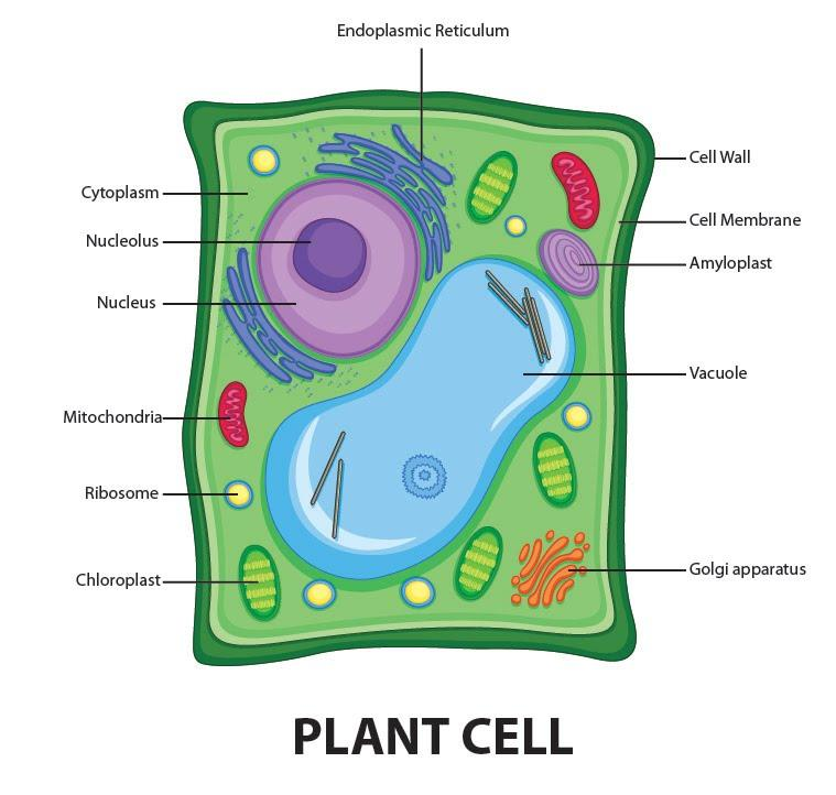
Draw A Welllabelled Diagram Of A Plant Cell Class 11 Biology Cbse
Answered Feb 6 2020 by KumariJuly 536k points selected Feb 6 2020 by.

Neat labelled diagram of plant cell class 11. How to draw animal cell labelled diagram Animal cell diagram for class 910 and 11 - YouTube. Compare The Location Of Nucleus In Animal Cell And Plant Cell Draw A. Draw a labelled diagram of a plant cell.
Labelled diagram of a plant cell. Find an answer to your question 1 Draw a neat labelled diagram of plant cell and list the function of each organalle. Energy is produced in the form of ATP in the process.
Compare Plant cell and Animal cell 4 Why Animal cell do not have cell wall for class 8 plz answer fast. The diagram given below represents a plant cell after being placed in a strong sugar solution. Link of our facebook page is given in sidebar.
If playback doesnt begin. Photosynthesis occurs in the chloroplasts of the plant cell. Draw a neat labelled diagram of animal cell.
Define plant cell and animal cell. Draw A Neat Diagram Of Plant Cell And Label Any Three Parts Which. ExplanationDiagram of plant cell and thier three parts Diagram of plant cell and thier three parts.
Study the diagram and answer the questions that follow. Answer verified by Toppr. Photosynthesis is the major function performed by plant cells.
Get Instant Solutions 24x7. 1 Answer 1 vote. Aaryansharmasml aaryansharmasml 20112020 English Secondary School answered Draw a neat labelled diagram of a plant cell.
The membrane is selectively permeable and allows only certain molecules to pass through. It is the process of preparing food by the plants by utilizing sunlight carbon dioxide and water. Ncert Solutions For Class 8 Science Chapter 8 Cell Structure And.
Cells under the microscope. Share It On Facebook Twitter Email. Rajubasak49041 rajubasak49041 28042020 Biology.
Class 11 Oscillations Redox. Cell Structure And Functions Class 11 Notes Biology Mycbseguide. Draw a labelled diagram of a plant cell.
Draw a neat diagram of the structure of chromosome and label the parts. A Centromere b p-arm. Plant and animal cell diagram class 8 NCERTHi friends In this video we will learn how to draw diagram of plant and animal cell this diagram is.
Draw a neat labelled diagram of the following cell organelles and explain their structure. Draw a neat and labelled diagram of a Plant cell and animal cell. It is very thin delicate elastic and selectively permeable membrane.
Class-11 Welcome to Sarthaks eConnect. Further plant cells are green in color due to the presence of special pigments that aid in photosynthesis. 13 Plant Cell Diagram Class 9Th PicturesThe class students can check below the diagram of plant cell and animal cell which can help them in understanding how to draw a cell diagram concept.
1st lab exam material biology 440 with dr. The plant cell is rectangular and comparatively larger than the animal cell. The cell membrane is a double-layered membrane made up of phospholipids that surrounds the entire cell.
Plant cells kept in hypertonic solution will get. Draw a neat diagram of plant cell and label any three parts which differentiate it form animal cell. The unit of life.
A Mitochondri b Centrioles. Students upto class 102 preparing for All Government Exams CBSE Board Exam ICSE Board Exam State Board Exam JEE MainsAdvance and NEET can ask questions from any subject and get quick answers by subject teachers. Draw a neat labelled diagram of an animal cell.
Class 8th 9th 10th 11th and 12th can use this diagram. Upvote 0 Was this answer helpful. 1 Draw the concept Map for the Cell 2.
How to draw a animal cell easy and step by step. Cytosol is the fluid present within a cell that is made up of water and. A unique platform where students can interact with teachersexpertsstudents to get solutions to their queries.
The cell wall is made up of cellulose which provides support to the plant. It is responsible for coordinating many of the important cellular activities such as protein synthesis cell division growth and a host of other important functions.
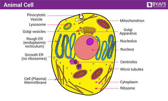
A Well Labelled Diagram Of Animal Cell With Explanation
Class 9 ScienceHome Page.

Labelled diagram of eukaryotic cell class 9. Draw well-labeled diagrams of any one. They are present in eukaryotic cell but absent in prokaryotic cells. A Draw a neat and labelled diagram of a prokaryotic cell.
Question Bank Solutions 7801. 9th Grade Prokaryotic Cell Diagram Class 9. Genetic material enclosed in nuclear envelope Genetic material not enclosed in nuclear envelope 3 Nucleolus and one or more chromosomes present.
Topic 1 2 Ultra Structure Of Cells Amazing World Of Science With. Viruses are smaller than prokaryotic cells. Name the kingdom in which they are placed.
Example plant cellanimal cell. Explain the structure and function of the nucleus. V Draw a labelled diagram of a prokaryotic cell.
These neat and well labelled diagram will ma. Draw a neat diagram of plant cell and label any three parts which differentiate it from animal cell. B Differentiate between a prokaryotic and eukaryotic cell any 4 points of difference.
A diagram of an eukaryotic nucleus is as given. Prokaryotic Cells Types Of Prokaryotic Cells Cell. B Differentiate between plant cell and animal cell.
It is responsible for storing the cells hereditary material or the DNA. Compare and contrast distinguishing characteristics of all these cell types only on the basis of diagrams. A well labeled diagram of mitochondria Return to CBSE Class 9 Chapter Wise Notes Structure and Fundamental Unit of Life - Cell Posted on February 14 2018 by Manoj Saxena Posted in No Comments A well-labelled diagram of mitochondria is given below for your better understanding of the structure.
A Well Labelled Diagram Of Animal Cell With Explanation. Significantly bigger than the prokaryotic cells eukaryotic cells have diameter ranging from 10µm -100µm. Start studying 9th grade.
A labeled diagram of the plant cell and functions of its organelles we are aware that all life stems from a single cell and that the cell is the most basic unit of all living organisms. Chioroplast and a large centrally placed vacuole. You can draw flowcharts wherever necessary.
Genetic material in the form of single chromosome. Example plant cellanimal cell. Eukaryotic cells are exclusively found in plants animals fungi protozoa and other complex organisms.
Draw a labelled diagram of mitochondria. Long Answer Type 1. Advertisement Remove all ads.
Found in eukaryotic cell. Neither of the two. Explain the structure and functions of the chloroplast.
The examples of eukaryotic cells are mentioned below. Draw a well labelled diagram of mitochondria class 9. Cell wall is the non-living protective layer outside the plasma membrane in the plant cells bacteria fungi and algae.
ICSE Class 9 Biology Sample Question Paper 6 with Answers. They are the building block or smallest unit of life of organisms as simple as amoeba and protozoa to the most complicated plants and animals. Test your Knowledge on Nucleus -.
2018 in Class IX Science by saurav24 Expert 14k points the fundamental unit of life 1 vote. Found in prokaryotic cell. Draw a labelled diagram of a animal cell and Plant cell.
Genetic material in the form of single chromosome. Genetic material enclosed in nuclear envelope Genetic material not enclosed in nuclear envelope 3 Nucleolus and one or more chromosomes present. Prokaryotic and Eukaryotic Cell.
CBSE CBSE English Medium Class 9. Download free cbse sample paper for class 9 biology. How can you differentiate between prokaryotic cell and eukaryotic cell.
Draw a well labelled diagram of typical prokaryotic cell. Inside it are various cell organelles which performs individual functions. Write the functions of mitochondria.
8th Grade Science Class. Discuss the main components of a typical cell. Class 9 Cell Basic Unit of Life Long Questions.
Large collection of high quality biology pictures photos images illustrations diagrams and posters on marine biology cell biology microbiology. Animal Cell Diagram For Class 10In pairs discuss the different organs in. 4 Cell divides by mitosis and meiosis.
A bacteria diagram basically facilitates us to profit more approximately this single cell organisms that have neither membrane. The nucleus has 2 primary functions. Important Science Diagrams From All Chapters For CBSE Class 8.
Draw a neat diagram of plant cell and label any three parts which differentiate it from animal cell. Eukaryotic cell are the developed advanced and complex forms of cells. Ncert Solutions For Class 9 Science Chapter 5 The Fundamental Unit.
Comparison of chart of all class mates can be done. Found in eukaryotic cell. Draw a neat well-labelled diagram of an animal cell.
Well Labelled Diagram Of An Animal Cell. Sketch Plant Cell Diagram For Class 9. Draw a labelled diagram of mitochondria.
A list any two structural differences and two similarities between animal cell and plant cell. A Draw a well labelled diagram of a plant cell and label any 4 parts. 4 Cell divides by mitosis and meiosis.
Prokaryotic and Eukaryotic Cell video tutorial 000715. It initiates and regulates cell division. Draw a neat labelled diagram of an animal cell.
I It is plant cell as it contains cell wall. Asked Feb 5. Prokaryotic And Eukaryotic Cells Read Biology Ck 12 Foundation.
Found in prokaryotic cell. Also Read Different between Plant Cell and Animal Cell Though this animal cell diagram is not representative of any one particular type of cell it provides insight into the primary organelles and the intricate internal structure of most animal cells.
This allows the 3 billion base pairs in each cell to fit into a space just 6 microns across. This allows the 3 billion base pairs in each cell to fit into a space just 6 microns across.

Dna Strand An Overview Sciencedirect Topics
How DNA is packaged in cells influences the activity of our genes and our risk for disease.

How small is a strand of dna. Select all the observations of Chargaffs rule. Unlike the small cages in London the cages in Italy were much larger and mimicked the environment in sub-Saharan Africa including temperature humidity and even the timing of sunrise and sunset. Elucidating this process will help researchers in all areas of health care from cancer and heart.
If you stretched the DNA in one cell all the way out it would be about 2m long and all the DNA in all your cells put together would be. The four types of nitrogen bases found in nucleotides are. Elucidating this process will help researchers in all areas of health care from cancer and heart.
Click to see full answer. A strand of human DNA is 25 nanometers in diameter There are 25400000 nanometers in one inch A human hair is approximately 80000- 100000 nanometers wide A single gold atom is about a third of a nanometer in diameter. Although each individual nucleotide is very small a DNA polymer can be very large and may contain hundreds of millions of nucleotides such as in chromosome 1.
Replication Bubbles _____ chromosomes have many bubbles. A strand of DNA Im assuming you mean double-stranded DNA rather than single-stranded is 2 nm in diameter - thats 2 billionths of a metre. The human genome comprises the information contained in one set of human chromosomes which themselves contain about 3 billion base pairs bp of DNA in 46 chromosomes 22 autosome pairs 2 sex chromosomes.
This mitochondrial DNA is more like bacterial DNAa single long circular piece of DNA made up of two strands of DNA. Get FREE solutions to all questions from chapter SAMPLE PAPER 2019. - Base pairs project toward the middle of a DNA strand.
As the TWO DNA strands open what forms. Watch complete video answer for A small stretch of DNA strand that codes for a poly of Biology Class 12th. Unlike the small cages in London the cages in.
A new strand of DNA. Adenine A thymine T guanine G and cytosine C. The total length of DNA present in one adult human is.
The leading strand is a new strand of DNA that is synthesized in a single continuous chain that starts at the 5 end and finishes at the 3 end. Chromosome 1 is the largest human chromosome with approximately 220 million base pairs and would be 85 mm long if straightened. How long is a single strand of DNA.
DNA exists as a double-stranded structure with both strands coiled together to form the characteristic double-helixEach single strand of DNA is a chain of four types of nucleotidesNucleotides in DNA contain a deoxyribose sugar a phosphate and a nucleobaseThe four types of nucleotide correspond to the four nucleobases adenine cytosine guanine and thymine commonly. How DNA is packaged in cells influences the activity of our genes and our risk for disease. How An Altered Strand Of DNA Can Cause Malaria-Spreading Mosquitoes To Self-Destruct.
If you stretched the DNA in one cell all the way out it would be about 2m long and all the DNA in all your cells put together would be about twice the diameter of the Solar System. Once the DNA has been seperated into 2 strands another enzyme _____ reads the DNA strand and attaches the correct base nucleotide. Your DNA is arranged as a coil of coils of coils of coils of coils.
You cant resolve something that fine with your eyes or even a light microscope even one of the latest super-resolution microscopes would struggle to resolve down to. The order or sequence of these bases determines what biological instructions are contained in a strand of DNA. Eukaryotic _____ chromosomes have a single bubble.
Select all of the following that are small single-ringed organic bases known as pyrimidines in DNA. - The amount of A equals the amount of T. Aptamers Apts are small single-stranded DNA or RNA sequences of oligonucleotides 2080 nucleotides that can fold into unique tertiary conformations capable of binding proteins phospholipids acid nucleic or CHs expressed by target cells.
How an altered strand of DNA could cause malaria-spreading mosquitoes to self-destruct. To form a strand of DNA nucleotides are linked into chains with the phosphate and sugar groups alternating. Listen 347 347.
DNA polymerase reads the 3-5 template strand to synthesize the complimentary leading strand from 5-3. - cytosine - thymine.
Also know that the membrane is not a rigid cell wall like in plant cells. Label each organelle on the diagram and draw each using a different color.
Draw A Diagram Of An Animal Cell And Label At Least Eight Organelles In It Sarthaks Econnect Largest Online Education Community
To draw an animal cell start by drawing an oval shape for the cell membrane.

Draw a neat diagram of an animal cell and label the following organelles. Draw a neat diagram of plant cell and label any three parts which differentiate it from animal cell. Asked Jan 2 2019 in Class X Science by navnit40 -4937 points. Draw a well labelled diagram of an animal cell label the following organelles.
C The organelle that forms cytoplasmic framework. There are two types of cells - Prokaryotic and Eucaryotic. D The organelle that helps in expelling excess water in amoeba.
Draw A Neat Diagram Of Plant Cell And Label Any Three Part Which. Draw a neat diagram of Plant and Animal cell and label its important cell organelles. A Labeled Diagram of the Animal Cell and its Organelles.
The cell membrane is a double-layered membrane made up of phospholipids that surrounds the entire cell. Draw a neat labeled diagram to show the metaphase stage of mitosis in an animal cell having 6 chromosome. B The organelle that has its own DNA.
Click here to get an answer to your question Draw a neat diagram of animal cell and label any three parts which differentiate it from plant cell. As observed in the labeled animal cell diagram the cell membrane forms the confining factor of the cell that is it envelopes the cell constituents together and gives the cell its shape form and existence. Cytosol is the fluid present within a cell that is made up of water and ions such as potassium proteins and small molecules.
Click hereto get an answer to your question Draw a neat labelled diagram of animal cell. 1 the organelle called the machinery of protein synthesis 2 the ATP synthesiser 3 the organelle that is selectively permeable 4 the organelle that aids in cell division - Science - Cell - Structure and Functions. Asked Jan 2 2019 in Class X Science by navnit40 -4938 points.
Fluid content inside the cell. A Plasma membrane b Golgi apparatus c Centroile d Rough endoplasmic - 41313324. Draw a neat diagram of the stomatal apparatus found in the epidermis of leaves and label the Stoma Guard cells Chloroplast Epidermal Cells cell wall and Nucleus.
Listed below are the Cell Organelles of an animal cell along with their functions. The membrane is selectively permeable and allows only certain molecules to pass through. 46 Draw The Diagram Of An Animal Cell And Label The Following ImagesIllustrate only a plant cell as seen under electron microscope.
Draw a neat diagram of an animal cell and label the following organelles. Where prokaryotes are just bacteria and archaea eukaryotes are literally everything else. Next draw the nucleus by adding a circle inside the membrane with a smaller circle inside it.
A The organelle that contains powerful digestive enzymes. Draw this animal cell by following this drawing lesson. Draw a neat diagram of the stomatal apparatus found in the epidermis of leaves and label the Stoma Guard cells Chloroplast Epidermal Cells cell wall and Nucleus.
Find an answer to your question Draw a neat diagram of an animal cell and label the following partsD. Eukaryotic cells are larger more complex and have evolved more recently than prokaryotes. Draw a neat diagram of a typical animal cell and label the following organelles.
Label the parts of the microscope. All microscopes share features in common.

Labeling Parts Of A Microscope Worksheet Parts Of A Microscope Labeling Worksheet And Kudotest Reading Worksheets Microscope Parts Parts Of Speech Worksheets
See answer thanks in advance kassygonzales29 kassygonzales29.

Label the parts of the microscope answers. 2 on a question. It is the topmost part of the microscope. Write your answer inside the rectangular shape with numbers.
If you want to download the image of Microscope Parts and Use Worksheet Answer Key or A Study Of the Microscope and Its Functions with A Labeled Diagram simply right click the image and choose Save As. Microscope World has PDF printable versions of both microscope activities shown below that. Add your answer.
High power objective 6. Download the Label the Parts of the Microscope PDF printable version here. Low power objective 4.
872018 2 Simple Compound Stereoscopic Electron Simple Microscope Similar to a magnifying glass and has only one lens. Below you will find both the label the microscope activity worksheet as well as one with answers. Draw and label of the Microsoft PowerPoint.
Handphone Tablet Desktop Original Size Back To Microscope Parts and Use Worksheet Answer Key. Label the parts of a microscope. Its magnification capacity ranges between 10 and 15 times.
Label parts of the Microscope. Let us take a look at the different parts of microscopes and their respective functions. Write the answers in your - 10393467 aungonclare0908 aungonclare0908 04022021 Science Elementary School answered.
3 question Label the parts of the microscope. Each microscope layout both blank and the version with answers are available as PDF downloads. Microscope Parts Power Point.
Use this with the Microscope parts activity to help students identify and label the main parts of a microscope and then describe their functions. Parts Of A Microscope Labeling Functions Worksheet Science Tpt. If you want to redo an answer click on the box and the answer will go back to.
Through the eyepiece you can visualize the object being studied. Answers answer6TRUE7TRUE8FALSE9TRUE10TRUE11TRUE12TRUE13TRUE14TRUE15TRUE Label the parts of a microscope worksheet. Https Www Biologyjunction Com Microscope 20lab Pdf.
Draw and label the parts of PowerPoint. Some of the worksheets shown are part microscope and use microscope mania microscope laboratory work part name of the student microscope from the light microscope review work filling the blank Wanganui High School. Handphone Tablet Desktop Original Size Back To Microscope Parts and Use Worksheet Answer Key.
Worksheet identifying the parts of the compound light microscope. Draw the label parts of microscope. Microscope parts worksheet answers The rough adjustment knob b is the bigger one on your microscope.
Drag and drop the text labels onto the microscope diagram. This activity has been designed for use in homes and schools. In this interactive you can label the different parts of a microscope.
If you want to download the image of Microscope Parts and Use Worksheet Answer Key Along with Labeling the Parts Of the Microscope Blank Diagram Available for simply right click the image and choose Save As. Labeling the parts of the microscope is a common activity in schools. Microscope Parts and Functions Microscope One or more lenses that makes an enlarged image of an object.
872018 3 Compound Microscope. Label the different parts of the microscope. Coarse adjustment knob 13.
You can view a more in-depth review of each part of the microscope here. Medium power objective 5. Label The Parts Of The Microscope Worksheet Answers Written By MacPride Monday April 10 2017 Add Comment Edit.
Labeling the Parts of the Microscope.
The stinger may be the most unpopular part of bee anatomy for most of us. This no prep interactive resource will help students learn parts of a bee.
One super that is deep sized or also called Hive Body and one shallow super or medium for the bees for Winter food storage.

Label parts of bee. Most beekeepers who live in the Southern US can over-winter bees in a standard configuration of 2 boxes. Join the popular membership section. Eye Antennae Body Wing Sting Leg.
Thorax legs and wings are connected here mostly contains muscles used for flight Label Your Own Honey Bee Honey Bee Anatomy Quiz. Explore more than 10000 Label Parts Of A Bee resources for teachers parents and pupils. They have a pair of antennae that are attached to their head.
Label and color the parts of bumble bee. Parts of a bee Complete the spaces with the following parts. Only female honey bees have stingers.
Parts of a Bee Label the bee. Honey Bee Unit Study Resources. Candy Khan Created Date.
Stinger Honey bees are usually not aggressive and only use the stinger for defense. They have three main body parts. See 12 Best Images of Parts Of A Bee Worksheet.
Bees find flowers using their _____. But youll get more out of beekeeping if you understand a little bit about the other various body parts that make up the honey bee. A quality educational site offering 5000 FREE printable theme units word puzzles writing forms book report formsmath ideas lessons and much more.
This Kindergarten 1st grade drag and drop activity is perfect for practicing reading and labelling especially during remote learning. To get started I wanted to learn more about honey bee anatomy. You can download this 4 page honey bee packet for free and use it in your honey bee study.
There are 2 different types of stingers in the colony. They have three pairs of. Label Parts Of A Bee Label parts of a bee in the correct order using the word bank below.
Colour the bee Word Bank antenna head abdomen body wings eye legs. 7312020 40320 PM. Abdomen - the segmented tail area it has nine segments of a bee that contains the heart reproductive organs wax glands and most of the digestive system.
The sting is actually a modified ovipositor. Bees use their four _____ to fly to flowers. The body parts include the _____ top _____ middle and _____bottom of the insect.
Great for new teachers student teachers homeschooling and teachers who like creative ways to teach. Bottom Board Base of a Hive. Explore more than 10000 Label Bee Parts resources for teachers parents and pupils.
Label the parts of a bee ELA Non fiction. Antenna - one of two sensory appendages attached to the head of adult bees. One of my Langstroth hives with the parts of the beehive labeled.
Insects have ____ body parts. I made a worksheet with a diagram of the bee and a bee anatomy vocabulary sheet plus I added a quiz and a diagram my son can label himself. That is just a technical term for a structure involved in egg laying.
Cut out the boxes with the words below. Because the end of a workers stinger is. Label Insect Body Parts Bumble Bee Connect Dots Worksheet Johnny Appleseed Worksheets Human Body Printable Worksheets Tree Stump Clip Art.
These quality downloadable worksheets are developed to help. T 04 2016 y. Label the Parts of a Bee Abdomen Antennae Head Thorax Wings Bees are insects.
Honey bees skeleton Like all insects the honey bees skeleton is on the outside. This is a free printable activity worksheet on labelling and coloring objects for preschools kindergartens and first graders. Paste them in the correct boxes next to the bee.
Inspiring Parts of a Bee Worksheet worksheet images. Everyone knows about at least one part of the honey bees anatomy. More Honey Bee Resources.
But without this defense mechanism the hive could not survive.
Answer verified by Toppr. Reabsorbs water and reminantsts of digestive secretions and formsstores feces.
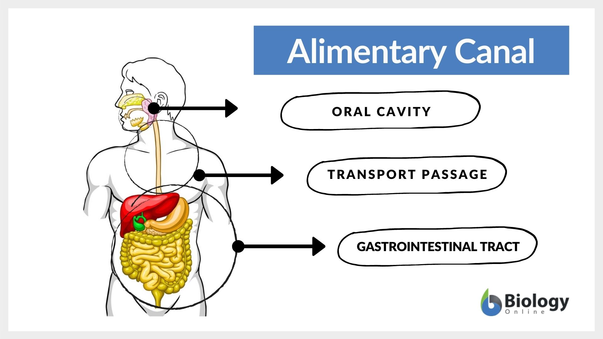
Alimentary Canal Definition And Examples Biology Online Dictionary
Watch 1000 concepts tricky questions explained.

Name the part of the alimentary canal that reabsorbs most water. Share It On Facebook Twitter Email. Thus Option B is correct. 402 K views 370 K people like this Like Share.
Name the part of the alimentary canal where symbiotic microbes live. Loading DoubtNut Solution for you. THIS SET IS OFTEN IN FOLDERS WITH.
It helps in the digestion of leftover food matter and absorption of water. Upvote 0 Was this answer helpful. The large intestine is a part of the alimentary canal beginning at the right iliac portion of the pelvis lying just at the right waist or below it.
The ileum is the last section of the small intestine that precedes the large intestine. Can you explain this answer. Alimentary canal - Mouth pharynx esophagus stomach small intestine large intestine rectum anus Describe the wall of the alimentary canal.
Iv Bile juice is produced. In which part of the alimentary canal - 21452362 siva149783630 siva149783630 24082020 Science Secondary School answered. Name the parts of the alimentary canal where- - 40057024.
Digestion of proteins begin in the. Iii Taste of the food is perceived. 1 Answer 1 vote.
Name the part of the alimentary canal where major absorption of digested food takes place. Answered Feb 10 2020 by Ritik01 481k points. Ii Digested food gets absorbed.
Name the parts of the alimentary canal where i water gets absorbed from undigested food. Asked Apr 26 2020 in Random by Joshua Mwanza Double Platinum 246k points 35 views. The alimentary canal is part of the reproductive system in pigs.
Mucosa - protects tissues beneath it and. More for absorption of water simple sugars and alcohol is aStomachbSmall intestinecLarge intestinedMouthCorrectanswer is option A. The large intestine comprises of the following parts.
Prev Question Next Question 0 votes. Correct option is. Name the part of the alimentary canal where the digestion of proteins begins.
Water gets absorbed from undigested food b. Absorbs ingested water and electrolytes remaining in alimentary canal. What are the absorbed forms of different kinds of food materials.
The pancreas and the liver are the body organs that are not part of the alimentary canal. Jul 072021 - That part of alimentary canal which is responsible. Name the parts of the alimentary canal in a pig in the proper order.
Asked Feb 10 2020 in Biology by KumariJuly 536k points Name the part of the alimentary canal where symbiotic microbes live. Name the part of the alimentary canal where major absorption of digested food takes place. Iv bile juice is produced ii.
EduRev NEET Question is disucussed on EduRev Study Group by 219 NEET Students. The liver and the pancreas are the organs that are not part of the alimentary canal. Click here to get an answer to your question Name the parts of the alimentary canal where a.
Name the part of the alimentary canal where 1 water gets absorbed from undigested food 2 Digested food gets absorbed3 Taste of the food is - 17599747. Click hereto get an answer to your question Name the parts of the alimentary canal wherei Water gets absorbed from undigested food. The part of alimentary canal that absorbs maximum amount of water and minerals is A.
Apne doubts clear karein ab Whatsapp par bhi. According to the International Foundation for Functional Gastrointestinal Disorders the ileum is responsible for absorbing water bile salts and vitamin B12. In which part of the alimentary canal the water from undigested waste is reabsorbed.
The major function of the large intestine is to absorb water from the remaining indigestible food matter and transmit the useless waste material from the body. What are the absorbed forms of different kinds of food materials. Share your questions and answers with your friends.
However the foundation explains that water absorption occurs in the upper small intestine as well. 2 See answers.
In what direction does blood flow through the heart. This prevents blood from flowing backward into the atria while the ventricles contract squeeze.

Circulatory Systems In Animals Transport Systems In Animals Siyavula
In systemic circulation oxygenated blood moves from heart to all parts of the body.

What is the order of blood flow in the heart. After the blood oxygenation process. First area of heart blood flows through. 55k views Reviewed 2 years ago.
When the ventricles are full the tricuspid valve shuts. Right atrium right ventricle left atrium left ventricle Humans have double circulation. Blood moves in two directions simultaneously.
It comes from the superior and inferior venae cavae. Blood Flow Sequence Activity The purpose of this activity is to understand the sequence of blood flow through the heart lungs and body. Click card to see definition.
Capillaries are small blood vessels that connect the arteries and veins. The order of blood vessels is great vessels arteries arterioles capillaries venules veins and back to the cavae. Blood flows from your right atrium into your right ventricle through the open tricuspid valve.
It flows into the lungs. Then it goes into the tricuspid vale and then to the right ventricle. To aorta to the body.
Blood enters the heart through two large veins the posterior inferior and the anterior superior vena cava carrying deoxygenated blood from the body into the right atrium. THIS SET IS OFTEN IN FOLDERS WITH. Through the aortic SL valve.
Leading into the right atrium is the superior ve. As the atrium contracts blood flows from your right atrium into your right ventricle through the open tricuspid valve. Superior and inferior vena cavae and the coronary sinus 2.
De-oxygenated blood enters the right side of the body and is pumped towards the lungs to pick up oxygen and start the process again. When the ventricle is full the tricuspid valve shuts. In pulmonary circulation deoxygenated blood is pumped by heart to get oxygenated in lungs.
Tap again to see term. Second area of heart blood flows through. The blood from the lower body is forced upward in the veins of the legs and then into the inferior vena cava where it eventually goes to the heart.
V veins Toward Blood is moving toward the heart. Blood flows from the right atrium into the right ventricle through the tricuspid valve. The blood flow in the heart begins from the right atrium that receives deoxygenated blood from the body to the right ventricle which pumps blood to the lungs.
In any case the heart is a 4 chamber organ which comprises of the right atrium and ventricle and the left atrium and ventricle. First the blood comes into the right atrium of the heart. The Aorta is the main highway that receives blood from the heart.
Figure 15 illustrates different parts of the heart involved in the blood flow. Oxygen-rich blood enters the heart on the left side and is pumped out to the cells of the body. 1 body 2 inferiorsuperior vena cava 3 right atrium 4 tricuspid valve 5 right ventricle 6 pulmonary arteries 7 lungs 8 pulmonary veins 9 left atrium 10 mitral or bicuspid valve 11 left ventricle 12 aortic valve 13 aorta 14 body.
Blood flows through the heart in the following order. Blood flows from the right atrium into the right ventricle through the tricuspid valve. Blood enters the heart through two large veins the inferior and superior vena cava emptying oxygen-poor blood from the body into the right atrium of the heart.
Pathway of the Heart - 9th - 2017. The process of the flow of blood through the heart is a complicated one. Click again to see term.
It requires a joint functioning of all the cardiac chambers to make proper circulation possible. Tap card to see definition. Blood enters the heart through two large veins the posterior inferior and the anterior superior vena cava carrying deoxygenated blood from the body into the right atrium.
It splits into Arteries these split into smaller arterioles then even smaller capillaries. Depends if you are talking about the heart or the circulatory system. That blood then goes out the left ventricle to the body and returns via the vena cavae.
Similarly one may ask what is the correct order of blood flow through the heart. Beginning with the superior and inferior vena cavae and the coronary sinus the flowchart below summarizes the flow of blood through the heart including all arteries veins and valves that are passed along the way. The left and right sides of the heart function jointly in pumping the de-oxygenated and oxygenated blood.
From the heart it is pumped back through the aorta into the body and the whole process repeats. One is systemic circulation and other is pulmonary circulation. The blood from upper body flows into the superior vena cava where it eventually goes to the heart.
Blood Flow Through the Heart. Then it flows through the pulmonary semilunar valves and into the pulmonary trunk.
Surrounded by the cerebral hemispheres the diencephalon forms the central core of the brain. See how quickly you can do it with 100 accuracy.

Brain Parts Of The Brain Thalamus Cf7zmfsb Brain Diagram Brain Anatomy Human Brain Anatomy
Brain diagram with labels hypothalamus vector brain diagram pons cerebrum and cerebellum brain pons brain anatomy amygdala brain labelled amygdala brain human midbrain diagram pons.
Label parts of a brain. The Cerebrum can also be divided into 4 lobes. It contains 8 proteins 1 carbohydrates 2 soluble organics and 1 insoluble salts. The nervous system - an image of the nerves of the lower body with blank labels attached.
It regulates balance posture movement and muscle coordination. The brain is a 3-pound organ that contains more than 100 billion neurons and many specialized areas. See labeled brain anatomy stock video clips.
There are 3 main parts of the brain include the cerebrum cerebellum and brain stem. Start studying Label the parts of the Brain. Get the answers you need now.
Of the three main parts of the brain the cerebrum is considered the most recent to develop in human evolution. The cerebrum is responsible for all voluntary actions eg. Learn vocabulary terms and more with flashcards games and other study tools.
Located in the front and middle part of the brain it accounts for 85 of the brains weight. It regulates balance posture movement and muscle coordination. Cerebellum - the part of the brain below the back of the cerebrum.
Label the parts of the brain. The parietal lobe takes care of sensation and perception managing how we react to sensory input. 12 cerebrum cerebellum brain stem spinal cord this part of the nervous system moves messages between the brain.
The cerebrum the largest part handles vision movement hearing language and touch. 2653 labeled brain anatomy stock photos vectors and illustrations are available royalty-free. Click on the link to the left to label the parts of the limbic system.
Consisting of largely of three paired structures the thalamus hypothalamus and epithalamus the diencephalon plays a vital role in integrating conscious and unconscious sensory information and motor commands. Home label 3 parts of the brain label parts of brain label parts of the brain psychology label parts of the brain quiz label the parts of human brain label the parts of the brain and spinal cord label the parts of the brain and spinal cord fetal pig label the parts of the brain identified in the diagram label the parts of the brain worksheet label. The brain is composed of 77 to 78 water and 10 to 12 lipids.
Read the definitions below then label the brain anatomy diagram. The nervous system - a PDF file of the nerves of the upper and lower body for printing out to use off-line. Label the Brain Diagram The Brain.
Motor skills communication emotions creativity intelligence and personality. The brain and its parts can be divided into three main categories. The frontal lobe is involved in reasoning motor control emotion and language.
Frontal lobes parietal lobes temporal lobes and. Click on the link to the left to label the lobes of the brain as well as the very basics of a neuron. Corpus Callosum - a large bundle of nerve fibers that connect the left and right cerebral hemispheres.
The frontal lobe is located in the forward part of the brain extending back to a fissure known as the central sulcus. Lobes and Neuron Diagram. The forebrain midbrain and hindbrain.
Medulla Regulates breathing and heart rate hanging a person works bc if done correctly it breaks this in half Pons Involved in sleeping waking and dreaming Cerebellum The lesser brain coordinates balance and coordination Thalamus Relays all sensory information to specific perception areas of the brain with the exception of smell Hypothalamus Part of the old brain it. Label all the parts of the brain of the central nervous system. This science quiz game will help you learn the 7 parts of the brain.
Lobes of the Brain The four lobes of the brain are the frontal parietal temporal and occipital lobes Figure 4.
The radioactively labeled phages are. After introducing to the phage culture to the bacterial sample they used a Waring blender to violently disturb the infected bacteria causing the protein shells to detach from their hosts.

The Hershey Chase Blender Experiments Paulingblog
The Hershey-Chase experiments settled the long-standing debate about the composition of genes thereby allowing scientists to investigate the molecular mechanisms by which genes function in organisms.
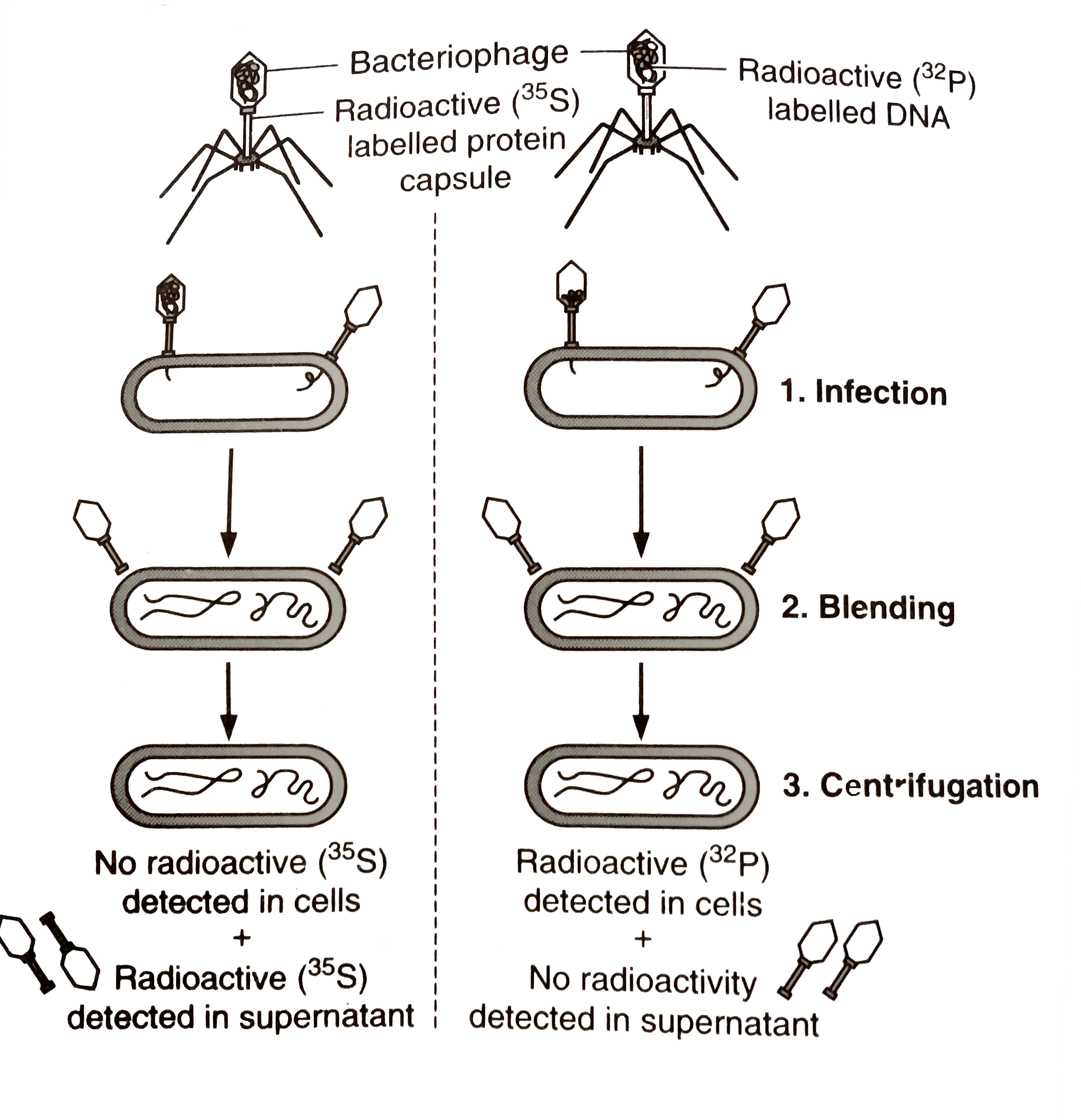
What is blending in hershey and chase experiment. Theirexperiment was thus a powerful new proof that DNA is the primarygenetic materialThe Double Helix by James Watson page 72. Then using a centrifuge they separated the bacterium from the phages and protein. The experiment began with the culturing of viruses in two types of medium.
The Hershey-Chase Blender Experiment. The most well-known Hershey-Chase experiment was the final experiment also called the Waring Blender experiment through which Hershey and Chase showed that phages only injected their DNA into host bacteria and that the DNA served as the replicating genetic element of phages. Hershey and Chase Experiment Steps Radioactive Labelling of Bacteriophage.
Hershey and Chase conducted an experiment to discover whether it was protein or DNA that acted as the genetic material that entered the bacteria. Diagram by Eric Arnold. Blending- As infection proceeds- the viral coats are removed from the bacteria by agitating in a blender Centrifugation - virus particles were seperated from.
Hershey and his research partner American geneticist Martha Chase at Cold Spring Harbor Laboratory New York. Describe the blender experiments of Hershey and Chase and what the results revealed about DNAs function. The phage works by attaching itself to.
Coli and so is a bacteriophage. Figure 521 shows the essential elements of the infective cycle of DNA. It has gotten 27 views and also has 49 rating.
A Hershey and Chase carried their experiment in three steps. The most well-known Hershey-Chase experiment was the final experiment also called the Waring Blender experiment through which Hershey and Chase showed that phages only injected their DNA into host bacteria and that the DNA served as the replicating genetic element of phages. After radioactive labelling of the phage DNA and protein Hershey and Chase infected the.
Infection blending centrifugation NEET Video EduRev video for NEET is made by best teachers who have written some of the best books of NEET. The most well-known Hershey-Chase experiment called the Waring Blender experiment provided concrete evidence that genes were made of DNA. Hershey and Chase have grown T-2 bacteriophages in the two batches.
What was the blender experiment. In 1952 seven years after Averys demonstration that genes were DNA two geneticists. They worked with a DNA virus called T2 which infects E.
They used a certain type of virus called the T-2 bacteriophage in their experiment a virus made up of DNA and a protein shell pictured below. DNA as Genetic Material. In order to decipher what the genetic material is Hershey and Chase needed to isolate the two variables.
The most well-known Hershey-Chase experiment was the final experiment also called the Waring Blender experiment through which Hershey and Chase showed that phages only injected their DNA into host bacteria and that the DNA served as the replicating genetic element of phages. The HersheyChase experiment often called the Waring Blender experiment was conducted in 1952 by American bacteriologist and geneticist Alfred D. The Hershey - Chase Experiments.
Illustration of the 1952 experiment connecting DNA and heredity. This experiment comes under the chapter molecular basis of inheritance About Press Copyright Contact us Creators Advertise Developers Terms Privacy Policy Safety How YouTube works Test new. Hershey and Martha Chase provided further proof.
Side by side experiments are performed with separate bacteriophage virus cultures in which either the protein capsule is labeled with radioactive sulfur or the DNA core is labeled with radioactive phosphorus. Al Hershey had sent me a long letter summarizing the recentlycompleted experiments by which he and Martha Chase establishedthat a key feature of the infection of a bacterium by a phage wasthe injection of the viral DNA into the host bacterium.
This is thoroughly answered here. Is between the forearm and the upper arm Forearm.
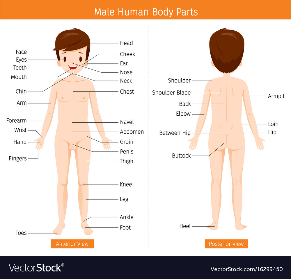
Male Human Anatomy External Organs Body Royalty Free Vector
It is a vital organ and provides outer covering which protects from external elements.

What are the external parts of the human body. Read about the different systems in the human body made simple for kids. Human Bosy Parts Meaning and Example Sentences. The head contains the brain which is the control center of the body.
The head and neck house the controlling organ brain that extends throughout the body. Sense organs are important parts of our body. In both animals and humans skin functions as a barrier between the outside and inside the environment.
The head the trunk and the limbs extremities. Human Body Parts For Kids The main structure of a human body. You have legs knees feet and toes.
These external body parts are easy to see on the average person. The human body consists of a bony skeleton and muscles. The body uses different systems to work properly.
Each major part has a specific function. Scroll down and take a look at the list and some interesting facts about the human body. Whats harder to see are the different body systems working under the skin in each body part.
Is between the wrist and elbow. The commentary makes the point that some people need glasses. Throat - the part of an animals body that corresponds to a persons throat.
The main organ systems of the human body are the. Our body has symmetry that means it looks the same on the left side as it does on the right side. Many translated example sentences containing the external parts of the human body German-English dictionary and search engine for German translations.
Hood - zoology an expandable part or marking that resembles a hood. Body Parts are Pieces in a Puzzle. Your skin is the biggest organ of your body and its.
Vital organs are the parts of our body that we need to stay alive. We can see these organs. Most of us have the same kind of body parts in the same places.
This post is for curious minds who want to learn about the mysterious human body and its functioning. The legs are used for locomotion like walking running jumping and swimming. The skin acquires an area of 19 to 20 square feet on our body.
See external human body parts stock video clips. The part of the body in humans between the ribs and the hips. 286 external human body parts stock photos vectors and illustrations are available royalty-free.
On the outside our bodies are separated into the following obvious parts head torso arms and legs. You have arms elbows hands and fingers. The front of the trunk from the neck to the abdomen.
Skin is the largest external organ of the human body. The study of the macroscopic morphology and function of the human body is called gross anatomy. These include the heart brain lungs kidneys liver and pancreas.
Head torso and limbs. Read on to know more about the fascinating facts and insights about the human body for kids. External organs side front nose elbow knee beauty cartoon ear listening outer ear elbow cartoon vector external organ female ears naked cartoon ear images.
In addition to the physical external parts the human body can also be divided by organ system and the parts that compose those systems. These external organs are our sense organs. An external body part that projects from the body.
You have a head and neck. Try these curated collections. Is rear surface of the body from the shoulders to the hips.
The uppermost part of the body containing the brain and the eyes ears nose mouth and jaws. Part of the body just above hips. The arms are used for holding things lifting pushing and pressing.
The level of this lake water is coming up to my waist. The three main parts of the body are. It also functions by protecting our internal organs from invading pathogens regulates our body temperature and pH prevents dehydration and also functions as the main sense organ.
Is the lower part of the back. We are familiar with the exterior body parts like ear eye nose hands and legs but we might not be knowing about all the internal human organs. The external organs of our body are eyes ears nose tongue and skin.
In general the human body can be divided into 3 main anatomical areas. In this way what are the external parts of the human body. The torso or body trunk the area between the head and legs is used to bend and twist our bodies around.
Small of the back. Breast - the part of an animals body that corresponds to a persons chest. You have a chest and a tummy.
The external part of a human body comprises the head neck and trunk forelimbs and hind limbs. External body part - any body part visible externally. An external body part consisting of feathers or hair about the neck of a bird or other animal.
Nerves muscles veins bones how do these body parts keep you going. The skeleton skull skin head neck arms elbows arms fingers chest tummy legs knees feet and toes are all visited.
A generalizedtypical eukaryotic cell as seen under Electron Microscope EM consists of cell wall absent in animal cells and some protists plasma membrane. Also involved in cell signaling and recognition.
Labelled diagram of plant and animal cell are as follow--- Both plant and animal cells belong to eukaryotic cells.

Labeled diagram of eukaryotic animal cell. Cytoskeleton provides structure and organisation to the cytoplasm and shape to the cell. Significantly bigger than the prokaryotic cells eukaryotic cells. Eukaryotic cells are distinguished mainly from prokaryotic cells in that they do not possess cell walls or chloroplasts.
Find Structure Animal Cell Labeled Parts Biology stock images in HD and millions of other royalty-free stock photos illustrations and vectors in the Shutterstock collection. Cytosol is the fluid present within a cell that is made up of water and ions such as potassium proteins and small. Except nucleus and plastids all other cytoplasmic structures can be seen under EM.
These extensions of the cell are covered with plasma membrane and supported internally with a structural system of microtubuleskind of like a bone covered in skin. Well-Labelled Diagram of Animal Cell The cell membrane is a double-layered membrane made up of phospholipids that surrounds the entire cell. Eukaryotic cells are larger more complex and have evolved more recently than prokaryotes.
Therefore it was decided to only have a few copies of each structure populating the cell. Its main characteristics are a well differentiated nucleus and a nuclear membrane. The information can be in the form of.
Wide collections of all kinds of labels pictures online. Eukaryotic cells 231 Draw and label a diagram of the ultrastructure of a liver cell as an example of an animal cell. CellPlasma Membrane a lipid bilayer made of lipids proteins and carbs that is semi-permeable and determines what can enter and exit the cell.
There are two types of cells - Prokaryotic and Eucaryotic. Make your work easier by using a label. It also helps to produce motion of organelles or of the whole cell.
Also contains many enzymes and is the site of many biochemical reactions. The nucleus is the site of most cellular genetic material DNA. In animal cells the MTOC is called the centrosome which consists of two centrioles.
Eukaryotic cell labeled eukaryotic cell structure animal. They are the building block or smallest unit of life of organisms as simple as amoeba and protozoa to the most complicated plants and animals. Found either floating free in the cytoplasm or attached to the surface of the rough endoplasmic reticulum and in mitochondria and.
Wide collections of all kinds of labels pictures online. Eukaryotic cell are the developed advanced and complex forms of cells. The endoplasmic reticulum is the site of protein and lipid production.
Genetic material enclosed in nuclear envelope Genetic material not enclosed in nuclear envelope 3 Nucleolus and one or more chromosomes present. Found in prokaryotic cell. Labels are a means of identifying a product or container through a piece of fabric paper metal or.
The Animal or eukaryotic cells Are the type of cells that form all the tissues of animals. The plasma membrane composed of a phospholipid bilayer controls cellular traffic. Genetic material in the form of single chromosome.
Cytoskeleton is the. Eukaryotic Cell Diagram With Labels. The gel-like substance that fills the cell and suspends the organelles.
A Labeled Diagram of the Animal Cell and its Organelles. Prokaryotic cells are those that form the plant kingdom. These tiny powerhouses of the cell are double-membrane bound organelles that extract energy from food to produce ATP adesnosine-5- triphosphate a multi-purpose molecule that carries energy for use within the cell.
Eukaryotic cells are one which have organised nucleus with a nuclear membrane and genetic material is organised into chromosomes. Figure 231 - Annotated drawing of an animal cell. Labels are a means of identifying a product or container through a piece of fabric paper metal or plastic film onto which information about them is printed.
In plant cells the nuclear envelope appears to function as the main MTOC. Plant and fungal eukaryotes show some differences in structure and components. 4 Cell divides by mitosis and meiosis.
The cell is the structural and functional unit of life. This eukaryotic cell is modelled after animal cells. Make your work easier by using a label.
Example plant cellanimal cell. The Golgi Apparatus processes and packages proteins. However they differ as animals need to adapt to a more active and non-sedentary lifestyle.
Plant cells and animal cells share some common features as both are eukaryotic cells. These cells differ in their shapes sizes and their structure as they have to fulfil specific functions. - Eukaryotic Cell - 3D model by The Center for BioMedical Visualization at SGU SGUMedArt b7d84e5.
Where prokaryotes are just bacteria and archaea eukaryotes are literally everything else. Thats distinct from prokaryotic cells which have a nucleoid a region thats dense with cellular DNA but dont actually have a separate membrane-bound compartment like the nucleus. Found in eukaryotic cell.
Thousands of new high-quality pictures added every day. Cytoplasm nucleus other organells and inclusions. Flagella singular flagellum which are longer than cilia aid in cell movement.
Eukaryotic cells include animal cells including human cells plant cells fungal cells and algae. Eukaryotic cells are characterized by a membrane-bound nucleus. 232 Annotate the diagram from 231 with the functions of each named structure.
The mitochondrion is the site where energy stored. There is an interlocking three dimensional network of protein filaments throughout the cytoplasm of eukaryotic cell which is called as cytoskeleton and is visible under the electron microscope.
The tibia is the larger weight-bearing bone located on the medial side of the leg and the fibula is the thin bone of the lateral leg. Peoples legs and feet can also swell if there are problems with the lymphatic system.

Developing Strength Stability In The Foot Ankle And Lower Leg Mountain Peak Fitness
The smaller lateral bone of the lower leg.
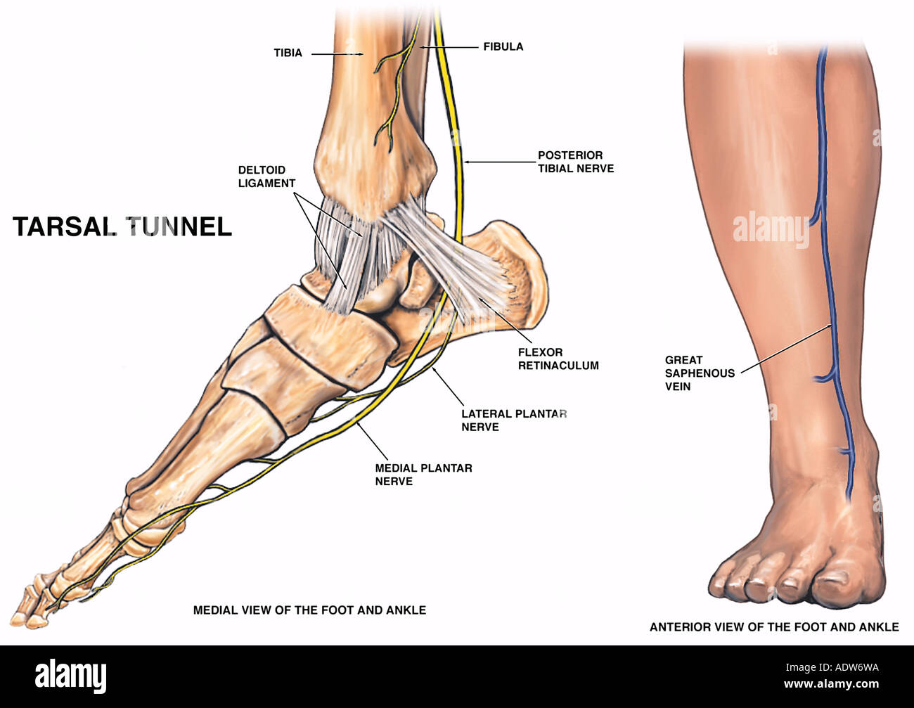
Parts of foot and lower leg. The swelling can occur in one particular part of the body for example the feet or ankles or may be more general depending on the cause. Front of the leg below the knee Heel. The proximal portion of the tibia is tibial plateau which acts as a cusp for the knee the distal portion tapers into the medial malleoli and the concave surface which articulates with the talus at the ankle joint.
Common peroneal nerve dysfunction is a type of peripheral neuropathy damage to nerves outside the brain or spinal cord. The cardiovascular system of the leg and foot includes all of the blood vessels that provide blood flow to and from the tissues of the lower limb. Brain or spinal cord.
In a typical foot the tibia. Connects the lower and upper leg. The bones of the foot are divided into three groups.
A numbness and tingling in your lower leg or foot is extremely common if youve been sitting down for a long stretch of time. Problems in the lower spine may affect the spinal nerve roots causing pain to radiate into the leg andor foot radiculopathy. Processes in the brain and spinal cord can result in lower leg numbness but often these conditions present with serious symptoms as well such as a loss of bladder control or consciousness.
The legs are the two lower limbs of the body. Theyre known as the. The peroneal nerve is a branch of the sciatic nerve which supplies movement and sensation to the lower leg foot and toes.
It lies directly under the tibia and fibula and is connected to them by strong ligaments. Spinal Causes of Leg Pain. The muscles of the lower leg called simply the leg by anatomists largely move the foot and toes.
The medial larger bone of the lower leg. Posterior segment ankle is the connection between foot and leg. The nerves of the leg and foot serve to propel the body through the actions of the legs feet and toes while maintaining balance both while the body is moving and when it is at rest.
This image shows a chronic case. These blood vessels supply vital oxygen and nutrients to support cellular metabolism in the lower limb while transporting carbon dioxide and metabolic wastes back to the trunk to be removed from the. The ankle is the joint of the connection between the lower portion of the foot and also the leg.
Oedema is a buildup of fluid in the tissues of the body. The major muscles of the lower leg other than the gastrocnemius which is cut away are shown in Figure 9-12. This condition can affect people of any age.
Issues within the central nervous system can result in lower leg numbness. The legs are one of the most common parts of the body impacted by PAD. Behind the ankle and connecting the foot to the rear side of the leg is known as the Achilles tendon.
They provide support and a range of movements. It is the largest tendon of the parts of leg. Joints of the hindfoot.
One of the first warning signs you have an inflamed Achilles tendon is pain in your lower calf near the back of your heel. The gastrocnemius muscle has two large bellies called the medial head and the lateral head and inserts into the calcaneus bone of the. Common causes of leg and foot pain that originate in the spine include.
A series of seven ligaments from a connection around the ankle. However with the exception of the bottom of the foot they play a lesser role here than in the upper extremities. This segment contains the talus in the mortice between the fibula and the tibia and the calcaneus.
Sensory nerves are of course present throughout the lower extremities. Tibia and Fibula long bones The foot is connected to the body where the talus articulates with the tibia and fibula. Most people with PAD experience pain and cramping in their legs and hips when they are walking or going upstairs.
This article provides a guide to the potential causes specific diagnostic procedures and the different types of treatment approaches available for leg and foot pain. Each leg contains five regions. Bones of the lower leg and hindfoot.
A primary condition in the brain or spinal cord that is resulting in leg numbness is most likely a vascular injury in. Muscle at the back of the lower leg Shin. Where the bottom of the foot curves.
Is the back part of the foot below the ankle. The head of the fibula serves as an attachment point for the lateral collateral ligament of the knee and a number of. The padded portion of the sole of the human foot.
Its a common injury that makes the tendon swell stretch or tear. Tibia Fibula Talus Calcaneus. The nerves in that part of.
Draw the diagram of the organelle which involves in photosynthesis - 27890009. Photosynthesis Diagram According to the diagram of photosynthesis the process begins with three most important non-living elements.

Photosynthesis Diagram Drawing Youtube
This reaction is catalysed by the enzyme Rubisco.

Draw the diagram of photosynthesis. With the digrams that are circles. Photosynthesis photos-light synthesis-putting together is an anabolic process of manufacture of organic compounds inside the chlorophyll containing cells from carbon dioxide and water with the help of sunlight as a source of energy. Draw the Diagram of Photosynthesis.
Write two functions of stomata. Photosynthesis is essentially the only mechanism of energy input in the living world. These are my choices.
Plants begin making their food which basically includes large quantities of sugars and carbohydrate when sunlight falls on their leaves. Draw a schematic diagram showing representation of the ofa stomatab write photosynthesis explained with vector stomata c3 and c4 definition equation given below is to. Photosynthesis tree tree oxygen photosynthesis process photosynthesis process of photosynthesis leaf photosynthesis minerals plant plant body photosynthesis vector earth science formulas.
Alternatively you can also click the tile from the right window to create a blank document and then use the available tools in the symbol library to start drawing your custom illustration from scratch. Below is the flowchart of photosynthesis process that shows the steps involved in the Light reaction and dark reaction of photosynthesis created on EdrawMax a powerful flowchart software that can help draw flowcharts within a few steps. During light reaction oxygen is evolved and assimilatory power ATP and NADPH 2 are formed.
Photosynthesis Flowchart A flowchart is a way to show the steps in a process. With the venn diagram for the photosynthesis and the respiration it says the terms may be use more than once. In the Photosynthesis circle it shows.
Rubisco can also catalyse a reaction between RuBP and oxygen to form one molecule of GP and one molecule of phosphoglycolate. The Process Of Photosynthesis Explained With Diagrams. Schematic draw a diagram showing write photosynthesis the ofa stomatab representation of vector plant stomata explained with transfer carbon in most.
See photosynthesis diagram stock video clips. 1 Light reaction or Light phase or Light-dependent phase or Photochemical phase 2 Dark reaction or Dark phase or Light independent phase or Biochemical phase. Could you please help me.
Absorbs Calvin cycles Chlorophyll CO 2 H-2-O Krebs cycle Mitochondria Releases. The Electron Transport Pathway from Water H2O to NADP the Nicotinamide Adenine Dinucleotide Phosphate oxidized form. Many versions of the Z-scheme are available in the literatureThis particular diagram was developed by Wilbert Veit and Govindjee 2000 and can be also found at.
During dark reaction assimilatory power is. The Z-Scheme Diagram of Photosynthesis. Water soil and carbon dioxide.
Draw a labelled diagram of stomata. Blackmann 1905 pointed out that the process of photosynthesis consists of two phases. In the non-cyclic reaction the photons are captured in the light-harvesting antenna complexes of photosystem II by chlorophyll and other accessory pigments see diagram at right.
This is what I needd help with. Vector Diagram Plant Photosynthesis Science Education Botany Poster Ilration Studying Image By Elina33 Stock 242853340. The antenna system is at the core of the chlorophyll molecule of the photosystem II reaction center.
The absorption of a photon by the antenna complex frees an electron by a process called photoinduced charge separation. Draw A Schematic Diagram Showing Photosynthesis Brainly In Schematic Draw A Diagram Showing Photosynthesis Brainly In. 817 photosynthesis diagram stock photos vectors and illustrations are available royalty-free.
Click the Photosynthesis template icon from the right pane to automatically create a new document and add the photosynthesis diagram in the template to it. During photosynthesis carbon dioxide reacts with ribulose bisphosphate RuBP to form two molecules of glycerate 3-phosphate GP. Draw A Diagram That Represents The Interdependence Of Photosynthesis Easy Drawing Draw A Diagram That Represents The Interdependence Of Photosynthesis.
This diagram depicts Nervous System. Nervous System Nervous system diagram showing various nerves in the human body including sciatic nerve lumbar plexus ulnar nerve etc.

Outline Of The Human Nervous System Wikipedia
Picture central nervous system picture human brain picture human circulatory system picture human digestive system picture human muscular system picture human respiratory system picture human skeletal system picture peripheral nervous system Inner Body picture central nervous system picture.

Pictures of the human body nervous system. Nervous System Human Body Nerves Structure Nervous System Anatomy Human Body Medical Art Watercolor Print 1204 A1 84x59cm Poster Of Illustration Human Nervous System Full Human Body The Nervous System More Than 90 000 Miles Of Sensations Human Body Parts Nervous System Man Anatomy Hand Drown Vector 1970 Vintage Poster Xl Anatomical Human Body Nervous System Medicine Wall Art Nervous System. Neuron system - human nervous system stock pictures royalty-free photos images. Find the perfect Human Body Nervous System stock photos and editorial news pictures from Getty Images.
14 photos of the Picture Human Nervous System. Find human nervous system stock images in HD and millions of other royalty-free stock photos illustrations and vectors in the Shutterstock collection. Some of the most vital systems of the body include the nervous system which controls and coordinates the body and includes the brain spinal cord and nerves.
Free for commercial use High Quality Images. Search from Pics Of The Human Body Nervous System stock photos pictures and royalty-free images from iStock. In the fall of 1925 two medical students in Kirksville Missouri received a cadaver and a challenge.
The circulatory system which pumps blood throughout the body and includes the heart blood vessels and blood itself. Pictures Of The Human Body Nervous System Read Pictures Of The Human Body Nervous System PDF on our digital library. PICTURES OF THE HUMAN BODY NERVOUS SYSTEM PDF The writers of Pictures Of The Human Body Nervous System.
Neural network on a blue background with light effects. Thousands of new high-quality pictures. Vector illustration of complete chart of different human organ system.
To dissect the bodys nervous system beginning at the base of the brain. You can read Pictures Of The Human Body Nervous System PDF direct on your mobile phones or PC. Read PICTURES OF THE HUMAN BODY NERVOUS SYSTEM PDF direct on your iPhone iPad android or PC.
Select from premium Human Body Nervous System of the highest quality. 500 Vectors Stock Photos PSD files. Development of the nervous system.
Nervous system Stock Photos and Images. Find high-quality stock photos that you wont find anywhere else. Find high-quality stock photos that you wont find anywhere else.
- Diagram - Chart - Human body anatomy diagrams and charts with labels. Browse 21450 human nervous system stock photos and images available or search for human nervous system anatomy to find more great stock photos and pictures. Pictures Of The Human Body Nervous System Charts Human Human Body Nervous System Anatomic Model Plastic Human Model Buy Human Body Nervous System Anatomic Model Nervous System Model Anatomical Model Peripheral Nervous System Wikipedia Nervous System Images Stock Photos Vectors Shutterstock Amazon Com Human Body Nervous System Anatomy Modern Wall Nervous System.
Download Human Body Nervous System Images Pics. 11508 Human nervous system Stock Photos Images Download Human nervous system Pictures on Depositphotos. Search from Pics Of Human Body Nervous System stock photos pictures and royalty-free images from iStock.
Medical Education Chart of Biology for Human Body Organ System Diagram. As per our directory this eBook is listed as POTHBNSPDF-145 actually introduced on 10 Jan 2021 and then take about 2158 KB data. Pictures Of The Human Body Nervous System - PDF-POTHBNS14-5 Download full version PDF for Pictures Of The Human Body Nervous System using the link below.
Download stock pictures of Human nervous system on Depositphotos Photo stock for commercial use - millions of high-quality royalty-free photos images. And the respiratory system which regulates oxygen exchange and includes the lungs diaphragm bronchi larynx and trachea. Find Download Free Graphic Resources for Nervous System.
Human anatomy diagrams show internal organs cells systems conditions symptoms and sickness information andor tips for healthy living. In biology the nervous system is a highly complex.
This quiz has tags. What is Anatomy and Physiology.

Map Quiz 3 33 Anatomy Of The Heart
Human Heart Anatomy Multiple Choice Quiz.
(230).jpg)
The heart anatomy quiz. MJK7221995617AM on February 14 2015 660 plays. From the quiz author. The Human Anatomy and Physiology course is designed to introduce students pursuing careers in the allied health field to the anatomy and physiology.
Human Heart Anatomy 15. The cells that carry oxygen to body cells are. With more than 55-questions on such topics your.
Anatomy of the Kidneys Regulation of Urine Concentration Urethra Quiz. Test your knowledge about the anatomy of the Human Heart and blood. Science Quiz Human Heart Anatomy Random Science or Anatomy Quiz Can you find the different parts of the human heart.
Random Quiz Create A Quiz. Heart Anatomy Quizzes Trivia Elvis sang that he didnt have a wooden heart showing that not only was he the king of rock and roll he also had a passable knowledge of heart anatomy. Heart Anatomy Test Answers.
This is an online quiz called Heart Anatomy Quiz. Inner layer of the pericardium. Anatomy And Physiology Quiz.
Test your knowledge of the location shape and size of the heart as well as the anatomy of surrounding features in this quiz. 11th grade Anatomy Quiz on The Anatomy of the Heart created by Shannan Muskopf on 24022015. The number of valves in the heart is.
There is a printable worksheet available for download here so you can take the quiz with pen and paper. You are comparing audio recordings of the heartbeats of three different patients. Hence you can not start it again.
The major blood vessel that carries oxygenated blood out of the heart is the. You have already completed the quiz before. 0 of 10 Questions completed.
9th - University grade. Quiz Chapter 11 Cardiovascular System. Based on this information blood from the right ventricle is on its way to the _____.
Free interactive quiz for students biology anatomy and physiology. The human heart has taken on an almost mystical quality with its associations to love and bravery but it is also an essential and functional part of our physiognomy. Click on the tags below to find other quizzes on the same subject.
The distal end of the heart Preview this quiz on Quizizz. Play this game to review Human Anatomy. The Human Bunker 29.
By timmylemoine1 Plays Quiz Updated Jan 21 2016. The sac of tissue that protects the heart is the. An online interactive study guide to tutorials and quizzes on the anatomy and physiology of the heart using interactive animations and diagrams.
Respiratory System Map 18. Forced Order Support Sporcle. Test prep cardiovascular system.
This quiz is about the Cardiovascular System or says the lifeline of the body which consists of vessels called arteries veins and capillaries. This is a free printable worksheet in PDF format and holds a printable version of the quiz Heart Anatomy QuizBy printing out this quiz and taking it with pen and paper creates for a good variation to only playing it online. In Patient Three you notice that the second.
How many chambers are in a human heart. The Heart Previous The Heart. Anatomy of the Heart.
Blood Flow and Parts. Structure of the heart. This quiz is all about the Heart and the passage of Blood through the heart.
How Blood Type is Determined The blood type test is used to determine which antigenic molecules are present on RBCs. The layer of simple squamous epithelium that lines the inside of the heart is called myocardium. Atoms Molecules Ions and Bonds Quiz.
Top Quizzes with Similar Tags. The fluid that surrounds the heart and prevents friction when it beats is. 1 The right ventricle is the chamber of the heart that pumps blood for the pulmonary circulation.
Rate 5 stars Rate 4 stars Rate 3 stars Rate 2 stars Rate 1 star.
There are bones joints muscles tendons and ligaments in each section. There are three major muscles in the foot.
Different Anatomical Parts Of The Human Foot Foot Pad Complex Download Scientific Diagram
Get 19 answers for Parts of the foot crossword clue in the Crosswords Dictionary.

Parts of the foot. It is made up of over 100 moving parts bones muscles tendons and ligaments designed to allow the foot to balance the bodys weight on just two legs and support such diverse actions as. Download Worksheet Include correct answers on separate page. 26 bones 33 joints 107 ligaments and 19 muscles.
Nails protect the tips of the toes. To play the game online visit Parts of the Foot. The foot is traditionally divided into three regions.
Additionally the lower leg often refers to the area between the knee and the ankle and this area is critical to the functioning of the foot. The midfoot is a pyramid-like collection of bones that form the. In humans the foot is one of the most complex structures in the body.
The foot can be divided into three sections. The average person takes 8000 to 10000 steps a. Parts of the Foot 1.
These include the three cuneiform. The calcaneus connects with the talus and cuboid bones. Regions of the Foot.
The foot contains. Hind means posterior so it. The soleus is the lower muscle of the calf.
The feet are divided into three sections. The anatomy of the foot The foot is divided into three sections the forefoot the midfoot and the hindfoot. Phalanges are the toe bones.
Bones of the human foot Bones of the foot showing the calcaneus heel bone talus and other tarsal bones ankle bones metatarsal bones bones of the foot proper and phalanges toe bones. Ligaments are fibrous strands that connect bones. The human foot is a strong and complex mechanical structure containing 26 bones 33 joints 20 of which are actively articulated and more than a hundred muscles tendons and ligaments.
What is another name for toes. The calcaneus also called the heel bone is a large bone that forms the foundation of the rear part of the foot. When these bones are out of alignment so is the rest of the body.
Forefoot The forefoot contains five phalanges or toes and the five metatarsals which are longer bones. The hindfoot the midfoot and the forefoot Figure 2. The connection between the talus and calcaneus forms the subtalar joint.
What are different parts of the foot called. 14 of all the bones in the human body are down in your feet. Midfoot The midfoot is the part of the foot that forms the arches of your feet.
Anatomically the foot is divided into 3 sections. This is an online quiz called Parts of the Foot. The hindfoot midfoot and the forefoot.
The forefoot contains the five toes phalanges and the five longer bones metatarsals. Nerves travel throughout the foot providing feeling. The forefoot midfoot and hindfoot.
The structure of the foot. The gastrocnemius is the large muscle of the calf. There is a printable worksheet available for download here so you can take the quiz with pen and paper.
The quadratus plantae are the. Anatomy of the foot Calcaneus heel bone Talus ankle bone Transverse tarsal joint Navicular bone Lateral cuneiform bone Intermediate cuneiform bone Medial cuneiform bone Metatarsal bones Proximal phalanges Distal phalanges Tarsometatarsal joint Cuboid. The joints of the foot are the ankle and subtalar joint and the interphalangeal articulations of the foot.
The hindfoot is the posterior part of the foot. The foots complex structure contains more than 100 tendons ligaments and muscles that move nearly three dozen joints while bones provide structure. Crossword clues from LA Times Universal Daily Celebrity NY Times Daily Mirror Telegraph NZ Herald and many more.
You can modify the printable worksheet to your liking before downloading.
The part of the foot which joins it to the leg is called the heel. Foot part 4 Back of the foot 4 Command to Rover 4 Part of foot 6 INSTEP.

Anatomy Of The Foot And Ankle Orthopaedia
The word that solves this crossword puzzle is 6 letters long and begins with A.

Parts of feet called. Covers the end of the top of the toes. Curve over 4 Curved structure 4 Something curved 4 Footwear insert 4 Curved span 4 Part of foot 4 HEEL. The hard foot of an ungulate is a hoof.
The outsole of the foot is the part on the bottom of the shoe that touches the ground. Is the back part of the foot below the ankle. Unlike calluses corns have a central core or spot in the middle that is surrounded by dead skin.
Additionally the lower leg often refers to the area between the knee and the ankle and this area is critical to the functioning of the foot. This consists of five long metatarsal bones and five shorter bones that form the toes phalanges. The talus forms a joint with four bones.
Best Answer for Parts Of Feet Crossword Clue. In humans the foot is one of the most complex structures in the body. The insides of your feet correlate to your spine.
The padded portion foot between the toes and the arch. Foot dysfunctions like heel spurs are most common in people who have plantar fasciitis inflammation of the fascia on the sole of the foot. The foot is divided into three sections - the forefoot the midfoot and the hindfoot.
It is made up of over 100 moving parts bones muscles tendons and ligaments designed to allow the foot to balance the bodys weight on just two legs and support. The foot is a part of vertebrate anatomy which serves the purpose of supporting the animals weight and allowing for locomotion on land. Where the bottom of the foot curves.
Tibia fibula calcaneus and navicular. A softer sole provides greater ability to absorb shock. Parts of your foot correlated with the stomach are found above the waistline.
Thus they define the boundaries of the three muscle compartments of the sole see below. The foot contains a lot of moving parts - 26 bones 33 joints and over 100 ligaments. A common one is the development of bony growths on the underside of the calcaneus called heel spurs that cause severe pain when standing or walking.
The feet of monkeys are much like the hands. The foot diagram has a complex structure made up of bones ligaments muscles and tendonsUnderstanding the structure of the foot is best done by looking at a foot diagram where the anatomy has been labeled. Parts correlated with the intestines are found below.
Most land vertebrates have feet and there are many different sorts of foot. The cuboid is on the lateral side of the foot outer foot and sits in front of the calcaneus. See below flat feet or high arches.
Part of foot 4 ARCH. The other bones of the foot. A higher heel places more pressure on the balls of the feet and toes.
Foot part 6 Part of foot 3 TOE. Calluses are thickened areas of skin over parts of the feet where excessive amounts of pressure or friction occur. The heel gives the shoe elevation.
The thinnest part of your foot usually found towards its center is known as the waistline. These make up the toes and broad section of the feet. The foot is traditionally divided into three regions.
The bones of the foot are organized into rows named tarsal bones metatarsal bones and phalanges. The navicular is on the medial inner side of the foot between the talus and the cuneiform bones in front. The bottom of the foot is called the sole.
The central component of this tissue extends to the supporting bones and gives two divisionsthe medial component and lateral component. Corns occur on the toes where they rub against the shoe. The sole and the longitudinal arch of the foot are supported by a thick connective tissue the plantar fascia.
The 26 bones of the foot consist of eight distinct types including the tarsals metatarsals phalanges cuneiforms talus navicular and cuboid bones. The bottom back part of the shoe is called the heel. The padded portion of the sole of the human foot between the toes and the arch.
The area just underneath your toes corresponds to the chest. The hindfoot the midfoot and the forefoot Figure 2. If you would like to learn all the parts of.
Pedal digit 3 Foot part 3 Foot digit 3 Shoe part 3 Gout target usually 3 Body part with a nail 3. The skeletal structure of the foot is.

