The bacterial nucleus is known as nucleoid. The proteins function in infection and to protect the nucleic acid from nucleases in the environment.
Bacteriophage Definition Structure Life Cycles Applications Phage Therapy
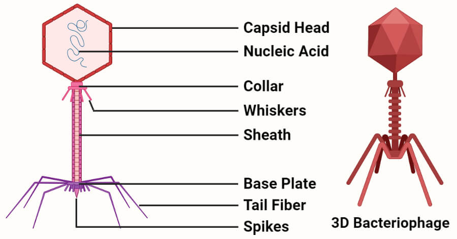
It has central hollow core or tube through which viral DNA is injected into the host.

Name the parts of bacteriophage and their functions. They then destroy or lyse the cell releasing new phage particles. The structure at top is the head which contains DNA inside a protein coat. Lysogenic phages incorporate their nucleic acid into the chromosome of the host cell and replicate with it as a unit without destroying the cell.
Other parts of bacteriophage are the base plate and the tail fibers. The viral genome within the host cell takes over the cell machinery. The distal end consists of a basal plate with tail fibres at each corner.
It first attaches to the susceptible bacterial cell and injects its genetic material into the host cell. The core is surrounded by sheath proteins. A capsid where the genome is packed a tail that serves as a pipe during infection to secure transfer of genome into host cell and a special adhesive system adsorption apparatus at the very end of the tail that will recognise the host cell and penetrate its wall.
Is the narrow cylindrical part. This is a T4 bacteriophage virus. Bacteriophage A bacteriophage informally phage is a virus that infects and replicates within bacteria.
Is the hexagonal plate with tail fibres usually 6 in number and used for attachment to the host cell wall during infection. A Bacteriophage possesses viral proteins which can disrupt their host cell ie. The term is d.
The sheath then contracts injecting the contents of the head DNA. The parts of bacteriophage are the head collar core and sheath. It lacks all membrane bound cell organelles such as mitochondria lysosome golgi endoplasmic reticulum chloroplast peroxisome glyoxysome and true vacuole.
The virus attaches itself to the host bacteria cell wall by its tail fibres. Bacterial cell have simpler internal structure. The tailed phages have three major components.
What are the different parts of bacteriophage. Newly acquired DNA thus provides a reservoir of genetic information for potential future use rather than being selected for immediate utility. The simplest phage have many copies of only one or two different proteins while more complex phages may have many different kinds.
Bacteriophage definition Parts And Function 1d47w7qo32n2. Under certain conditions lysogenic phages can be induced to follow a lytic cycle. Find an answer to your question Name the parts of the flower and their functions bottttttty bottttttty 4 weeks ago Geography College answered Name the parts of the flower and their functions 2 See answers lol i didnt mean to put geography.
The bacteriophage attaches to the bacteria with the help of these tail fibres. Attached to this is the tail consisting of a tube-like sheath and tail fibres at bottom. Bacteriophage Definition Parts and Function - Free download as Word Doc doc docx PDF File pdf Text File txt or read online for free.
Download View Bacteriophage definition Parts And Function as PDF for free. All about Bacteriophage in your BIOTECHNOLOGY ASSIGNMENT READ ANALYZE and UNDERSTAND. The term is derived from bacteria and the Greek φαγεῖνphagein to.
Bacteria also lacks true membrane bound nucleus and nucleolus. Tail- The tail consists of an inner hollow tube which is surrounded by a contractile sheath with 24 annular rings. This selection for genome size plays an important role in bacteriophage evolution providing a mechanism for DNA gain and loss that is independent of gene function.
Bacteriophage Definition Parts and Function Bacteriophage A bacteriophage informally phage is a virus that infects and replicates within bacteria.
They cause motion and produce a force that the body uses to move and manipulate the body. Grasshoppers breathe through a series of holes called spiracles which are located along the sides.
![]()
Leg And Knee Anatomy Bones Muscles Soft Tissues Kenhub
Body is flat and covered by.

Well labelled diagram of the leg. Health diagram bone skeleton leg knee science anchor chart human human body. They have a body divided into three regions called tagmata head thorax and abdomen have three pairs of legs and. This will help you to understand the mechanism as well as the working.
Insect morphology is the study and description of the physical form of insectsThe terminology used to describe insects is similar to that used for other arthropods due to their shared evolutionary history. Their body is covered with a hard exoskeleton. Labelled Diagram Of Long Bone.
A gasketed cover or a gasketed lid except for leg sleeves automatic bleeder vents rim space vents column wells ladder wells sample wells and stub drains. The diagram would also include the legs and antenna of the cockroach. The Internal Organs.
Ventral View of Thigh Muscles. Otherwise note the leg color and the overall strength of the color to help identify the duck but be aware that dirty water or mud may obscure the true color. Read the definitions then label the grasshopper anatomy diagram below.
12 photos of the Labelled Diagram Of Long Bone draw a well labelled diagram of a long bone labelled diagram of a bone cell labelled diagram of bones in the body labelled diagram of hip. Bone August 3 2016 August 22 2016. Most ducks have relatively short legs though whistling-ducks have much longer legs and that length can help with identification.
Three physical features separate insects from other arthropods. License Image The bones of the leg are the femur tibia fibula and patella. The femur or the thigh bone is closest to the body.
The knee joint you need a perfectly labeled diagram of the knee. Each muscle also has. The ilium is the big bone of the hip the ischium is the bone on which one sits and the pubis forms the lower frontal hip bone as seen in the diagram.
These are commonly known as crabs and are found buried under rocks wood pieces and in sand along sea shore. Join the popular membership section. The Internal Organs.
A Labeled Diagram of the Knee With an Insight into Its Working. Head - the head is at the front end of the grasshoppers body and is the location of the brain the two compound eyes the mouth parts and the points of attachment of its two antennae. Ventral View of Thigh and Leg Muscles.
The cleidocervicalis is labeled clavotrapezius in your book. The Internal Organs. Labelled Diagram Of Long Bone Find out more about Labelled Diagram Of Long Bone.
They have six jointed legs two pairs of wings and two antennae. As these muscles contract and relax they move skeletal bones to. This figure illustrates the position of the transversus abdominus in relation.
To understand one of the most complex joints of our body ie. A quality educational site offering 5000 FREE printable theme units word puzzles writing forms book report formsmath ideas lessons and much more. In this article we will discuss about the structure of crabs with the help of suitable diagrams.
May 28 2016 - The human body muscles are the main contractile tissues of the body involved in movement. The majority of muscles in the leg are considered long muscles in that they stretch great distances. In humans the neck of the femur connects the shaft and head at.
The bones of the leg are the femur tibia fibula and patella. Each penetration of the internal floating roof for the purpose of sampling must have a slit fabric cover that covers at least 90 percent of the opening. The longest and the strongest bone in the human skeletal system as you can observe in the labeled skeleton diagram of the human body.
Great for new teachers student teachers homeschooling and teachers who like creative ways to teach. The head forms a ball-and-socket joint with the hip at the acetabulum being held in place by a ligament within the socket and by strong surrounding ligaments. A bacteriophage b æ k ˈ t ɪər i oʊ f eɪ dʒ also known informally as a phage ˈ f eɪ dʒ is a virus that infects and replicates within bacteria and archaeaThe term was derived from bacteria and the Greek φαγεῖν phagein meaning to devourBacteriophages are composed of proteins that encapsulate a DNA or RNA genome and may have structures that are either.
Long bones are found in the arms humerus ulna radius and legs femur tibia fibula as well as in. Kenyon College Cat Anatomy. A diagram of a cockroach would include labeled segments of the insect including the head and thorax.
Femur upper bone of the leg or hind leg. Grasshopper Anatomy Like all insects the grasshoppers have three main body parts the head the thorax and the abdomen.
There is no distinct branch from the cephalic vein to the basilic vein. Heart diagram using - Labelled diagram.
Labelled Diagram Of Vein Valves And Vein Artery Circuit
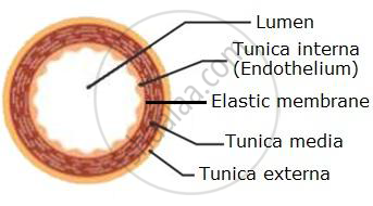
Lining the core of each is a thin layer of endothelium and covering each is a sheath of connective tissue but an artery has thick intermediate layers of elastic and muscular fiber while in the vein these are much thinner and less developed.
Labelled vein diagram. Right Atrium Right Ventricle Left Atrium Left Ventricle Tricuspid Valve Mitral Valve Pulmonary Valve Aortic Valve Superior Vena Cava Inferior Vena Cava Pulmonary Trunk Right Pulmonary Artery Left Pulmonary Artery Pulmonary Veins. Diagram Iv Vein Diagram Full Version Hd Quality Vein Diagram. Free Artery Vein Diagram Templates.
State how each of these are adapted for its functions. Aorta Pulmonary artery Pulmonary vein Right atrium Left atrium Right ventricle. The veins carry the blood towards the heart which consists of the waste.
Human Body Vein Diagram In Detail. Both arteries and veins are types of blood vessels in the cardiovascular system. 20 5 Circulatory Pathways Anatomy And Physiology.
In this image you will find a facial vein internal jugular vein superior vena cava hepatic vein renal vein gonadal vein inferior vena cava in human body vein diagram in detail. Fully Labeled Arteries Diagram Jpg Superficial Temp Oral Artery. The vein is the blood vessel which carries the deoxygenated blood from different parts of the body back to heart.
Want to learn more about it. Labeled Diagram Of Arm Veins Diagram Labels Label Gallery Get some ideas to make labels for bottles jars packages products boxes or classroom activities for free. Venipuncture And Arterial Puncture Policy.
A labeled diagram of the human heart you really need to see. Labels are usually small in size so you should carefully choose the font of the texts to make sure it is readable. Arteries and veins have the same layers of tissues in their walls but the proportions of these layers differ.
Labeled diagram of arm veins. Labelled Diagram Of Vein Valves And Vein Artery Circuit. There are three veins most commonly used in venipuncture or phlebotomy.
The major nerves and veins start in your neck and run the length of your arms often into your hands. Try to label the following structures yourself and then check to see if you are correct. It is the outermost coat which is formed of connective tissues.
The placement of these three veins forms a letter H as in the diagram below. Draw neat labelled diagram of a cross section of an artery and a vein. Tunica externa is also called tunica adventitia.
Anatomy Label Major Arteries And Veins Major Arteries Of The Head And Neck Labelled Medical Stock Images Company - Related posts of anatomy veins arteries diagram. Labeled Arteries And Veins Diagram Written By JupiterZ Tuesday July 18 2017 Add Comment Edit. Arteries dont have valves in them with the exception of the semilunar valves found in the pulmonary artery and the aorta.
These three veins are found in the antecubital area. Major arteries veins and nerves of the body. This diagram shows the major veins in human body venas.
They are the cephalic median cubital and basilic veins. Arteries and veins diagram 205 circulatory pathways anatomy and physiology. As you can see there are also have a common iliac vein internal iliac vein external iliac vein deep femoral vein femoral vein.
With the help of labelled diagrams describe the structure of an artery a vein and a capillary. Arterial And Venous Network In The Forearm Download Scientific. Lab Practicum 2 Circulatory System Anatomy.
You can also put your logo at the top or bottom corner of the label. How to draw labelled diagram of TS of artery vein and capillaries step by stepHello Friends in this video I tell you about how to draw labelled diag. A Simple Schematic Of The Layers Within An Artery.
Below is a blank diagram followed by the labeled diagram with the answers. Venous system diagram Use this interactive 3-D diagram. Circulatory Pathways Anatomy And Physiology Ii.
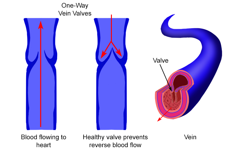
It is a middle coat which is chiefly formed of elastic connective tissue and smooth muscle fibres. Valves in these veins allow blood to flow from the superficial veins to your deep veins but not the other way. Most of the times we put the labels to show some specific information.
Veins have valves or more precisely non return valves preventing reverse blood flow. Diagram Showing The Venous Anatomy Of The Leg Vascular. Arteries Of The Body Diagram Untpikapps.
This diagram shows the major veins in the human body. Cephalic Vein Anatomy And Clinical Points Kenhub. The wall of an artery and a vein consists of three coats.
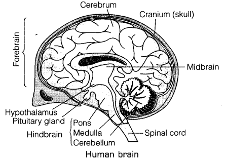
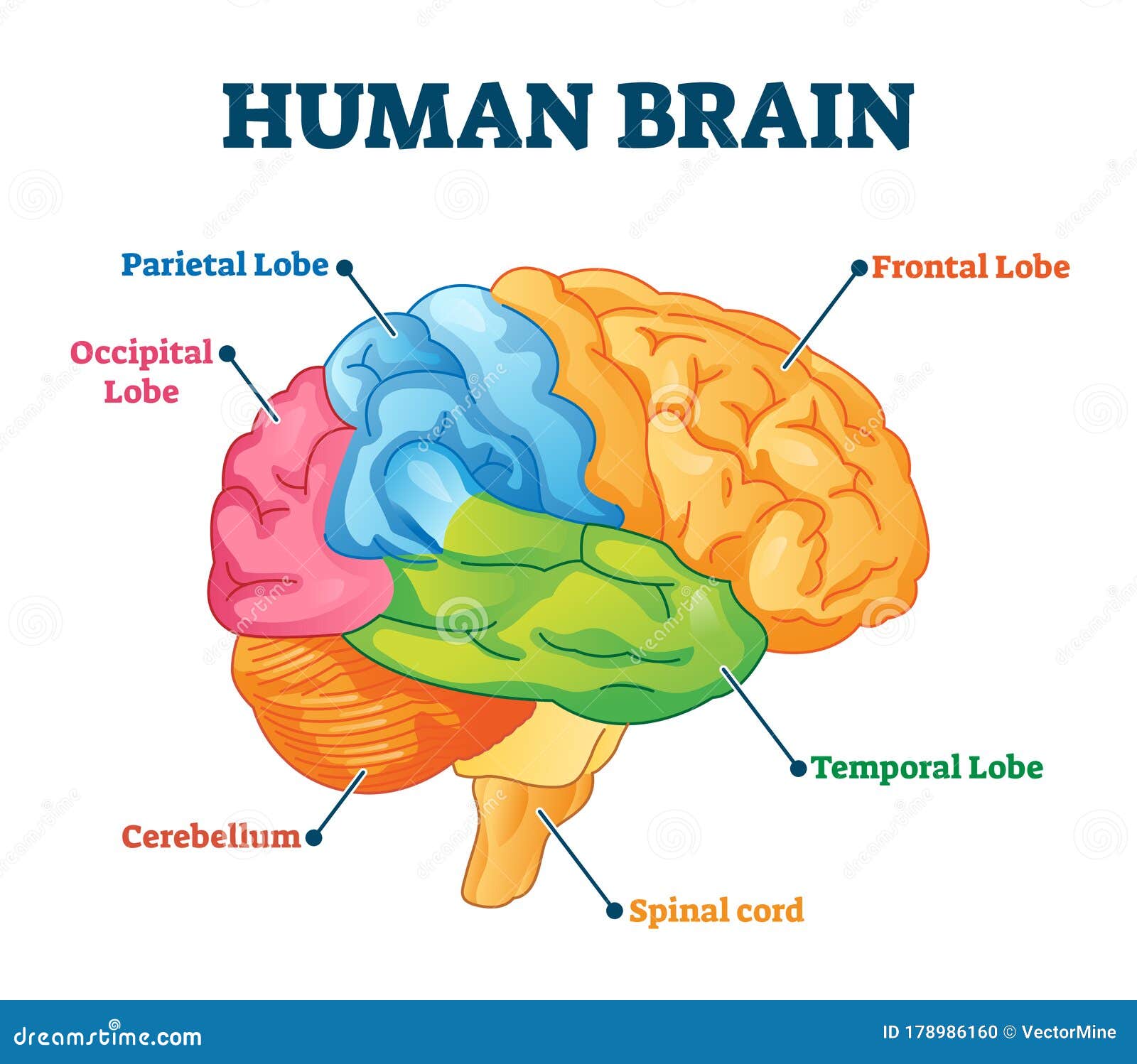
The frontal lobe is located in the forward part of the brain extending back to a fissure known as the central sulcus. It is the anterior part of the brain.
Draw A Labelled Diagram Of Human Brain And Mention The Functions Cbse Class 10 Science Learn Cbse Forum
Consisting of largely of three paired structures the thalamus hypothalamus and epithalamus the diencephalon plays a vital role in integrating conscious and unconscious sensory information and motor commands.
Labeled parts of human brain. The forebrain the midbrain and the hindbrain are the three main parts of the brain. The brainstem is made of three regions. The three regions of the hindbrain coordinates.
The parietal lobe takes care of sensation and perception managing how we react to sensory input. Midbrain Pons and Medulla oblongata. Nervous System - Label the Brain.
The forebrain is responsible for a number of functions related to thinking perceiving and evaluating sensory information. The forebrain is the anterior part of the brain which comprises the cerebral hemispheres the thalamus and the hypothalamus. See labeled brain anatomy stock video clips of 27 brain diagram with labels hypothalamus vector brain diagram pons cerebrum and cerebellum brain pons brain anatomy amygdala brain labelled amygdala brain human midbrain diagram pons.
It also consists of two subdivisions called the telencephalon and diencephalon. 2 frontal occipital parietal temporal. The medulla oblongata the pons and the midbrain.
Choose the correct names for the parts of the brain. The cerebrum the largest part handles vision movement hearing language and touch. The brain is a 3-pound organ that contains more than 100 billion neurons and many specialized areas.
The forebrain has two major parts called the diencephalon and the telencephalon. There are 3 main parts of the brain include the cerebrum cerebellum and brain stemThe Cerebrum can also be divided into 4 lobes. Lobes of the Brain The four lobes of the brain are the frontal parietal temporal and occipital lobes Figure 4.
Surrounded by the cerebral hemispheres the diencephalon forms the central core of the brain. Smallest and central part of the brain. It has the following parts.
The temporal lobes are involved with memory and hearing. Consisting of or divided into three major partsmedulla pons and midbrainit serves to develop the connection of spinal cord with the cerebellum and cerebrum. Frontal lobes parietal lobes temporal lobes and occipital lobesThe brain stem consists of three major parts.
The brain and its parts can be divided into three main categories. It helps the body survive by controlling the basic functions like breathing digesting food sleeping as well as heart rate. Which performs thinking reasoning speech intelligence and usage of information.
A net-like structure of mixed gray and white matter known as the reticular. The brain is surrounded by a layer of tissue called the meninges. The lower part of the brain.
The brain stem is located in front of the cerebellum and connects to the spinal cord. The part of the brain that makes up the spinal cord is called the brain stem. Where lobes are responsible for detecting the smell from different receptors.
Parts of Human Brain Forebrain Largest part of the brain. The forebrain midbrain and hindbrain. Many of the most basic survival functions of the brain are controlled by the brainstem.
The brain is so complex that even to simply discuss it certain distinctions have to be made regarding its structure and for that reason scientists divide the brain up into three major portions with each of these portions divided into many subregions and structures. It consists of three major parts. 1 cerebrum cerebellum brain stem spinal cord.
The occipital lobes contain the brains visual processing system. This science quiz game will help you learn the 7 parts of the brain. The human brain is the central organ of the human nervous system and with the spinal cord makes up the central nervous systemThe brain consists of the cerebrum the brainstem and the cerebellumIt controls most of the activities of the body processing integrating and coordinating the information it receives from the sense organs and making decisions as to the instructions sent to the.
The main portions of the brain are the cerebrum the cerebellum and the brainstem. 3 frontal occipital parietal temporal. The brainstem is the place from which ten of the twelve cranial nerves originate.
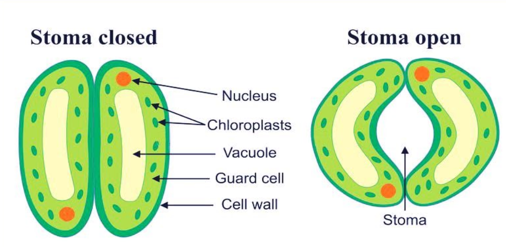
It maintains the moisture balance according to weather by opening and closing. The inner walls of guard cells are thick while the outer walls are thin.
Draw A Labelled Diagram Of Stomata Write Two Functions Class 9 Biology Cbse
They regulate the process of transpiration and gaseous exchange.
Draw the labelled diagram of stomata define transpiration. They regulate the process of transpiration and gaseous exchange. Draw a neat diagram of the stomatal apparatus found in the epidermis of leaves and label the Stoma Guard cells. Stomata facilitate carbon dioxide uptake and release of oxygen during the process of photosynthesis.
It is the responsibility of stomata for transpiration and the movement of guard cells via actions. This water evaporates by the process of transpiration. It is one among the few important topics and is majorly asked in the board examinations.
V The movement of air bubble in graduated tube in a minute gives the rate of transpiration. Draw label and annotate a diagram of a potometer to show how it can measure transpiration rate Stem must go in water immediately after being cut to maintain the transpiration stream through the plant and avoid introducing air bubbles. 1 Theory of Photosynthesis in Guard Cells 2 Starch Sugar Inter-conversion Theory 3 Theory of Glycolate Metabolism and 4 Active K Transport or Potassium Pump Theory and Role of Abscisic Acid or Active Potassium Pump Theory.
The following four points will highlight the four important theories of stomatal movement. Stoma Guard cells Chloroplast Epidermal cells. Ii They do not have striations.
Stoma Guard cells Chloroplast Epidermal cells and Cell wall. Write two differences between striated and smooth muscles. Stomata are small pores present in the epidermis of leaves.
Draw a labelled diagram of stomata. Stomata are the tiny pores present on the surface of the epidermal leaves. Labelled Diagram Of Stomata.
It helps the plant to get rid of excess water through transpiration. The diagram of the Stomata is useful for both Class 10 and 12. Its two functions are-These helps in the gaseous exchange.
What are stomata draw a labelled diagram of stomata. A brief description of the Stomata along with a well-labelled diagram is given below for reference. I Exchange of gases ii Transpiration b The conditions in which photosynthesis takes place are.
Sketch and Label the Diagram Question 1. Draw a well labelled diagram of cardiac muscle found in the human body. ICSE solutions for Class 10 Biology Chapter 4 TranspirationSketch and Label the Diagram.
The figure for stomata is given below. Draw a labelled diagram of unstriated muscle tissue and mention its occurrence features and functions. Stomata are present in leaf epidermis.
The opening and closing of the stomata is due to the help of guard cells. It has been seen that stomata show periodic opening and closing during the day diurnal variation depending upon the heat and light water content of the cell and humidity. Draw a labelled diagram of stomata.
I The cells are long and spindle-shaped. List two functions of stomata. Draw a labeled diagram of the stomatal apparatus and label the following in it.
Answer verified by Toppr. Upvote 0 Was this answer. Thus due to transpiration water is pulled upward which creates an upward suction force called transpiration pull.
Chemical equation for photosynthesis. Write two functions of stomata. Draw a diagram of transpiration under your definition.
The main two functions of Stomata are. Stomata are open during the day and close during nightStomata take in carbon dioxide required for. What are stomata draw the diagram of stomata.
Water in the mesophyll cells of leaves cells located below the stomata is in contact with water or sap in xylem of leaf petiole stem and root. Thus it regulates the water content in plant cells. Draw a neat diagram of the stomatal apparatus found in the epidermis of leaves and label the Stoma Guard cells Chloroplast asked Feb 20 2019 in Biology by Amita 885k points transpiration.
6CO 2 6H 2 O C 6 H 12 O 6 6O 2. They are enclosed by two bean-shaped guard cells. It helps in the process of transpiration.
The stomatal pore is enclosed between two bean-shaped guard cells. A Diagram of stomata. Role of Stomata in Transpiration.
Tomato helps in the exchange of gases and transpiration. Write two functions of stomata. Draw a labeled diagram of the stomatal apparatus and label the following in it.
Carbon dioxide water chlorophyll and sunlight. Since most of the water is lost through stomata plants regulate the degree of stomatal opening and closing to reduce the water loss. The guard cells control the opening and closing of stomata.
The four important theories of stomatal movement are. The stomata also close down to prevent loss of water. Stomata are the pores present on epidermis layer of plant.
In most people the cranium is made up of 8 bones and the face is made up of 14. Sutures connect cranial bones and facial bones of the skull.
![]()
Skull Human Anatomy Human Body Human Skeleton Skull Label Anatomy Human Body Png Pngwing
Inspiring Skull Labeling Worksheets worksheet images.

Labeled diagram of the skull bones. The remainder of the bones in the skull are the facial bones. Dodge Durango Wiring Diagram. The Respiratory System Diagram.
They protect and surround the brain. The cranium and mandible was exported from CT data. Use this facial bone mnemonic list to remember the anatomy names and structure of each of the facial bones of the skull.
Weve created a blank skull diagram free for you to download as A PDF below. Pictures of skulls that are unlabeled and has empty boxes for students to add labels. The following anatomy diagrams of the skull contain the parts of bones and lateral view of the skull.
Discover and save your own Pins on Pinterest. Simple Boat Wiring Diagram. You can also download the labeled version and use this to make some notes.
It is comprised of many bones which are formed by intramembranous ossification and joined by sutures fibrous joints. It was then cleaned adapted and polypainted in ZBrush. Skull Bones Unlabeled Worksheet Rib Cage Anatomy Worksheet Unlabeled Bones of the Head and Face Hand and Wrist Bones Diagram Unlabeled Skull Bones Anatomy.
Those of the cranium which consist of the cranial roof and cranial base and those of the face. Blank Skull Diagram. In the CT data the upper teeth and lower teeth were joined as one mesh therefore I completely remodelled some of the teeth so that the mandible could be detached from the cranium and can be.
Learn the major cranial bone names and anatomy of the skull using this mnemonic and labeled diagram. The facial bones consist of two Maxillae forming the centre of the face the Zygomatic bones forming the cheekbones and the Mandible which is the lower jaw. Develop a good way to remember the cranial bone markings types definition and names including the frontal bone occipital bone parietal bone temporal bone sphenoid bone and ethmoid bone using this mnemonic and labeled.
Uses labeled diagrams to show the structure and anatomy of each facial bone. Browning Buckmark Parts Diagram. Labeled Human Skeleton Diagram Study guide for students and teachers.
A large PNG version of the human skeleton diagram. Draw a Skull - Halloween Special. SkullBonesUnlabeled Anatomy bones Human skull anatomy Skull anatomy.
The Mandible is the only bone in the skull that can move. Includes discussion about features and functions of the maxillary mandible nasal conchae zygomatic vomer lacrimal and palatine bones. See 15 Best Images of Skull Labeling Worksheets.
This quiz will test your knowledge on how to identify these bones. The skull is a bony structure that supports the face and forms a protective cavity for the brain. This is a model of the Human Homo sapiens skull.
Skull anatomy unlabeled In this image you will find Skull anatomy in it human anatomy bones skull 82 90 label the lateral features the skull - Human Anatomy Charts i feel like something cool could be done with this- like maybe a placemat or something. Oct 15 2017 - This Pin was discovered by Taylor Cox. Browning Buckmark Parts Diagram.
Once youve done that its time to learn anatomy with our skull labeling quiz. The bones that make up the cranium are called the cranial bones. In Figure 69 some of the bones of the hard palate forming the roof of the mouth are visible because the mandible is not present.
Oct 15 2017 - This Pin was discovered by Taylor Cox. The Cranium bones are composed of the Frontal Parietals Occipital and Temporal bones. The human skull can be divided into two sections the cranium and the face.
The bones of the skull can be considered as two groups. To view a high res version of an image click on the thumbnails below. We also cover the ear bones and the hyoid boneTranscriptnotesSkull bonesY.
In this video we discuss the locations of the bones of the skull and label them. Figure 67 and Figure 68 show all the bones of the skull as they appear from the outside. Human Heart Bone Diagram.
Skull bones quiz of the cranial and facial bones for anatomy and physiology. Images adapted from images in the public domain. Skull is pictured from many anglesHuman Skull No Text No Color free clip art text color coloring blank human pages skull diagram label skeleton without bones anatomy labels unlabeled diagrams.
The bone that forms the back of the head and connects with the occipital condyles and foramen magnum skeletal structures located on the underside of the skull. Often the ossicles of the ear and the hyoid bone are counted as part of the skull. Dodge Durango Wiring Diagram.
When you are taking anatomy and physiology you will be required to know the location of the cranial and facial bones.
Huge collection amazing choice 100 million high quality affordable RF and RM images. Bones and Joints of the Foot and Ankle The Ankle Lateral side of the ankle Joint capsule.
Parts Of The Foot Back To Health Wellness Centre
Hinge joints typically allow for only one direction of motion much like a door-hinge.
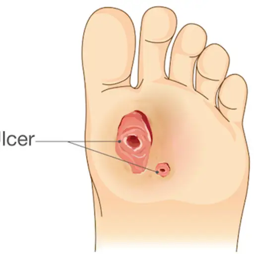
Parts of the foot pictures. Even the basics can lead you to a very simple self-treatment. The talus connects to the tibia which is the main bone in your lower leg. Gout of the foot is a type of arthritis that often affects the joint at the bottom of the big toe causing severe pain even when there is no pressure on the foot.
Athlete taping foot pressure points pair of feet anatomical leg pressure zones foot girl foot bottom human medicine icon boys soles foot isolated boy sole. Columns of the Foot. The ankle joint is both a synovial joint and a hinge joint.
Pictures of Common Foot Problems. The anatomy of the foot. Believe it or not the feet absorb more than 100000 pounds of.
Try these curated collections. Tips to Avoid Foot Pain From High Heels. The midfoot is made up of 5 tarsal bones.
45699 foot anatomy stock photos vectors and illustrations are available royalty-free. The end of the leg on which a person normally stands and walks. Its made up of 4 bones.
Laura Evans Full fat dairy products can exacerbate gout. 11139 bottom of foot stock photos vectors and illustrations are available royalty-free. The calcaneus which is the bone in your heel.
See foot anatomy stock video clips. The foot is sometimes described as having two columns Figure 3. The ankle joint or tibiotalar joint is formed where the top of the talus the uppermost bone in the foot and the tibia shin bone and fibula meet.
Find the perfect human foot anatomy stock photo. Picture of Foot Anatomy Detail. This condition is caused when uric acid builds up in the body.
No need to register buy now. In many animals with feet the foot is a separate clarification needed organ at the terminal part of the leg made up of one or more segments or bones generally including claws or nails. The end of the leg on which a person normally stands and walks.
This consists of five long metatarsal bones and five. Parts of your foot correlated with the stomach are found above the waistline. At the same time the foot must be strong to support more than 100000 pounds.
The midfoot Bones Anatomy. The thinnest part of your foot usually found towards its center is known as the waistline. The tendons ligaments and muscles in the feet number more than 100.
Search from Sole Of Foot stock photos pictures and royalty-free images from iStock. Parts Of Foot Bones Anatomy Skeleton Pictures Hindfoot Bones Anatomy. Bottoms of feet ankle muscle foot muscle anatomy human anatomy foot bones of the foot foot muscles 3d illustration foot foot muscle human foot anatomy anatomy foot vintage.
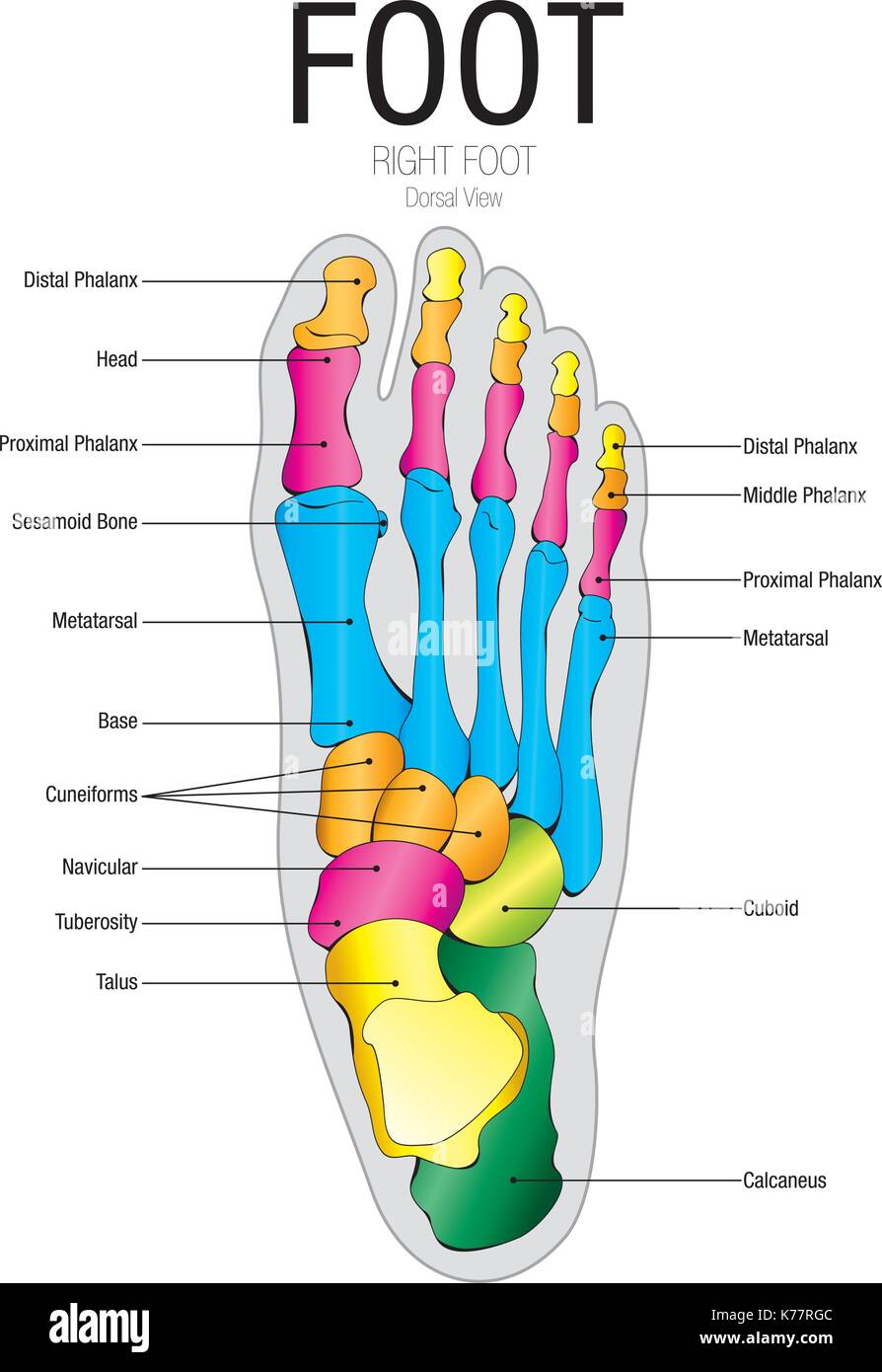
The foot is an extremely complex anatomic structure made up of 26 bones and 33 joints that must work together with 19 muscles and 107 ligaments to execute highly precise movements. Feet are very complex comprised of 28 bones and 30 joints. Feet is an anatomical structure found in many vertebrates.
The lateral column is stiffer and includes the calcaneus cuboid and the 4th and 5th metatarsals. The foot is divided into three sections - the forefoot the midfoot and the hindfoot. The cuneiform bones the navicularis and the cuboid all of which function to give your foot a solid yet somewhat flexible foundation.
The talus which is the bone in your ankle. The feet are located at the end of the legs and are used to stand and walk. See bottom of foot stock video clips.
The bottom of your foot is connected with your pelvic area. The foots shape along with the bodys natural balance-keeping systems make humans capable of not only walking but also running climbing. The foot is an extremely complex anatomic structure made up of 26 bones and 33 joints that must work together with 19 muscles and 107 ligaments to execute highly precise movements.
Like already mentioned the hindfoot is the posterior part of the foot. It is the terminal portion of a limb which bears weight and allows locomotion. The foot is the lowermost point of the human leg.
Parts correlated with the intestines are found below. Bones Tendons Ligaments and More. The foot contains a lot of moving parts - 26 bones 33 joints and over 100 ligaments.
Find high-quality stock photos that you wont find anywhere else. The medial column is more mobile and consists of the talus navicular medial cuneiform 1st metatarsal and great toe.
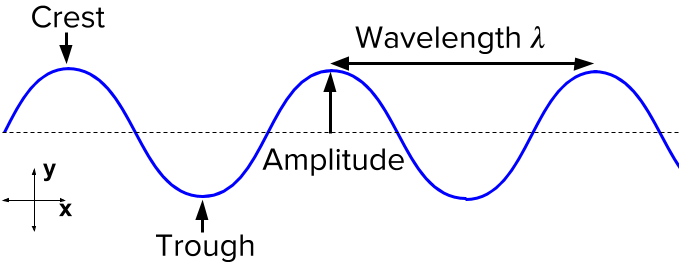
The crest and the trough of a wave are always twice the waves amplitude which is the height apart from each other. The crest is the top of the wave.
Wave Characteristics Review Article Khan Academy
What is the lowest part of a transverse waves.

What are the parts of a transverse wave. A transverse wave like all the other waves have also its parts. The crest is the top of the wave and the trough is the bottom. The amplitude refers to the height of the wave from the midpoint or rest point of the wave.
Displacement is the distance moved by. The highest point on these waves is called the crest. Is the top of a transverse wave called.
Parts of waves The crest is the top of the wave The trough is at the bottom of the wave The wavelength is the length of the wave The amplitude of a wave is the highest amount of vibration that the medium gives from the rest position The rest position is the position where a wave would be if there. The particles move up and down as. The wavelength is the length of the wave.
The amplitude of a wave is the highest amount of vibration that the medium gives from the rest position. The wavelength is the length of the wave. Compression which are areas of high molecular density and rarefactions which are areas of low molecular density.
Transverse waves have two parts. Parts of a transverse wave include the crest trough amplitude and wavelength. Click to see full answer.
The crest is the top of the wave. Parts of a Transverse wave. The trough is at the bottom of the wave.
The amplitude of a wave is the highest amount of vibration that the medium gives from the rest position. 5 points safinafarhodova123 Asked 05202020. Transverse waves have what are called peaks and troughs.
The wavelength is the length of the wave. Parts of a Transverse wave. What is the height of a transverse wave called.
The amplitude of a wave is the highest amount of vibration that the medium gives from the rest position. Parts of a Transverse wave. Parts of a transverse wave include the crest trough amplitude and wavelength.
We will be considering the parts of a wave with the wave represented as a transverse wave as in the following diagram. What are the highest parts of a transverse wave called. What are the highest parts of a transverse wave called.
Examples of transverse waves include. Parts of A Transverse Wave - YouTube. Energy is transferred from left to right.
Parts of a Transverse wave. If playback doesnt begin shortly try restarting your device. However none of the particles are transported along a transverse wave.
Longitudinal waves also have two parts. Some waves such as transverse waves have crests and troughs. The trough is at the bottom of the wave.
See answers 2 Ask for details. ShowHide Sub-topics Waves A LevelTypes of WavesParts of A Transverse WaveLongitudinal WavePhase DifferenceIntensityPolarization. In some other ways of defining it is that it is a moving wave which vibrates at right angles to the direction of the propagation.
Wavefronts show the position of points of a wave which are in phase. Transverse waves are waves where the vibration is at right angles 90 degrees to the direction of motion. What is the highest part of a wave.
The lowest point is called the trough. This medium could be imagined as a rope fixed at one end a few feet above the ground and held by you at the other end. The wavelength is the length of the wave.
In the above diagram the white line represents the position of the medium when no wave is present. The amplitude of a wave is the highest amount of vibration that the medium gives from the rest position. Electromagnetic waves eg light waves microwaves radio waves.
A Mexican wave in a sports stadium. The peak is the crest or top point of the wave and the trough is the valley or bottom point of the wave. A crest which is the highest point of the wave and the trough which is the lowest part of the wave.
Parts of A Transverse Wave. Ripples on the surface of water. On such a wave we can label lots of features including peaks troughs amplitudes and.
The crest is the top of the wave and the trough is the bottom. The amplitude refers to the height of the wave from the midpoint or rest point of the wave. The crest is the top of the wave.
A TRANSVERSE WAVE is a wave moving which is made up of oscillations that are perpendicular to the direction of the transferred energy. Vibrations in a guitar string. The trough is at the bottom of the wave.
The trough is at the bottom of the wave. The crest is the top of the wave.
Best offers for your Garden - httpsamznto2InnD0w-----Male Female Reproductive Parts of a Flower. A long-tube that connects stigma and ovary.

The Male And Female Reproductive Parts Of A Flower Brighthub Education
Parts of the flower include petals sepals one or more carpels the female reproductive organs and stamens the male reproductive organs.

What are the male and female reproductive parts of a flower jiskha. What are the male and female reproductive parts of a flower 6886 results page 2 science help. Seahorses change color and swim together in predetermined ways before mating. The testes and sperm are located within the scrotum which is located on the outside of the male body to regulate a temperature for sperm production.
In a flower the female reproductive part is called the Pistil. There are often several stamens for every one pistil. Floral reproduction is bisexual and flowers have male and female partsThe male or pollen-bearing part is called the stamen and is composed of the filament and the antherThe female or seed-bearing part is called the pistil and is composed of the ovary the stigma and the style.
What are the male and female reproductive parts of a flower - 18179862 baileypeters2007 baileypeters2007 10072020 Biology College answered What are the male and female reproductive parts of a flower 1 See answer baileypeters2007 is waiting for. The anther is the part of the organ that produces pollen and the filaments hold up the anthers. Female reproductive parts of a flower.
Stamen is the male reproductive organ and is also known as Androecium. Genitalia allow males and females to mate fertilize internally and support. Pollination is the process in which the pollen is transferred from the male part of a flower anther to the female part of a flower stigma of the same flower or of another flower.
The transfer of pollen grains from anther to stigma takes place by a pollinating agent like insect. The Female parts consists of one ovary. The female or seed-bearing part of a flower is called the.
Male reproductive part of flower consists of anther and filament. Seed formation takes place in. It consists of two parts.
Click to see full answer. Male and female reproductive structures and their functions Topic. Correct answer - What are the male and female reproductive parts of a flower.
The female reproductive part consists of stigma style and ovary. A narrow and threadlike section of the stamen functions by supporting the pollen-bearing anther. These behaviors are called 1 point asexual reproduction.
The gonads are the precursors of the genitalia in males that mostly include external penis scrotum epididymis and testes and in females that mainly include internal vagina uterus fallopian tubes cervix and ovary organs. Then devin215 devin215 06022017 Biology Middle School answered Describe the structure and function of the male and female reproductive parts of a flower 1 See answer devin215 is waiting for your help. This is because having multiple stamens increases the number of pollen.
It has three main parts called stigma style and ovary. Generally the reproductive parts of a plant are located in its flowers. A flower may have exclusively male parts exclusively female parts or commonly both.
A ductless reproductive gland holding ovules. Of these the male reproductive organ is the Stamen while the female reproductive organ is the Pistil which is the collective term for carpels. The topmost part of the flower.
Flower parts vary tremendously in number. Anther contains pollen grains which contain male gamete. A function of the female reproductive.
The pollen-bearing body of the stamen exhibits a wide variety of forms and means of attachment. Stamen is the male reproductive part of a flower. The ovary contains ovules which are the small egg part of the plant which contains the female reproductive cells.
A male peacock displays his colorful tail feathers in order to. Called stamens these reproductive organs are made up of two parts. The male reproductive parts of a flower are much simpler than the female ones.
The male or pollen-bearing part is called the stamen and is composed of the filament and the anther. The female part of a flower and forms the innermost part of the flower. Add your answer and earn points.
Click here to get an answer to your question What is the male and female reproductive parts of a flower. The female or seed-bearing part is called the pistil and is composed of the ovary the stigma and the style. The flower is the reproductive unit of some plants angiosperms.
What is the male and female reproductive parts of a flower. Ovary stigma and style together make the pistil. Reproductive parts of the flower are the stamen male collectively termed the androecium and carpel often the carpel is referred to as the pistil the.
Pollination can occur through different ways such as by the wind or water by insect bird or bat. Petals sepals carpels and stamens form parts of the flower.
Also mark the point O about which the rotation takes place. Its entire process is carried through the body surface with the help of pseudopodia.

Name The Process By Which An Amoeba Reproduces Draw The Various Stages Of Its Reproduction In A Proper Sequence
C The process of obtaining food is called phagocytosis which means cell.
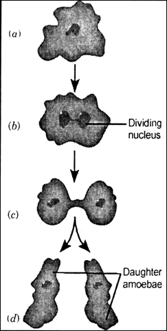
Draw a neat labelled diagram explaining the mode of reproduction in amoeba. The body rotates in anticlockwise direction. B The mode of nutrition in amoeba is holozoic. State where the sub-atomic particles are present in the atom.
Draw a neat labelled diagram showing forces acting on the meniscus of water in a capillary tube. After nuclear division the cell membrane divides into two parts by constriction and along with this. 1Starting with the zygote draw the diagrams of the different stages of embryo development in a dicot.
Two forces of magnitude F act at point A and point B. Amoeba does not have any specialized organ for nutrition. Springer Precision P320 Magazine Extension Avengers Watch Spider-man Far From Home Fanfiction Wattpad Merlin And Arthur Mpreg Relationship.
Draw a labelled diagram of human heart. Amoeba is unicellular and hence it does not have mouth Amoeba takes the food into the body by forming structures called pseudopodia around the food particle. Draw a neat labelled diagram to show the direction of two forces acting on a body to produce rotation in it.
Mention a purpose served by this part other than nutrition. State the event with this reproduction starts. Name the three sub-atomic particles in the atom and represent them symbolically showing the mass and charge of each.
A Diagram of Nucleosome b Histones are positively charged because they are rich in the basic amino acid residues like lysine and arginine which carry positive charges in their side chains. Watch 1 minute video. Answered Feb 10 2020 by Ritik01 481k points selected Feb 10 2020 by KumariJuly.
Draw a neat labelled diagram representing the duct system of liver gall bladder and pancreas. These are called the daughter cells now. Asexual type of reproduction is found only in lower organisms.
This is accomplished by. The mode is the cell division. Amoeba is a unicellular organism and just like bacteria it reproduces through binary fission.
Draw a diagram of nutrition in amoeba. The mode of nutrition in amoeba is known as holozoic nutrition. It involves the ingestion digestion and egestion of food material.
During reproduction there is formation of new cells which must carry the same amount and type of hereditary information as present in the parent cell. Explain with a neat labelled diagram the process of nutrition in amoeba. The undigested food collects inside the cell and the cell membrane ruptures.
Draw a neat and well labelled diagram of male reproductive system of a frog. In this method two similar individuals are produced from a single parent cell. 2What aretthe possibleotypes of pollinations in chasmogamous flowers.
Share It On Facebook Twitter Email. Amoeba multiplies by asexual reproduction that is it requires only one parent. Differentiate between Fertilization is fusion of haploid male and female gametes to produce a diploid zygote.
Lower organisms produce same young ones by this method which are also exactly similar to parents. 3With a neat labelled diagram describe the parts of a mature angiosperm embryo sacMenionthe role f synergids. Draw Neat Labelled Coloured Sketches to Show the Characteristics of the Following Geographical Feature.
Draw neat labelled diagram explaining grade of organisation Share with your friends. During this the nucleous first elongates then the cytoplasm does after which they break into two equal parts. Write a Brief Description Alongside Explaining It Block Mountain.
In case of hydra again asexual reproduction is there. Amoeba is a microscopic single celled organism which is found in pond water. 1 Answer 1 vote.
Labelled Diagram Binary Fission In Amoeba Label Amoeba Diagram sparkeroding co uk April 12th 2019 - Label Amoeba Diagram Ebook Label Amoeba Diagram currently available at www sparkeroding co uk for review only if you need complete ebook Label Amoeba Diagram please fill out registration form to access in our databases your. This pseudopodia forms a vacuole around the food particle called food vacuole and the vacuole is taken inside the cell. After replicating its genetic material through mitotic division the cell divides into two equal-sized daughter cells.
This will also help you to draw the structure and diagram of amoeba. Through this the undigested food is thrown out of the body. Mention the information source of making proteins in the cell.
Draw a neat labelled diagram representing an atom. In amoeba the nucleus in the cell elongates and divide into two parts from the centre. To keep watching this video solution for.
Please find below the solution to the asked query Grades of. The main components of an amoebas diet are bacteria and algae.
The Papilionoidea comprises of 6 families. Butterfly eggs are surrounded by a layer called a CHORION which is surrounded by a thin layer of wax which prevents it from getting dry.
![]()
The Children S Butterfly Site
The Hesperioidea comprises of a single family Hesperiidae.

What are the parts of female butterfly. The female butterfly attaches the eggs to leaves or stems of plants that will also serve as a suitable food source for the larvae when they hatch. Eggs can be laid from spring summer or fall. A butterflys body is divided into three main sections the head the thorax and the abdomen.
Limenitis arthemis can be split into two major groups. Butterflies belong to a large group of insects called the Lepidoptera which includes both butterflies and moths. These plants will then become the food for the hatching caterpillars.
The vulva is the part of your genitals on the outside of your body your labia clitoris vaginal opening and the opening to the urethra the hole you pee out of. The mouthparts of female mosquito are piercing and sucking type. Its members are called Skippers and are generally thought of as being butterflies.
The butterfly stroke has three major parts the pull the push and the recovery. Perhaps one of the better examples that we have of sexual dimorphism in Ohio is the Black Swallowtail. The three most important parts of a butterfly are the head thorax and abdomen.
The males bottom picture of this species have a yellow band across the lower portions of the wings while in the female top picture the yellow band is replaced by a less extensive blue band. Eggs are laid on plants by the adult female butterfly. The larva or caterpillar that hatches from the egg is.
Simply put a butterfly is a winged insect that undergoes complete metamorphosis in other words goes from egg to caterpillar to pupa to adult. They have three body segments. A female butterfly lays a lot of eggs usually on leaves or stems of plants.
Female mosquitoes feed on the blood of warm blood vertebrates. Though we can often determine a butterflys sex by looking at the most obvious differences in its appearance - like the scent spots of the male monarch or wing patterncolor of tiger swallowtails - the best way to distinguish between males and females particularly if you arent intimately acquainted with each species is actually by looking at the butterflys genitalia. The head the abdomen and the thorax.
Five of these - the Papilionidae Lycaenidae Riodinidae Pieridae and Nymphalidae have always been regarded as butterflies. But the vulva has a lot more going on than just the vagina. Some butterflies are sexually dimorphic meaning males and females look considerably different.
1 - Head 2 - Thorax - legs abdomen 3 - Wings - venation scales 4 - Wing scales - scanning electron microscope images 5 - Hearing organs flight thermoregulation Head Antennae eyes palpi proboscis. The tiny antennae which are near the mouth parts sense smells. It has been studied for its evolution of mimicry and for the several stable hybrid wing patterns within this nominal species.
Butterfly eggs are very tiny and may be in different shapes like spherical oval or cylindrical. These types of mouth parts are present in almost all the bloodsucking insects like tse-tse fly bed bug etc. Limenitis arthemis the red-spotted purple or white admiral is a North American butterfly species in the cosmopolitan genus Limenitis.
From the initial position the arm movement starts very similarly to the breast stroke. Females lay a lot of eggs at once so that at least some of. A butterflys antennae palps legsand many other parts of the body are studded with sense receptors that are used to smell.
These parts contribute to their health and help them grow. Each body section has very different functions and all are needed for the butterfly to live. Butterfly eggs are tiny vary in color and may be round cylindrical or oval.
A butterflys head is full of extremely important organs that allow the butterfly to sense what is around it and to feed. While vaginas are just one part of the vulva many people say vagina when they really mean the vulva. On the head you will find.
The mouthparts of mosquito are modified for piercing the skin of the vertebrates and then sucking their blood. It is one of the most dramatic examples of hybridization between non-mimetic and mimetic populations. The sense of smell is used for finding food usually flower nectar and for finding mates the female smelling the males pheromones.
These can also be further subdivided. This depends on the species of butterfly.
Doctors do not know what causes this type of eczema but it has been linked to allergies and. Big toe is pointing outwards from the center of the foot.
What Can Your Feet Tell You About Your Health
Pain swelling bruising started after intense or repetitive exercise.
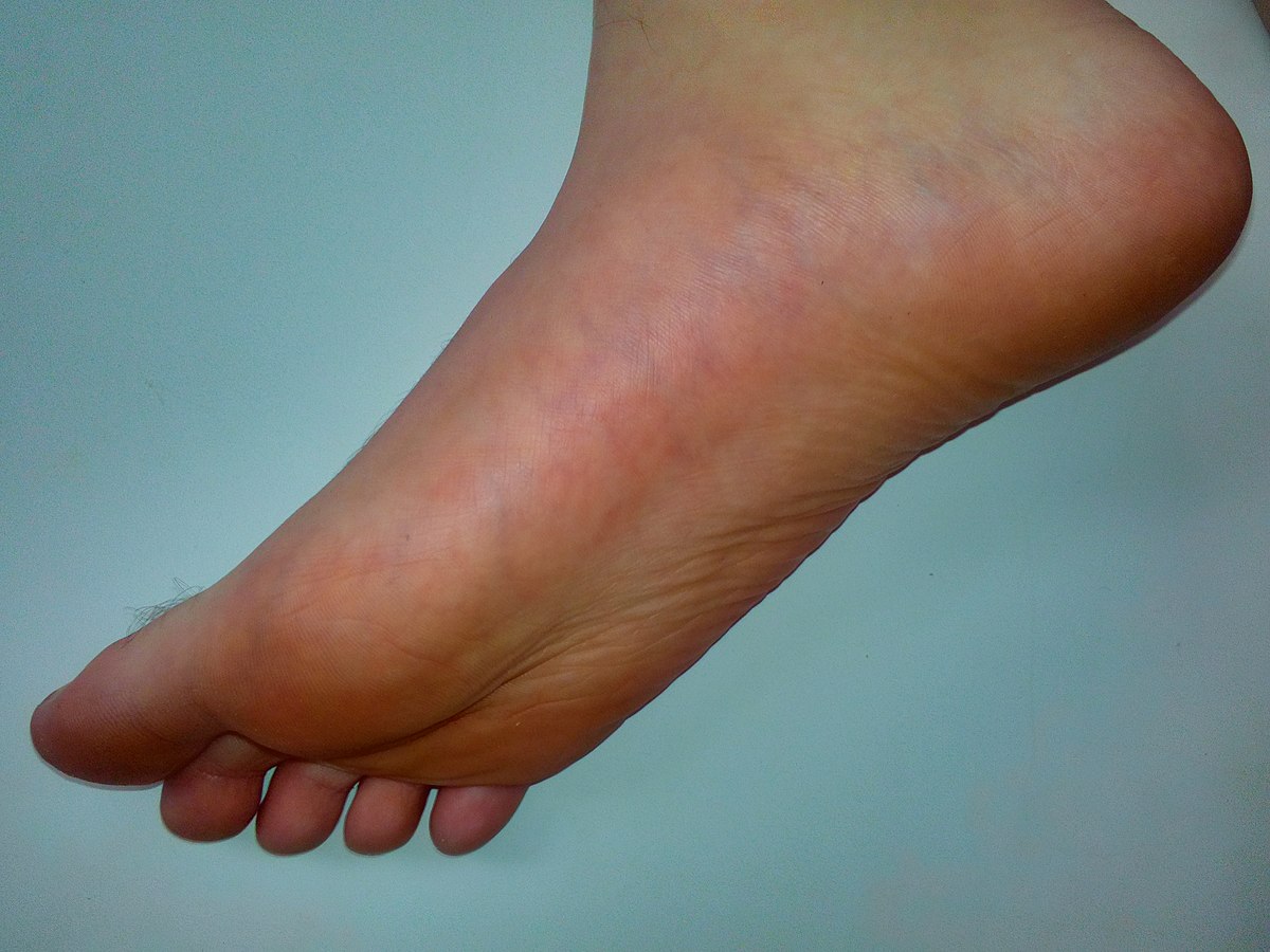
What is the bottom of my foot called. Dyshidrotic eczema may cause bumps on the bottom of the foot that are itchy and filled with fluid. Plantar calluses occur commonly on the. It is called the plantar fascia and it is a ligament that connects the heel to the front part or ball of your foot.
It is the formation of fibrous tissue around the plantar nerve that runs in the spaces between the second and third or third and fourth metatarsal heads. This tissues connects your heel to your toes and in some cases forms a growth called a fibroma. Plantar fasciitis is an inflammatory condition.
This is the area between the arches and toes on the bottom of the foot. You usually feel it on the bottom of your foot between your toes. The plantar fascia is the thick tissue on the bottom of the foot.
Bottom of the foot. Metatarsalgia centers under the five bones at the bases of the toes the metatarsals. Sharp burning or shooting pain near your toes ball of your foot feels like a lump or small stone under your foot.
Also called verruca plantaris. This growth in the plantar fascia can be very painful. The posterior tibialis tendon courses down your inner lower leg and attaches to the bottom of your foot near the inside of the arch.
Also called the sole. The tendon helps support your foots natural arch and irritation here may cause pain limited walking ability and flatfoot deformity. It connects the heel bone to the toes and creates the arch of the foot.
A viral epidermal tumor on the sole of the foot sensitive to pressure and painful during walking. The neuroma can feel painful and make it hard to walk. Bottom of the foot pain near toes may result from a condition called Mortons Neuroma named after Dr.
It also supports your arch. Pain in the sole of the foot. When you have Mortons neuroma the nerve between the bones of your toes may become swollen and inflamed.
Its also called intermetatarsal neuroma. It is not properly aligned. There are a number of medical conditions directly linked to the appearance of a callus hyperkeratosis Flat feet.
This does not have to cause pain or even very much in downtime at all. Metatarsalgia refers to pain and inflammation in the ball of the foot. It forms the arch of your foot and connects your heel to your toes.
The plantar fascia works like a rubber band. It occurs in the plantar fascia which is a band of tissue that runs along the bottom of your foot. The pain of metatarsalgia can be caused by a number of conditions and can have varied treatments.
There is one piece of tissue that runs along the bottom of the foot that is essential for your foot to function. Mortons neuroma affects your forefoot or ball of your foot between the metatarsal bones and toes. Symptoms can include.
Sometimes caused by going barefoot. Plantar calluses are tough thickened skin that form on the surface of the bottom part of your foot the plantar side. A table showing some of the possible causes of pain in the bottom of the foot with their associated symptoms.
Symptoms of hard spot on the bottom of the foot pain. Ideally in looking at preventing a callus on bottom of the foot it helps to understand what causes them. Removal surgery can be very effective in done by a podiatrist in the office within a few minutes.
Flexion of the foot caused by stroking or scratching the lateral border of the foots sole from the heel to the toes. When this tissue becomes swollen or inflamed it is called plantar.
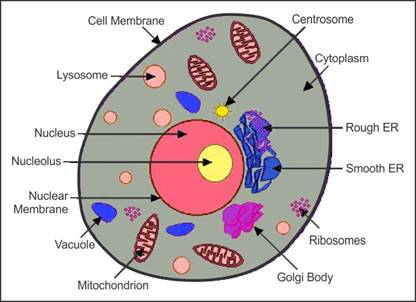
Answer verified by Toppr. Draw a labelled diagram of a plant cell.

Draw A Neat Labelled Diagram Of An Animal Cell
A brief explanation of the different parts of an animal cell along with a well-labelled diagram is mentioned below for reference.
Draw the neat labelled diagram of animal cell. Posted by Unknown at 2209. Ncert Solutions For Class 8 Science Chapter 8 Cell Structure And. Labelled diagram of plant and animal cell are as follow---.
Link of our facebook page is given. Printable Plant And Animal Cell Labelled Diagram Of A Parts Of A Plant Cell 7th Grade Images Galleries Cells Mr Plant Cell Diagram Cell Diagram Plant Cell. Compare The Location Of Nucleus In Animal Cell And Plant Cell Draw A.
Draw a labelled diagram of a animal cell. Draw A Neat Diagram Of Plant Cell And Label Any Three Parts Which. Draw a neat labelled diagram of animal cell.
Share to Twitter Share to Facebook Share to Pinterest. Draw a neat diagram of Plant and Animal cell and label its important cell organelles. CBSE NCERT Notes Class 9 Biology Fundamental Unit of Life.
It initiates and regulates cell division. Draw A Labelled Diagram Of A Animal Cell. Draw a Labelled Diagram to Show the Metaphase Stage of Mitosis in an Animal Cell Having 6 Chromosomes.
The plant cell is rectangular and comparatively larger than the animal cell. B The organelle which is found only in animal cell is Centrosome. Draw a labelled diagram of a animal cell.
Get Instant Solutions 24x7. A Centromere b p-arm. Chapter 8 The Unit of Life.
Plant and animal cells are different from each other as plant cell. Ii How many daughter cells are formed at the end of mitosis and at the end of meiosis. A Diagram of animal cell.
Forms spindle fibres with the help of asters. Eukaryotic cells are one which have organised nucleus with a nuclear membrane and genetic material is organised into chromosomes. Get the answer to this question and access a vast question bank that is tailored for students.
Draw a neat labelled diagram of animal cell. Draw a neat diagram of plant cell and label any three parts which differentiate it form animal cell. Cell Structure And Functions Class 11 Notes Biology Mycbseguide.
Question Bank Solutions 26084. Both plant and animal cells belong to eukaryotic cells. Draw a neat labelled diagram of an animal cell.
Draw A Neat Diagram Of Plant Cell And Label Any Three Parts Which Differentiate It From Animal Ce Cbse Class 9 Science Learn Cbse Forum. Upvote 0 Was this answer helpful. The cell theory that all the plants and animals are composed of cells and that the cell is basic unit of life was presented by two biologists M.
The animal cell diagram is widely asked in Class 10 and 12 examinations and is beneficial to understand the structure and functions of an animal. NCERT Class XI Biology. A draw a neat diagram of a plant cell and label the.
Diagram of the ultra-structure of a typical animal cell Prev Question Next Question Related questions 0 votes. Draw a diagram of the longitudinal section of a mature anatropous ovule and label any ten parts in it. Iii With reference to cell division explain the following terms.
Asked Feb 6 2020 in Biology by Ritik01 481k points cell. Concept Notes Videos 249. Asked Nov 28 2017 in Class IX Science by ashu Premium 930 points Draw a neat diagram of animal of an animal cell and label any four parts of it.
Click hereto get an answer to your question Draw a neat labelled diagram of animal cell. Draw a neat labelled diagram of the ultra-structure of a typical plant cell. It is very thin delicate elastic and selectively permeable membrane.
Class 12 Solved Question paper 2020 Class 10 Solved Question paper 2020. Animal Cell Diagram For Class 9 With Label. The plant cell can also be larger than the animal cell.
Draw a labelled diagram of a plant cell. Draw a labelled diagram of a animal cell. Draw a labelled diagram of a animal cell and Plant cell images of animal cell Images of Plant cell.
With a neat labelled diagram. The unit of life. The functions of the centrosomes are.
CISCE ICSE Class 10. Draw a neat diagram of animal of an animal cell and label any four parts of it. Draw a neat diagram of the structure of chromosome and label the parts.
Cells under the microscope. I Draw a neat labeled diagram to show the metaphase stage of mitosis in an animal cell having 6 chromosome.
The cerebrum is the large main part of the brain and serves as the thought and control center. Studies published here integrate data spanning from molecular cellular developmental and system architecture to the neuroanatomy of behavior and cognitive functions to clinical neuroanatomy and brain dysfunction.

Brain Structure And Function Home
Brain Structure and Function IF is decreased by a factor of 032 and approximate percentage change is -895 when compared.
Brain structure and function impact factor. Brain connectivity connectomics resting. 2018 Impact Factor 3709. The ISSN of Brain Structure and Function journal is 18632653 18632661.
It is thought that this determination is based on how huge an emotional response an event evokes. Brain Structure and Function is a bimonthly peer-reviewed scientific journal covering research on brain structure-function relationshipsIt was established in 1891 as Anatomische Hefte renamed first Zeitschrift für Anatomie und Entwicklungsgeschichte in 1921 and then Anatomy and Embryology in 1974 before obtaining its current name in 2007. 2019 Impact Factor 3622.
Volker Coenon Universität Freiburg Keywords. 2016 Impact Factor 3789. 2009 Impact Factor 4673.
By better understanding how different parts of the brain function you can also better appreciate how disease or injury may impact certain functions. Brain Structure and Function Impact Factor 371 IF number of citations detailed analysis and journal factor. Function of cortical microcircuits neuronal.
3 in the past 20 years our ability to quantify this atrophy has improved using structural brain imaging technology computed tomography and magnetic resonance imaging mri. The human brain is remarkably complex and researchers are still working toward understanding many of the mysteries of how the mind works. MFB human reward circuitry subthalamic nuclei networks.
The human brain is the most complex of all living constructions processing sensory information while. Brain Structure Function publishes research that provides insight into brain structurefunction relationships. Socioeconomic factors impact a childs brain structure.
Studies of the mammalian nervous system. An International Standard Serial Number ISSN is a unique code of 8 digits. Manuscripts with focus on the spinal cord or the peripheral nervous system are not accepted for publication.
The cerebral cortex is responsible for many higher-order brain functions such as sensation perception memory association thought and voluntary physical action. The Brain Structure and Function Impact Factor IF measures the average number of citations received in a particular year 2020 by papers published in the Brain Structure and Function during the two preceding years 2018-2019. 11 rows Brain Structure.
Neuroscience Peer Review ConsortiumBrain Structure and Function is a member of the Neuroscience Peer Review Consortium NPRC an alliance of neuroscience journals that have agreed to share manuscript reviews at the authors request. Studies published here integrate data spanning from molecular cellular developmental and systems architecture to the neuroanatomy of behavior and cognitive functions. Brain Structure Function publishes research that provides insight into brain structurefunction relationships.
SJR acts as an alternative to the Journal Impact Factor or an average number of citations received in last 2 years. The best quartile for this journal is Q1. If you think that you are experiencing symptoms of a brain.
18 the first two of these are most important with regard to ageing. The brain role as part of the Central Nervous System is to regulate most functions of human body including vital functions such as heart rate or breathing basic functions like being hungry sleeping or sexual instinct also complex functions like speaking thinking remembering etc. 2013 Impact Factor 3567.
Genomic imaging genetic factors structural and functional MRI. 2014 Impact Factor 3808. It is published by Springer ScienceBusiness Media.
Brain Structure And Function Impact Factor. Neuroanatomy diffusion tensor imaging DTI deep brain stimulation DBS medial forebrain bundle mfb rodent. The amygdala is also responsible for determining what memories are stored and where the memories are stored in the brain.
The impact factor IF 2019 of Brain Structure and Function is 3298 which is computed in 2020 as per its definition. 2011 Impact Factor 6759. It is involved in the processing of emotions such as fear anger and pleasure.
2012 Impact Factor 8598. 2478 Read 1100 articles with impact on ResearchGate the professional network for scientists. Studies published here integrate data spanning from molecular cellular developmental and systems architecture to the neuroanatomy of behavior and cognitive functions.
2017 Impact Factor 4107. Brain Structure and Function Citations. 2015 Impact Factor 4091.
Brain Structure and Function. The NPRC has been formed to expedite the review process to speed the publication of research reports and to reduce the overall burden on peer. This journal has an h-index of 82.
Studies published here integrate data spanning from molecular cellular developmental and systems architecture to the neuroanatomy of. Dirk Feldmeyer Forschungszentrum Jülich Keywords. Brain Structure And Function Impact Factor.
20202021 Impact Factor 327. Brain Structure Function publishes research that provides insight into brain structurefunction relationships. The Brain Structure and Function Impact Factor IF measures the average number of citations received in a particular year 2020 by papers published in the Brain Structure and Function during the two preceding years 2018-2019.
Function publishes research that provides insight into brain structure-function relationships. The amygdala is involved in autonomic responses. 2010 Impact Factor 5305.
The heart pumps enriched blood cells. The pattern mentioned earlier consists of eight hearts and eight arrows that can be seen in a diamond of superb cut quality.

Normal Cardiac Anatomy The Blue Arrows Give The Flow Of Oxygen Poor Download Scientific Diagram
Oxygen-rich blood cells travel to the heart from the lungs.

Heart diagram with arrows. Now see How to Draw a Love Heart Ribbon. Then the blood enters the right atrium chamber of the heart. Diagram of the cardiac anatomy showing the top chambers atria and bottom chambers ventricles.
The blood enters the heart from the body through the superior vena cava and the inferior vena cava. A diagram shows a cross-section of a heart between two lungs. The atria are smaller than the ventricles and have thinner less muscular walls than the.
This is what youll see by putting the diamond into a hearts and arrows. The right side of the heart has less myocardium in its walls than the left side because the left side has to pump blood through the entire body while the right side only has to pump to the lungs. A Atria comes before V Ventricles so the atria will be located on topbefore the ventricles.
It is located in the middle of the chest and slightly towards the left. They are cut to ideal proportions with good optical symmetry polish and a specific faceting pattern. As shown on the left image below each pavilion main facet shown in face down view when inverted becomes visible as both the shaft part of one arrow A directly below and is also reflected 180 to produce the arrowhead part B of a second arrows figure.
HttpyoutubecVe2YUfOAPkSee a simple way to draw a love heart with a ba. Arrows show the path of blood flow in the human heart. The small cardiac vein parallels the right coronary artery and drains the blood from the posterior surfaces of the right atrium and ventricle.
Find premium high-resolution illustrative art at Getty Images. Consult your anatomy book to draw arrows that show where the blood enters the heart the valves it moves through and the direction it exits the heart. Hearts and Arrows diamonds are precision-cut variations of the traditional 57 faceted round brilliant cut.
Heart Diagram Use this labelled diagram of a human heart major blood vessels to answer these questions. 55101315 - Heart symbol diagram for graph infographic presentation with. For the ideograph of a heart pierced by an arrow see Heart symbol.
Add to Likebox 79213210 - heart rate decrease. The heart pumps around 57 litres of blood in a day throughout the body. If you clench your hand into a fist this is approximately the same size as your heart.
115142712 - Cupid with heart arrows and bow vector greeting card of Valentines. A heart diagram labeled will provide plenty of information about the structure of your heart including the wall of your heart. The wall of the heart has three different layers such as the Myocardium the Epicardium and the Endocardium.
On the left side of the picture above is a flawless diamond displaying eight symmetrical hearts. The middle cardiac vein parallels and drains the areas supplied by the posterior interventricular artery. The heart contains 4 chambers.
Heres more about these three layers. Exterior of the Human Heart. The right atrium left atrium right ventricle and left ventricle.
View top-quality illustrations of Heart Diagram With Arrow. The coronary sinus is a large thin-walled vein on the posterior surface of the heart lying within the atrioventricular sulcus and emptying directly into the right atrium. How to Draw a Basic Heart with a n Arrow.
Make small arrows that show the flow of blood for another study aid. Blue arrows show the path of oxygen-poor blood. The average male heart weighs around 280 to.
The structure of the heart. Blue arrows show the path of oxygen-poor blood. Use the alphabet A before V to remember the atria are on top and the ventricles are on the bottom.
On average the heart beats about 100000 times a day ie around 3 billion beats in a lifetime. Though not as complex as the heart pattern the creation of arrows is no small task. Red arrows show the path of oxygen-rich blood cells.
They travel through the arteries to the body. A diagram shows a cross-section of a heart between two lungs. Red arrows show the path of oxygen-rich blood cells.
If youre learning about how blood circulates through the body and the heart draw tiny arrows within the hearts segments. The blood then moves through the tricuspid valve shown as two white flaps into the right ventricle chamber of the heart. Chambers of the Heart.
Black arrows indicate direction of blood flow Blue structures contain deoxygenated blood Red structures contain oxygenated blood A B С С D N M E K I O T. The heart is situated at the centre of the chest and points slightly towards the left.
Educators choose from black-and-white or color options. They occur worldwide where soil water and temperature allow.
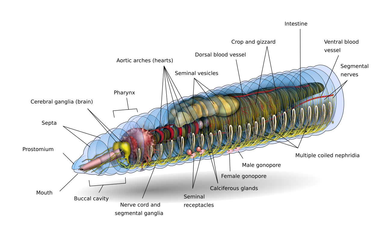
Earthworm Wikipedia
The body of the animal is divided into segments internally and externally called metameres.

Draw well labelled diagram and write the classification of earthworm. Locate the clitellum of a mature earthworm. Pharynx throat passes food from mouth to esophagus 4. Draw a labelled diagram of the longitudinal section of a flower.
Students cut and paste or write words in boxes to label an earthworm diagram. Color the reservoir grey and the flagellum black. The food is engulfed by the earthworm by rhythmic contractions of the pharyngeal wall which further leads to the buccal chamber.
Dissection of Nervous System 3. This will also help you to draw the structure and diagram of earthworm. Great for new teachers student teachers homeschooling and teachers who like creative ways to teach.
EARTHWORM Pheretima It belongs to phylum Annelida. Segments of ody rings around body body is segmented 3. Join the popular membership section.
Jul 12 2012 - Label Earthworm External Anatomy Diagram Printout. External Anatomy of Earthworm. An extended version of the black-and-white diagram is included.
Setae help in locomotion of earthworm. Ventral lood Vessel. Anus - the opening at the end of a worm through which waste exits.
Educators choose from black-and-white or color options. Explain the internal structure of Dicot root with the help of well-labelled diagram and also differentiate between asked May 19 2020 in Internal Structure of. Draw well labelled diagrams in practical notebook.
Earthworm Digestive System in Detail with Diagram. Dissection of Reproductive System. It requires students to draw food and castings while coloring the diagram in addition to labeling the.
Sep 6 2013 - Although not normally a subject I would draw earthworms have become curious creatures to me. A quality educational site offering 5000 FREE printable theme units word puzzles writing forms book report formsmath ideas lessons and much more. In this article we will discuss about the external and internal anatomy of earthworm.
A page on earthworms. Draw well labeled diagrams of amoeba euglena and April 25th 2018 - Draw well labeled diagrams of amoeba euglena and paramecium Discover The Secrets Of Drawing Realistic Pencil draw labeled diagrams amoebaeuglena describe with labelled diagram the structure of qs study may 11th 2018. It ingests food by the pumping action of its pharynx.
Mouth takes in food from soil 2. In this article we will discuss about the dissection of earthworm. Muscular layer in the body wall of an earthworm is made up of only circular muscles.
The clitellum of each species of earthworm has a distinct colour size and shape. An extended version of the black-and-white diagram is included. December 18 2019 by Ranganr.
EARTHWORM DISSECTION DIAGRAM 1. Open 1 answers 1769 views. The Alimentary System 2.
Dissection of Earthworm With Diagram Zoology. They may have any combination of shapes. In earthworm a single male genital pore is present.
Using the definitions listed below label the earthworm diagram. The diagram shows the shape and structure of the clitellum. Another key structure found on the clitellum is the tubercula pubertatis.
An earthworm Digestive System is quite similar to higher animals. They seem like creepy bottom-dwellers but without worms the soil would not be fertile for vegetation. The body of Pheretima is nearly circular in cross-section and varies from 7 to 8 inches 18-19 cms in length.
Draw neat and labelled diagram of longitudinal section of flower. Earthworms are commonly found in soil eating a wide variety of organic matter. Also learn about- 1.
It requires students to draw food and castings while coloring the diagram. Longitudinal section of flower. Clitellum - the enlarged part of the earthworm that contains the reproductive.
An earthworm is a terrestrial invertebrate that belongs to the phylum AnnelidaThey exhibit a tube-within-a-tube body plan are externally segmented with corresponding internal segmentation and usually have setae on all segments. The last segment which contains the anus is called the periproct. Earthworms are delicate animals Fig21 and need careful handling to avoid damage to internal organs.
Ventral Nerve ord like our spinal cord carries nerve impulses 5. Adaptive Features Earthworm lives inside a burrow in. In earthworms there is the presence of a pair of the male genital pore.
Draw a labelled diagram of the longitudinal section of a flower. Typhlosole is the part of the intestine of earthworm. Ive been collecting worms for fishing and composting.
Students cut and paste or write words in boxes to label an earthworm diagram. Chitinous setae which act as locomotory organ are present. Asked apr 16 2015 by shiv 2 208 points tags.
Castings - the waste produced by an earthworm. The general colour.
Helium is the region or the junction where ovule fuses with funicle. In seed plants the ovule is the structure that gives rise to and contains the female reproductive cells.

Sketch And Label V S Of Mature Anatropous Ovule Pre Fertilization In Plant Structure And Events Stamen Microsporangium And Pollen Grain Shaalaa Com
Draw a neat labelled diagram of a mature anatropous ovule before fertilization.
Labelled diagram of mature ovule. In this diagram of a panicle the spikelets are depicted as brown ovals. Draw a well-labelled diagram of a mature ovule showing its internal structure. The integument forming its outer layer the nucellus and the female gametophyte in its center.
It is attached to placenta by a stalk known as funicle. Scroll down for labeled diagrams of the cotton flower bud and the mature cotton flower. The mature ovary is a fruit and the mature ovule is a seed.
Loading DoubtNut Solution for you. 2 Hilum It is the point where the body of the ovule is attached to the funiculus. Draw a neat labelled diagram of a mature anatropous ovule before fertilization.
Draw a neat diagram of plant cell and label any three parts which differentiate it. Iv Chalaza is inverse to the micropylar end speaking to the basal piece of the ovule. When fully mature the graafian follicle breaks open and releases the ovumJun 01 During the ovarian cycle ovarian follicles mature at different stages from primordial follicle to the mature follicle or Graafian follicle at which ovulation occurs and the egg is expelled from the ovary into the fallopian tubes where they wait for fertilization.
Ovary style and stigma. Only a few are actually classified as being fleshy and sweet. The gynoecium represents the female reproductive whorl of the flower composed of carpels.
Ii Each ovule has a couple of defensive envelopes called integuments. Flower Parts Diagram Front And Back View With All Parts Labeled Useful For School Education And Botany Biolog Parts Of A Flower Flower Structure Flower Anatomy. It consists of three parts.
Nucellus is surrounded by outer and inner integuments. 1 Funiculus It is a stalk-like structure which represents the point of attachment of the ovule to the placenta of the ovary. Draw A Well Labelled Diagram Of A Mature Ovule Showing Its Internal Structure Mention The Fate Of All Brainly In.
The nutritive tissue enclosed inside the ovule is the nucellus. Each carpel has three parts viz. A typical ovule is composed of the following organs.
Step by step video image solution for Draw a well-labelled diagram of a mature ovule showing its internal structure. Public domain image at wikimedia commons you may have seen a diagram like this one describing the various parts of a hypothetical flower. The connecting slender stalk between ovule and placenta is known as Funiculus.
Each ovule consists of nucellus surrounded by two integuments and a stalk or funiculus. Apne doubts clear karein ab Whatsapp par bhi. The Pistil Megasporangium ovule and Embryo sac.
Draw a labelled diagram of the longitudinal section of a maize grain to show the structure of mature embryo. The ovule is made up of the nucellus which is the largest part of the ovule the integuments forming the tough outer protective layer of the ovule and the female gametophyte called an embryo sac in flowering plants. With a neat labelled diagram describe the parts of a typical angiosperm ovule.
Draw a labelled diagram of the longitudinal section of mature ovule which is ready forfertilization. Step by step solution by experts to help you in doubt clearance scoring excellent marks in exams. The angiosperm ovule is a small structure attached to the placenta by means of a stalk known as funicle.
Watch 1000 concepts tricky questions explained. With a neat labelled diagram describe the parts of a typical angiosperm ovule. It is a stalk-like formation which represents the point of the accessory of the ovule to the placenta of the ovary.
Find an answer to your question 21. 595 K views 30 K people like this. 3 Integuments They are the outer layers surrounding the ovule that provide protection to the developing embryo.
The female gametophyte specifically termed a megagametophyte is also called the embryo sac in angiosperms. I The hilum is an intersection among ovule and funicle. Answer the ovule is the structure that gives rise to and contains the female reproductive cells.
I Funiculus is stalk-like structure by which ovule is attached to the placenta. By Biology experts to help you in doubts scoring excellent marks in Class 12 exams. The megagametophyte produces an egg cell for the purpose of fertilization.
IiiMicropyle is an opening presentation at the tip where integument is missing. Each ovule consists of nucellus surrounded by two integuments and a stalk or funiculus. Ovule is the female gametophyte develops inside the ovary from a cushion like structure called placenta.
Mature flower diagram clip art wine grape diagram en clip art block diagram visio hierarchy clip art.
Simple typical plant cell diagram. Label diagram of a p.
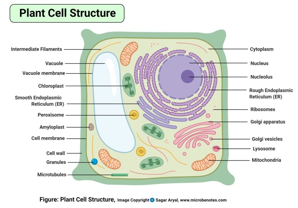
Plant Cell Definition Labeled Diagram Structure Parts Organelles
How To Draw A Plant Cell Plants Botany Easily Quickly Well Labelled Diagram Youtube Plant cell.

Labelled diagram of typical plant cell. About Press Copyright Contact us Creators Advertise Developers Terms Privacy Policy Safety How YouTube works Test new features Press Copyright Contact us Creators. The protoplasm is the living part of the cell. Structure of Plant Cells.
Labelled diagram of Plant Cell. The lipid molecules on the outer and inner part lipid bilayer. Animal and plant cel.
This is the reason why you need to use a microscope to observe a cell. Some plant cells build up very thick secondary cell. Plant cells are eukaryotic cells but unlike animal cells which have a cell membrane plant cells have cell walls.
Understanding Plant Cells Hanford Mills. All the best plant cell drawing with labels 37 collected on this page. And do tell me on.
A labeled diagram of the plant cell and functions of its organelles we are aware that all life stems from a single cell and that the cell is the most basic unit of all living organisms. Illustration of Labelled diagrams of typical animal and plant cells with editable layers. Cell wall is the non-living protective layer outside the plasma membrane in the plant cells bacteria fungi and algae.
A Typical Plant Cell Diagram Labeled Written By JupiterZ Monday October 5 2020 Add Comment Edit. It is made up of chitin lignin and other materials. A labeled diagram of the plant cell and functions of its organelles we are aware that all life stems from a single cell and that the cell is the most basic unit of all living organisms.
A cell is a complete functional biological unit with many different internal structures. A vacuole is a membrane bound structure found in the cytoplasmic matrix of a cell. The synthesis of cell wall in controlled by Golgi bodies.
As the central vacuole shrinks it leaves the cell wall unsupported. The typical characteristics that define the plant cell include cellulose hemicellulose and pectin plastids which play a major role in photosynthesis and storage of starch large vacuoles responsible for regulating the cell turgor pressure. Plant cells contain many organelles.
Stock vector labelled diagrams of typical animal and plant cells with editable layers 222613513 in cell diagram simple the structure and contents of a typical animal cell every has membrane cytoplasm nucleus but not all cells have rat liver cell a scheme of the typical cell membrane structure with lipid bilayer integral proteins. In bacteria the cell wall is composed of protein and non-cellulosic carbohydrates while in most algae fungi and all plant cells the cell-wall is formed of cellulose. Draw a labelled diagram of a typical plant cell.
The cell being the smallest unit of life is. The typical characteristics that define the plant cell include cellulose hemicellulose and pectin plastids which play a major role in photosynthesis and storage of starch large vacuoles responsible for regulating the cell turgor pressure. In this diagram the plant cell is surrounded by a cell wall in this picture just the primary cell wall is shown.
Plant Cell Diagram BasicFigure. Labelled Diagram Definitions and Structure Published by Admin on July 28 2021 July 28 2021. Plant Cell Diagram Clipart.
Plant Cell Diagram Vacuole. Cells Mr Scott S 6th Grade Class. Plant cell showing typical organelles and structures animal cells are somewhat different.
Plants have a rigid cell wall that surrounds the plasma membrane. A Typical Plant Cell Diagram Labeled Fresh Imaging Cell Structure Elodea Leaf Cell Diagram Vmglobalco Plant Cell Wall Diagram Plants Diagrams Exceptional Of A Plant Cell Diagram Labeled With Functions Or Plant And Animal Cell Animal Cell Parts And Functions Withcarbon Cheetah Labeled Diagram Elegant Chapter 5 Notes Plant Cell Structure With Plant Cell Mitosis Diagram Labeled. Download Typical Plant Cell Diagram Labeled Background.
Cell wall provides mechanical support and gives a definite shape to the cell. Simple Plant Cell Drawing At Paintingvalley Com Explore. Labeled plant cell diagram as you can see from the diagram below plant cells contain a nucleus just as labelled diagrams typical animal plant cells stock vector royalty.
A plant cell has a rigid cell wall which is the outermost of the cell. A bacteria diagram clearly enables us to learn extra approximately this single cell organisms that have neither membrane-bounded nucleolus or organelles like mitochondria and. Structure Of A Typical Animal Cell Animal And Plant Cell.
The cell wall is made of cellulose and lignin which are strong and. Labelled diagram of a plant cell under microscope posted on march 18 2011 by admin onion cells stained with methylene blue look at the images of onion cells as they would be seen under a microscope draw each magnification label appear high picture plant and animal cell diagrams vs science for kids. How To Draw Diagram Of Plant Cell Step By Step For Beginners Youtube.
Cytoskeleton golgi apparatus central vacuole chromatin nucleolus smooth endoplasmic reticulum ribosomes rough. Posted by unknown at 2209. Somewhat like an entire city in miniature.
The plant cell is the basic structural and functional unit found the plant cell is rectangular and. The structure of the plant cell is. Vector art clipart and stock vectors.