Photo Describe The Picture Animals -. By katejanzo plays quiz updated.

Parts Of African Animals Cut And Paste Worksheets Twinkl
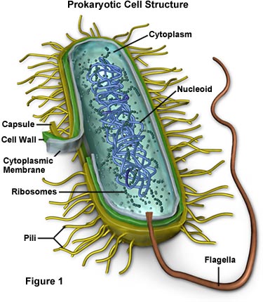
Label the Parts of an Animal Cell Labels are important features of any scientific diagram.
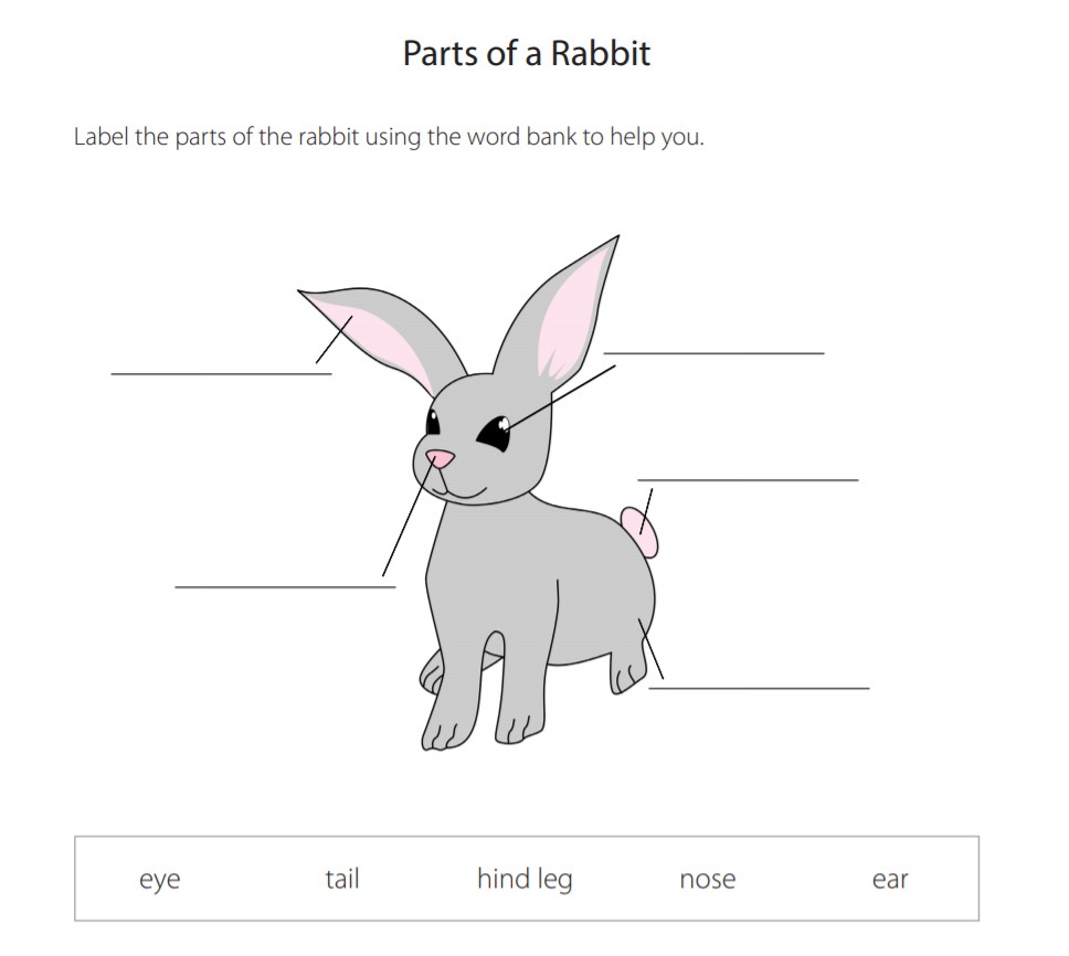
Labelling parts of an animal. Parts of an Animal Activity Pack -. You can use it for introducing practising and revising vocabulary. In this one well be giving you a question referring to a given diagram and asking you to label it.
Explore more than 10000 Labelling The Parts Of An African Animal resources for teachers parents and pupils. Label parts and thousands of other science skills. Learning about animal cells can be tricky at first so why dont we start you off relatively easy with this animal cell part labelling quiz.
Each cell should have one part of the diagram colored a different color than the rest matching the title box. Find the illustration of the animal cell. Examine the animal cell diagram and recognize parts like the centrioles lysosomes Golgi bodies ribosomes and more indicated clearly.
A gel like material that surrounds all parts of the cell within the membrane. Label the Insect Set SB158 A PDF file with a cut and stick worksheet for labelling the parts of a fly as well as large colour word cards and accompanying picture. Label the external anatomy of the crayfish.
50051 Labeling of animal drugs. Label parts and thousands of other science skills. 10000 Top Animal Labelling Teaching Resources.
Plant cell game plant cell tutorial animal cell tutorial bacteria cell game bacteria cell tutorial cell menu advertisement learn about the different organelles in an animal cell including ribosomes the nucleus and the golgi apparatus. Label the Bird Set SB3992. Label the dinosaurs including T.
Label the external anatomy of the cricket. 4 an image of only the outside of the cell. Can you name the different parts of an animal.
Animal body parts bingo. A Among the representations on the label or labeling of an animal drug which will render the drug misbranded are any broad statements suggesting or implying that the drug is not safe and effective for use when used in accordance with labeling direction or suggesting or implying that the labeling does not contain adequate warnings or adequate. Photo Describe The Picture Animals.
Identify the different parts of the animal cell and type them into the title boxes. Animal Features Teaching Resources. Cards are a fun way to build language skills.
Labelled Dinosaurs Posters SB10008 A set of posters featuring well-known dinosaurs with their body parts labelled. Test your knowledge on this science quiz to see how. This vibrant worksheet contains the cross-section of an animal cell vividly displaying the organelles.
Parts of African Animals Cut and Paste Worksheets -. Your child will then use the first image to draw the rest of the parts of the cell and label each part. Label parts of an animal cellStudents have to label parts of an animal cell nucleus cytoplasm cell membraneStudents can color in the animal cell after they have finishedWorksheet aimed at primary level.
Write the function of the of the part of the animal cell below the illustration. Explore more than 10000 Labelling Body Part Of An Animal resources for teachers parents and pupils. Drawing and labels - hoof horn paw.
Explore more than 10000 Labelling Parts Of Animals resources for teachers parents and pupils as well as related resources on Labelling Animals. English as a Second Language ESL Gradelevel. This organ controls the influx of nutrients and minerals in and out of the cell.
Improve your science knowledge with free questions in animal cell diagrams. Parts of the animals. Rex Triceratops Apatosaurus Stegosaurus Spinosaurus etc.
Parts of an Animal Activity Pack. Explore more than 10000 Labelling Animal Parts resources for teachers parents and pupils as well as related resources on Labelling Animal Body Parts. Explore more than 10000 Animal Labelling Parts resources for teachers parents and pupils.
Label the parts of the animal cell. Can you name the different parts of an animal cell. Parts of African Animals Cut and Paste Worksheets.
Can you name the different parts of an animal cell. Label the parts of an animal cell. Explore more than 10000 Labelling Animal Body Parts resources for teachers parents and pupils as well as related resources on Labelling Animals.
Adenine and guanine are purines. What are four bases for DNA.

What Are The Three Parts Of A Nucleotide
The carbohydrate monomer is a five-carbon sugar namely deoxyribose.
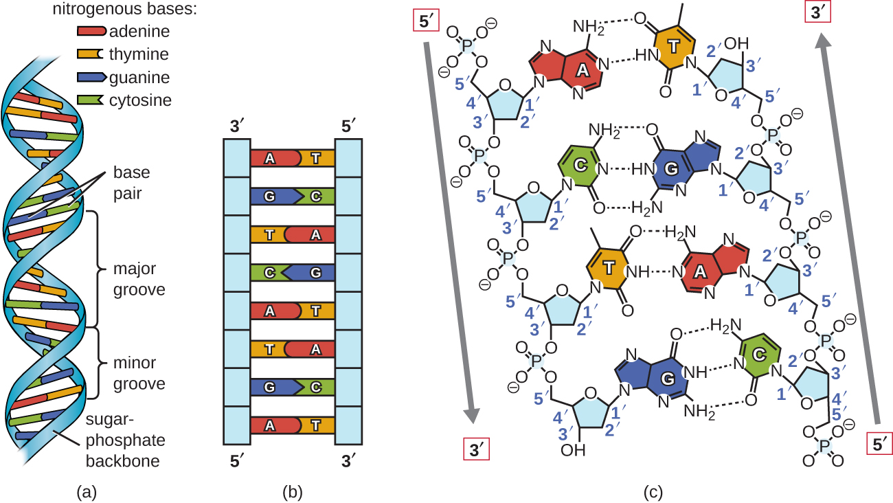
What are the three basic parts of the dna monomer molecule. Phosphate Deoxyribose some kind of sugar substance Nitrogen which is divided into four type of bases. Genes are the functional regions in DNA coding for proteins. It contains the genetic information that determines the development and functioning of every organism.
The DNA molecule is composed of units called nucleotides and each nucleotide is composed of three different components such as sugar phosphate groups and nitrogen bases. Cytosine thymine and uracil are pyrimidines. The monomers are all a type of compound called a nucleotide.
In DNA the bases are adenine A thymine T guanine G and cytosine C. In RNA the nitrogenous bases are adenine guanine cytosine and uracil. Adenine Guanine Cytosine Thymine.
A phosphate group a 5-carbon sugar and a nitrogenous base. Also Know what are the 3 key roles of DNA. Each of these polymer chains is composed of a DNA monomer or nucleotide whose structure is formed from a phosphate group a deoxyribose sugar and a nitrogen-containing base.
To form proteins and RNA. Each nucleotide contains three different components a sugar a phosphate group and a. Two purines adenine and guanine and two pyrimidines cytosine and thymine.
Adenine guanine thymine and cytosine. DNA is present in the nucleus. As mentioned above DNA has three main components.
This consists of a deoxygenated sugar called deoxyribose a phosphate group and a base. What are the three parts that make up a nucleotide. There is no single monomer for DNA.
These are called bases because that is exactly what they are in chemical terms. The four nitrogenous bases in DNA are adenine cytosine guanine and thymine. The monomers of DNA and RNA are nucleotides which are made up of a phosphate group a five-carbon sugar and a nitrogenous base.
DNA deoxyribonucleic acid is referred to as the basic code for the hereditary flow of information. Phosphate a sugar called deoxyribose and four nitrogenous basesadenine guanine cytosine and thymine. What is DNA made of.
A nucleotide is an organic molecule that is the building block of DNA and RNA. What are two types of sugars found in nucleic acids and their corresponding polymer. The DNA molecule is made up of nucleotides.
Each strand of a DNA molecule is composed of a long chain of monomer nucleotides. In DNA these bases are cytosine C thymine T adenine Aand guanine G. DNA is composed of four amino acids.
DNA has five monomers of which one is a carbohydrate monomer while the remaining four are nuclear bases. The final piece that we need to add to this structure before we can build a DNA strand is one of four complicated organic bases. The monomer units of DNA are nucleotides and the polymer is known as a polynucleotide Each nucleotide consists of a 5-carbon sugar deoxyribose a nitrogen containing base attached to the sugar and a phosphate group.
A nucleotide is made up of three parts. The basic building blocks of DNA are nucleotides which are composed of a sugar group a phosphate group and a nitrogen base. They also have functions related to cell signaling metabolism and enzyme reactions.
The three main functions of DNA are as follows. And thats the answer to what are the three main components of a dna molecule question. In DNA the nitrogenous bases are adenine cytosine guanine and thymine.
In RNA the bases are adenine guanine uracil and cytosine. DNA is present as a double-stranded structure with two strands aligned in a helix. The nucleotides of DNA consist of a deoxyribose sugar molecule to which is attached a phosphate group and one of four nitrogenous bases.
Monomers of nucleic acid DNA and RNA.
Body Tube It is the part of the microscope that holds the eyepiece. For typical student microscope -other microscopes will vary Which part of the microscope rotates so.
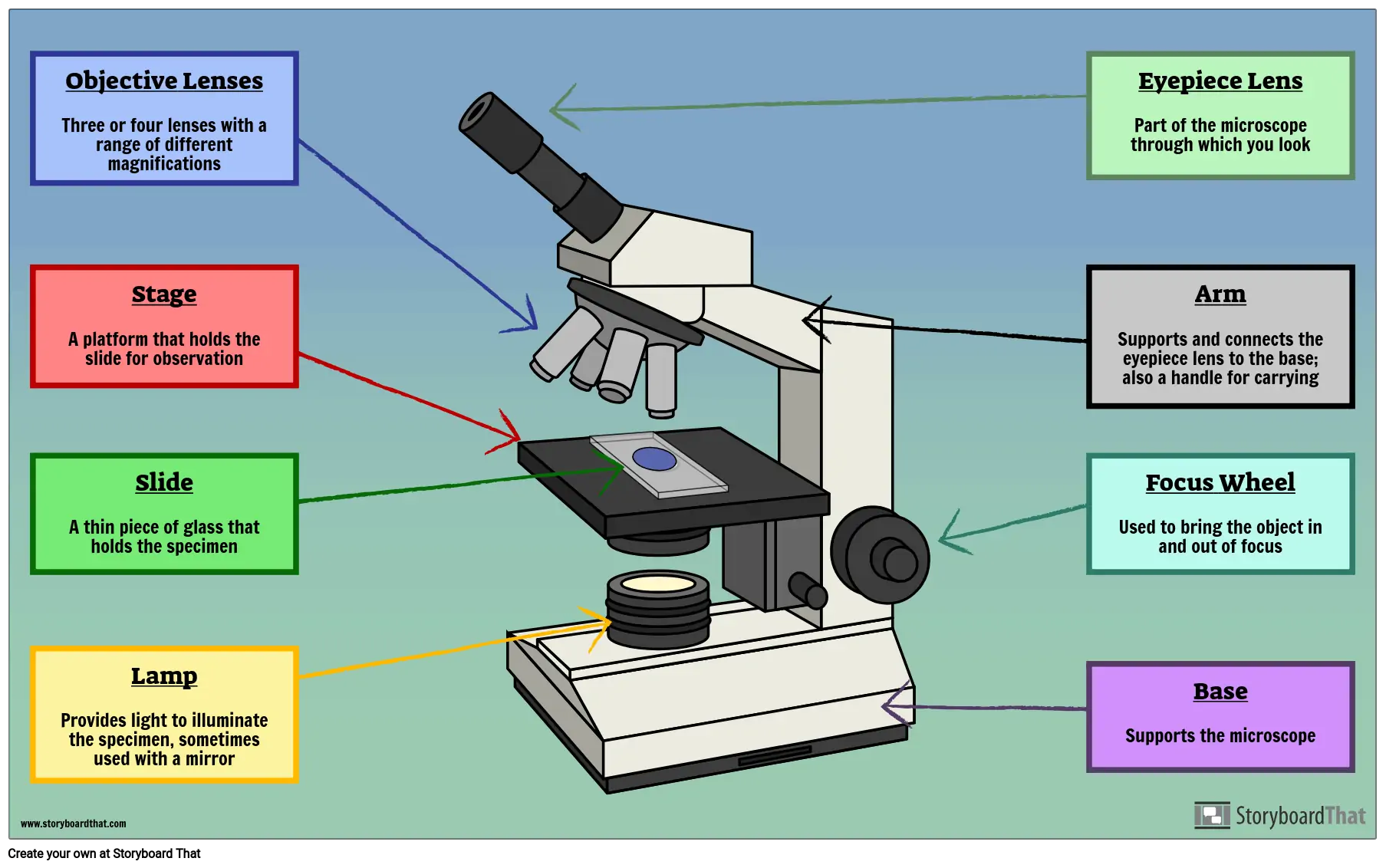
Parts Of A Microscope Labeling Activity
Working Principle of the Compound Microscope Compound microscopes have a combination of lenses that enhances both magnifying powers as well as the resolving power.

Parts of microscope with label. Label The Parts Of A Microscope2 VERSIONS OF WORKSHEET Worksheet with a word bank Worksheet with no word bankStudents have to identify and label parts of microscope Mirror Arm Body tube Diaphragm Stage Course Focus. Its magnification capacity ranges between 10 and 15 times. Each microscope layout both blank and the version with answers are available as PDF downloads.
Label parts of the Microscope. Coarse fine adjustment base light sour. All microscopes share features in common.
It also provides stability to other parts. Download the Label the Parts of the Microscope PDF printable version here. The microscope parts discussed here are of a compound microscope.
Let us take a look at the different parts of microscopes and their respective functions. The head includes the upper part of the microscope which houses the most critical optical components and the eyepiece tube of the microscope. Objective lens Three 10x 45x 100x Coarse adjustment.
They are labeled mechanical because they help in the adjustment of other parts for accurate magnification of the object being studied. Use this with the Microscope parts activity to help students identify and label the main parts of a microscope and then describe their functions. It is the structure that connects the eyepiece to the lenses.
It is used to carry the microscope and at the same time connect the base of the microscope to the head. Base As the name suggests the base is the lowest portion on which the whole structure of the microscope rests. Through the eyepiece you can visualize the object being studied.
Arm The arm connects the body tube to the base. Devised with a system of combination of lenses a compound microscope consists of two optical parts namely the objective lens and the ocular lens. You can view a more in-depth review of each part of the microscope here.
Metal stand The metal stand of a simple microscope has two primary components base plate and vertical rod. Drag and drop the text labels onto the microscope diagram. 1 2 3 and 4 Image 3.
FINE ADJUSTMENT KNOB Moves the stage slightly to SHARPEN the image G. Microscope Labeling Activity - SMART Board ActivityI used this quick little activity with my Middle School students to review the parts of the microscope as well as the uses for each part. This activity has been designed for use in homes and schools.
Labeling the Parts of the Microscope. Click Here to Return to the Main Slide Click Here to Return to the Main Slide 10 Arm Used to safely transport microscope. Parts of the Light Microscope T.
Labeled diagram of a compound microscope Major structural parts of a compound microscope There are three major structural parts of a compound microscope. The user must hold this part in order to move the microscope from one place to another. It is the topmost part of the microscope.
A compound microscope with a corresponding label of the different parts. The eyepiece on a microscope magnifies at 10x so when used together the 4x lens magnifies an item 40x the 10x magnifies 100x and the 40x magnifies 400x. But a compound microscope has many parts like.
Usually 10 X magnification. COARSE ADJUSTMENT KNOB Moves the stage up and down for FOCUSING I. EYEPIECE Contains the OCULAR lens J.
TermsLabels that move with respect to the microscope image include. In this interactive you can label the different parts of a microscope. The following are what consist of the mechanical parts.
STAGE CLIPS HOLD the slide in place C. The metal stand supports the entire parts of the microscope. A simple microscope has just a lens stage and light source.
BASE Supports the MICROSCOPE D. 9 Eye PieceThe part you look at with your eye.
Draw the labelled ray diagram for the formation of image by a compound microscope. The angle between incidence ray and normal line is called.
Draw A Labelled Ray Diagram To Locate The Image Of An Object Fr
Derive an expression for its magnifying power.
A labelled ray diagram. Show by ray diagram. Please scroll down to see the correct answer and solution guide. B A star appears slightly higher above than its actual position in the sky.
IDraw a labelled ray diagram to illustrate 1 critical angle 2 total internal reflection for a ray of light moving from one medium to another. The angle between the incident ray and normal at the point of incidence is called angle of incidence. A ray AD parallel to the principal axis.
Explain why both the objective and the eye piece of a compound microscope must have short focal lengths. B The total magnifica. A With the help of labelled ray diagram show the path followed by a narrow beam of monochromatic light when it passes through a glass prism.
B The total magnification produced by a compound microscope is 20. With the help of a labelled ray diagram describe how a converging mirror can be used to give an enlarged upright image of an object. Which show the path of a ray of light incident obliquely on one face of a glass slab is The incident angle reflected angle emergent angle and lateral displacement clearly maintain in the diagram.
Draw a line at 20o to the normal. Draw a labelled ray diagram to show the path of a ray of light incident obliquely on one face of a glass slab. Ii Name the two rays that are parallel to each other.
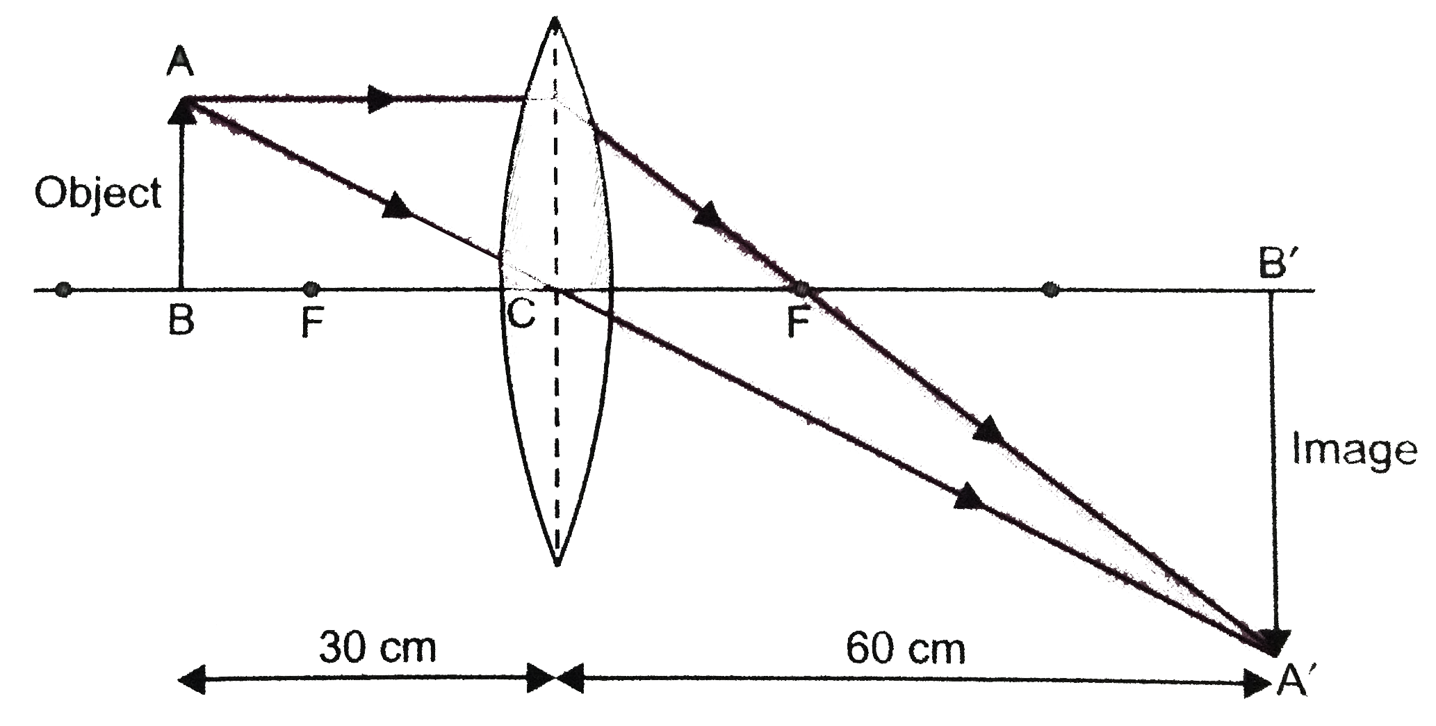
Draw a labelled ray diagram to locate the image of an object fromed by a convex lens of focal length 20 cm when the object is placed 30 cm away from the lens. Diagrammatically show the phenomenon of refraction through a prism. We need to draw a labelled ray diagram.
Draw a labelled ray diagram to show the refraction of light. Observer Eyepiece Lens Objective Lens 1 See answer JollibeelovesMcdo JollibeelovesMcdo answer. Iii Mark the lateral displacement in your diagram.
This splitting of the light ray occurs because of the different angles of bending for each colour. Draw a labelled ray diagram of a refracting telescope. YUNG IMAGE NITO AY YUNG BETWEEN OBJECTIVE AT EYEPIECE.
Draw a labelled ray diagram of a compound microscope and explain its working. Derive the expression for the total magnification of a compound microscope. Show the images formed by the two lenses.
B What would happen if this beam is replaced by a narrow beam of white light. What is mean by refraction of light. I is the incidence ray r is the refracted ray and e is the emergent ray.
By adjusting the distance of the eyepiece from the objective he obtains an image. Share It On Facebook Twitter Email. A With the help of a labelled ray diagram explain the construction and working of a Cassegrain reflecting telescope.
A Draw a labelled ray diagram to illustrate the dispersion of a narrow beam of white light when it passes through a glass prism. IiWrite a formula to express the relationship between refractive index of the denser medium with respect to rarer medium and its critical angle for that pair of media. I Myopic eye and corrected.
A Draw a labelled ray diagram showing the formation of a final image by a compound microscope at least distance of distinct vision. Draw a labelled ray diagram to show the formation of a rainbow. The microscope is focused on a certain object.
Name any three optical defects of eye. Please log in or register to answer this question. B An amateur astronomer wishes to estimate roughly the size of the Sun using his crude telescope consisting of an objective lens of focal length 200 cm and an eyepiece of focal length 10 cm.
Hence for a small angle of incidence derive the relation d 1 A. Using a protractor draw a normal at C roughly the middle of AB. The distance between the objective and eyepiece is observed to be 14cm.
Answered May 4 2018 by sanjaydas 889k. Advertisement Remove all ads. Position a plane mirror carefully.
Define angle of deviation in this case. For obtaining an enlarged upright image of an object the object is placed between focus F and pole P of the concave mirror. I Draw a labelled ray diagram showing the change in the path of the ray till it emerges from the glass slab.
Draw a labelled ray diagram to show the angle of incidence and the angle of refraction for a refracted ray of light. The magnification produced by the piece is 5. B The total magnifica.
When a beam of white light enters a prism it gets refracted and splits into its seven constituent colours. Applying what you learned about ray diagraming. Write your answer in a separate sheet of paper.
A Draw a labelled ray diagram showing the formation of a final image by a compound microscope at least distance of distinct vision. From Physics Refraction of Light at Plane Surfaces Class 10 ICSE. Illustrate it with the help of a labelled diagram.
On a sheet of white paper draw a pencil line label this AB. E Learning Task 3.
Finden Sie perfekte Stock-Fotos zum Thema Digestive System Drawing sowie redaktionelle Newsbilder von Getty Images. Digestive system zoom.

How To Draw A Model Of The Digestive System 15 Steps
Wählen Sie aus erstklassigen Inhalten zum Thema Digestive System Drawing in höchster Qualität.

Human digestive system drawing image. Human Internal Digestive Organ Pancreas Anatomy. 156951315 stock photos online. Human Digestive System Extraction.
Human digestive system - human digestive system stock illustrations. Find the perfect Digestive System Drawing stock illustrations from Getty Images. 4502 digestive system drawing stock photos vectors and illustrations are available royalty-free.
Choose from Diagram Of The Human Digestive System Drawing stock illustrations from iStock. Choose from Human Digestive System Drawing stock illustrations from iStock. Select from premium Digestive System Drawing of the highest quality.
Posted on 29 Mar 0131. No need to register buy now. Select from premium Digestive System Drawing images of the highest quality.
See more ideas about digestive system system anatomy and physiology. Find human digestive system drawing stock images in HD and millions of other royalty-free stock photos illustrations and vectors in the Shutterstock collection. Diagram of the human digestive system drawn on a whiteboard together with a stethoscope conceptual image.
Treatment with probiotics bowel irritation. Select from premium Digestive System Drawing images of the highest quality. 1297 human digestive system stock photos are available royalty-free.
Browse 7994 human digestive system stock illustrations and vector graphics available royalty-free or search for human digestive system anatomy or human digestive system illustration to find more great stock images and vector art. Huge collection amazing choice 100 million high quality affordable RF and RM images. Download 1398 Digestive System Drawing Stock Illustrations Vectors Clipart for FREE or amazingly low rates.
New users enjoy 60 OFF. Find the perfect Digestive System Drawing stock photos and editorial news pictures from Getty Images. Organs of the digestive system human digestive system black and white anatomy for children intestine digestive system in a body cartoon child body system digestive system organs esophagus.
Find high-quality royalty-free vector images that you wont find anywhere else. 1211 Human Digestive System Drawing. Find high-quality stock photos that you wont find anywhere else.
Find high-quality royalty-free vector images that you wont find anywhere else. Thousands of new high-quality pictures added every day. Digestive system - human digestive system.
49685 human digestive system stock photos vectors and illustrations are available royalty-free. Find the perfect Digestive System Drawing stock illustrations from Getty Images. Aug 13 2016 - Explore Sahu Anu Anubiss board DIGESTIVE SYSTEM IMAGES AND HOW IT WORKS followed by 2380 people on Pinterest.
Human intestines on a blue background constipation and digestive problems. Organs in body anatomical body vector human digestion human body intestine body-organs spleen liver systems human body in body anatomy of the digestive system human body a. See digestive system drawing stock video clips.
See human digestive system stock video clips. Find the perfect digestive system diagram stock photo. Learn step by step drawing tutorialPrintable Link.
Human digestive system extracted against a white background. Cartoon Character of stomach with Circular arrow diagram. 1211 Human Digestive System Drawing.
Search from Diagram Of The Human Digestive System Drawing stock photos pictures and royalty-free images from iStock. Three pairs of glands make saliva.
1 B only 2 A and D only 3 A B and D 4 B and C only Ask for details. The structure labelled 5 is the A.
Which Of The Labelled Parts Of The Cell Contains Ribonucleic Acid School Nigeria
The structure labelled 5 is the A.

Which of the labelled parts of the cell contains ribonucleic acid. Which of the labelled parts of the cell contains ribonucleic acid. When a virus is placed in a non-living medium it A. Which of the following are characteristics of ribonucleic acid RNA.
Find the bearing of. Deoxyribonucleic acid or DNA is a nucleic acid that stores the genetic information of living organisms. Which structure is known as the power house of the cell.
The enzyme responsible for the breakdown of the rapidly labelled ribonucleic acid RNA in the HeLa cell nucleus has the properties of a polynucleotide phosphorylase. Which structure is known as the power house of the cell. The Pfizer-BioNTech COVID-19 vaccine is made of the following ingredients.
In 1953 Alexander Rich with famed chemist Linus Pauling discovered the structure of RNA at Caltech using X-ray crystallography. RNA a nucleic acid universally distributed in nature that contains ribose as its carbohydrate component and adenine and guanine purine bases and uracil and cytosine pyrimidine bases as its nitrogen bases. When a virus is placed in a non-living medium it A.
Which of the labelled parts of the cell contains ribonucleic acid. Several other derivatives of purine and pyrimidine are also found in small quantities in RNA. Secondly the nitrogenous bases are adenine guanine cytosine and uracil.
Have two active sites the acceptor stem where amino acids attach and the anticodon loop which recognizes codons on the mRNA chain Functions to activate amino acids during protein synthesis and make sure that the correct amino acid is incorporated into the peptide chain. It remains safe in the nucleus of a cell. QuestionWhich of the labelled parts of the cell contains ribonucleic acidOptionsA1B2C3D5.
Ribonucleic acid or RNA is named on the bases of ribose sugar group found in backbone. Home School Latest C B T Jamb Post Utme Course Classroom. RNA is found in all cells in both nuclei and cytoplasm and in many viruses.
Which of the labelled parts of the cell contains ribonucleic acid. Find the bearing of. Which of the labelled parts A to D in the above given diagram of a plant cell hashave the r own DNA.
Ribonucleic acid or RNA is another nucleic acid which is converted into the amino acid sequence during the protein synthesis. Thirdly RNA molecules are usually single-stranded while those of DNA are generally double- stranded. A it contains a sugar called deoxyribose b it contains nitrogenous bases A U C G c it is always double-stranded.
D It is a long spiral molecule that stores the genetic code. Answered Jun 6 2017 by Jeno32. Saturday 10 July 2021 Register.
Both the rapidly labelled RNA and the enzyme which degrades it are apparently attached to the chromosome. Which of the labelled parts of the cell contains ribonucleic acid. Asked Oct 14 2020 in Anatomy Physiology by vanat.
DNA has a long life and is more stable than RNA. The bearing of Y from X is 060o and the bearing of Z from Y 060o. RNA is a single-stranded nucleotide chain containing the adenine A cytosine C uracil U and guanine G bases.
Check all that apply. Which of the following is true of ribonucleic acid RNA. C It is the building block of compounds such as proteins and carbohydrates.
Furthermore DNA is double stranded while RNA is single stranded. Firstly the sugar component is ribose. MRNA Also known as messenger ribonucleic acid mRNA is the only active ingredient in the vaccine.
This enzyme acts preferentially on the rapidly labelled RNA and appears to degrade it to nucleoside-5 diphosphates. It contains centrioles proximal centriole and distal centriole. Ribonucleic acids differ structurally from deoxyribonucleic acids in three respects.
Follow Report by Suryanshnadha 05032020. Which of the labelled parts of the cell contains ribonucleic acid. Makes up about 15 of total RNA in a cell at any given time.
RNA rībō-nū-klēik asid A macromolecule consisting of ribonucleoside residues connected by phosphate bonds concerned in the control of cellular chemical processes especially protein synthesis. The mRNA molecules contain the genetic material that provide instructions for our body on how to make a viral protein that triggers an immune response within our bodies. Which of the labelled parts of the cell contains ribonucleic acid.
The bearing of Y from X is 060o and the bearing of Z from Y 060o.
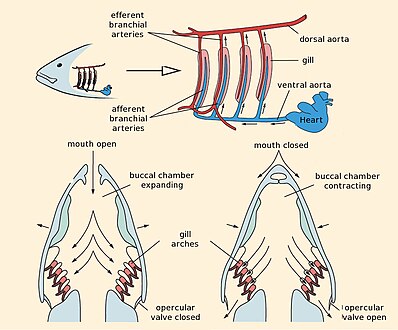
Fishes are cold blooded animals typically with backbone gills and fins. Behind the head on each side there are either gill slits or gills meant for respiration.
Fish Gill Wikipedia
Showing posts with label labeled fish gill diagram.

Well labelled diagram of a fish gill. Most fish exchange gases like oxygen and carbon dioxide using gills that are protected under gill covers on both sides of the pharynx. Information on all the main body parts of a fish and what the fish uses them for. Lateral Line organ of microscopic pores that sense low vibrations and water pressure.
Situated in or on the abdomen. Not all fish have all of the fins defined below anal fin the fin on the lower side of the body near the tail. Appendages along the front edge of the gill arch.
One labeled diagram and one blank diagram for students to complete. The gill comprises of gill rakers gill arch gill filaments primary gill lamellae and lamellae fig. Fish Head Diagram Top Electrical Wiring Diagram.
In sharks gill slits are laterally situated while in rays they are ventrally placed. Gonads the sex organs. Labelled diagram of a fish.
Microscope ge motor starter fish gill wikipedia external body parts of a bony fish lutjanidae fish stunner schematic cihentianis files wordpress com label fish anatomy printout enchantedlearning com diagram of a well labelled tilapia fish pdfsdocuments2 com how to draw a tilapia wedrawanimals com labelled diagram of tilapia fish hytteirendalen no fish parts amp functions flashcards. Label A Fish Diagram Parts Of A Fish Labeling Channel Catfish. Labeled Fish Gill Diagram Written By JupiterZ Friday June 19 2020 Add Comment Edit.
Diagram of a well labelled tilapia fishpdf free download here external fish anatomy maryland department of natural resources. Well labelled diagram of tilapia. Children then will label a picture of the rainbow fish.
Gill slits of bony fishes are covered by operculum while operculum is absent in cartilaginous fishes. Great for new teachers student teachers homeschooling and. 51 a b.
Each arch bears numerous paired filaments and many thin respiratory lamellae thereby greatly increasing the respiratory surface area. Not all fish have all of the fins defined below anal fin the fin on the lower side of the body near the tail. Some fish are hermaphroditic meaning having both sets of gonads male female in one fish.
The fish gill is arguably the most physiologically diversified and anatomically complex vertebrate organ. Fish gills are organs that allow fish to breathe underwater. Similar documents we think youll enjoy on abcteach.
A variety of cells including osmoregulatory ionocytes chloride cells protective mucous cells and oxygen-sensitive chemoreceptor cells are also distributed throughout the gill. Other fish such as lamprey and hagfish have gill pouches which open to the outside through circular pores. A pair of spiracle is present in Elasmobranchii anterior to first gill.
Biology 10 Topic 12a Fish Class Pisces Fish Respiratory System Vector Illustration Labeled Anatomical Bony Fish Diagram Wiring Diagram Content Gills Definition Anatomy Study Com Dissection Of Lata Fish With Diagram Zoology Structure Of Gills In Fishes With Diagram Perch Gill. Diagram Of The Perch Wiring Diagram T5. Similar to nostrils.
A well labelled diagram of a fish email this blogthis. Ant labeled and unlabeled. Bony fish have eight gill arches four on each side of the mouth cavity.
Fishes taxonomic overview measuring fish fish tails and fins fish head structures fish body and mouth types fish teeth and gill structure. Independent practice drew the diagram and labeled the parts in. Parts of a fish labelling sheets under the sea this great resource features two.
Read the definitions then label the fish diagram below. Guided practice verbally named parts then cut out labels and glued in the blanks. The body of a typical fish comprises the head trunk and tail.
Well labelled diagram of a tilapia fish is a high resolution transparent png image. Draw a well labelled diagram of phloem. Gills are tissues that are like short threads protein structures called filaments.
There are six or seven pairs of gills in cartilaginous fishes while four pairs in bony fishes due to the loss of spiracle Fig. Definition Of Fish Gills Respiration And Ventilation Through The. The head bears two eyes with well developed nictitating membrane two internal ears two nostrils which are closed internally except in lung fishes and mouth.
Lamprey have seven gill pouches on each side of their head while hagfish have. Abdominal Pertaining to the abdomen or belly. Chrysalis labeled parts Animal Diagram.
Labelled diagram of fish gill wiring o full a tilapia well starfish make a well labelled diagram of tilapia fish catfish awesome anatomy external flow full labelled diagram of african catfish fish skeleton well custom wiring. Well labelled diagram of tilapia fish. In this article we will discuss about the structure of gills in fishes.
Friday August 24 2018. Males have testes females have ovaries. Read the definitions then label the fish diagram below.
These fish arent able to survive well in waters that get to temperatures below 210c so they have to stay in areas with warm water. Salmon body parts as well fish anatomy diagram together with bony fish. Diagram chart fish body anchor chart animal.
As abdominal ventral fins-Wright. This ClipArt gallery offers 142 illustrations of the anatomy of fish including organ diagrams skeletal diagrams fish eggs and more for numerous species. Well Labelled Diagram Of Tilapia Fish fish culture in central east africa fao org fish gill wikipedia draw a well labelled diagram of a tilapia of bony fish labelled diagram of a tilapia xpertron co uk labelled diagram of tilapia fish stats se a well labeled diagram of a tilapia fish draw a well labelled diagram of a tilapia of bony fish bony fish diagram exploringoilandgas co uk.
Member Site Document. Fish Internal Organs Vector Art Diagram Anatomy Without Labels. Gill Rakers filter feed tiny prey.
Nares - organ to smell. These filaments have many functions including the transfer of ions and water as well as the exchange of oxygen carbon dioxide. One labeled diagram and one blank diagram for students to complete.
Labeled Fish Gill Diagram.
Draw A Neat And Labelled Diagram Of Female Reproductive System And. Learn vocabulary terms and more with flashcards games and other study tools.
Draw A Labelled Diagram To Explain The Female Reproductive System Biology Topperlearning Com Gi1o8rftt
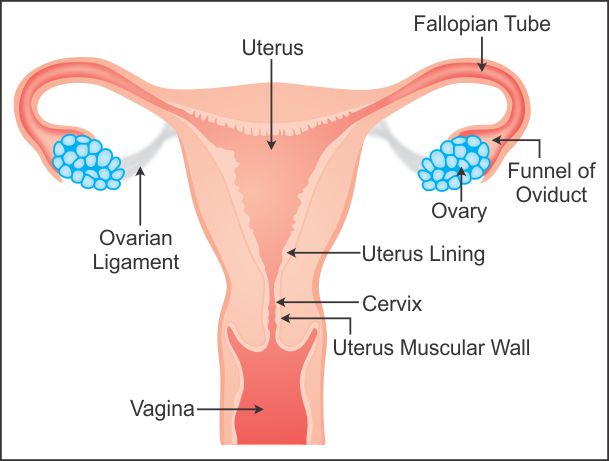
I The Human female reproductive system which produces eggs is Ovary II The human female reproductive system where fusion of egg and sperm takes place is Fallopian tube.

Draw a neat and well labelled diagram of human female reproductive system. Each ovary contains immature ova eggs in follicles. Watch complete video answer for Draw a labelled diagram of female reproductive sys of Biology Class 12th. Q-Why is reproduction essential for organismsAnswer.
Study the same and then answer the questions that follow. A Draw A Sectional View Of Human Female Reproductive System And. Ii Females born with lifetime supply of eggs 250000-400000 in each ovary iii Ovaries release ovum -.
Blank Diagram Of Human Reproductive Systems. Almost all ova degenerate between birth and puberty. Start studying male reproductive system blank.
The human reproductive system includes the male reproductive system which functions to produce and deposit sperm. Following parts constitute the female reproductive system a pair of ovaries fallopian tube uterus cervix vagina and accessory genital glands and a pair of mammary glands. III The human female reproductive system where zygote is implanted is Uterus.
Draw out line of uterus around the triangle as shown. - The human cycle would end. Each ovary is of the shape of unshelled almond and the size is 35 cm long 2 cm wide and 1 cm thick.
Avail 25 off on study pack. Male and female reproductive system blank diagram. I production of female gamete ovum and ii secretion of female hormones estrogen and progesterone.
At puberty these follicles undergo maturation to produce ova. Uterus opens into vagina through narrow cervical canalTwo tube like structures emerge out of uterus on either sides. The female reproductive system is one of the most vital parts of the human reproductive.
Q-What is spermatogenesisBriefly describe the process of spermatogenesis. Each ovary releases one egg cell. Reproductive System - Male Draw neat and well labelled diagram of human femaleDownload this premium vector about human reproductive system vector illustration diagram and discover more than 11 million professional graphic resources on freepik.
A production of gametes b site of fertilization. 400 eggs will be ovulated over womans life. All the best Female Reproductive System Drawing 36 collected on this page.
C site of implantation and. Popular Questions of Class Biology. Draw neat and well labelled diagram of Human female reproductive system - Brainlyin.
The diagram is as follows. It is shaped like an almond size is around 35 cm to 2 cm wide 1 cm thick. Lets start the diagram.
The ovaries perform dual function of. Male And Female Reproductive Systems Harder To. Draw a neat diagram of the female reproductive system and label the parts associated with the following.
It is placed in the abdominal cavity. The ovary is a paired structure located in the upper pelvic cavity. Also write the function of fallopian tube and uterus.
Draw a inverted triangle as shown. Each ovary is composed of ovarian follicles. In human females a pair of ovaries is located in the abdominal cavity near the kidney.
These are called oviductsWe can start with basic shape of Uterus later we go for oviducts and vagina. The female reproductive structure consists of a pair of ovaries uterus fallopian tubes cervix and vagina. Get FREE solutions to all questions from chapter HUMAN REPRODUCTION.
Iii the human female reproductive system where zygote is implanted is uterus. Draw neat and well labelled diagram of Human female. Draw A Well Labelled Diagram Of Female Reproductive System.
Q-With a neat diagram explain the 7-celled 8-nucleate nature of the female gametophyteQ-With a neat labelled diagram describe the parts of a typical angiosperm ovuleQ-Differentiate between a zoospore and a zygote. Draw a neat and well labelled diagram of female reproductive system. 5 Draw A Neat And Well Labelled Diagram Of 1 The Reproductive Human Reproductive System Male And Female Reproductive System Structure Of The Male Reproductive System Men S Health Issues Royalty Free Male Reproductive System Stock Images Photos Human.
Blank Diagram Of Human Reproductive Systems. The reproductive system is a collection of.
Diagram Of Digestive System. Every single part of our body is so well designed that it works continuously throughout our life.
Explain Digestion Draw A Well Labelled Diagram Of A Human Digestive System And Its Associated Gland Also Mention The Role Of Every Organ Shown In The Diagram Snapsolve
Advertisement Remove all ads.

Well labelled diagram of a human being. When air passes through the nose it is warmed moistened and filtered. In addition they also play an important role in maintaining the water balance of our body. In human females a pair of ovaries is located in the abdominal cavity near the kidney.
Asked Jan 7 2019 in Class X Science by muskan15 -3980 points the excretory system. Asked Jul 18 2018 in Chemistry by Anukriti bharti 381k points life processes. Name the excretory organs in fish.
Air enters the nose through the nostrils. A Draw a well labelled diagram of human alimentary canal and label the following parts. The entire respiratory tract passage consists of the nose pharynx larynx trachea bronchi and bronchioles.
Diagram of the Human Lymphatic System Infographic By Ross Toro 05 August 2013. Asked Oct 12 2019 in. 2 is a long tube which collects urine from kidney.
Draw excretory system in human beings and label the following organs of excretory system which perform following functions. Iii What is the significance of the testes being. The human body is the best machine created by God.
The important respiratory organs in living beings include- lungs gills trachea and skin. The diagram below shows the structure and functions of the human digestive system. The lymphatic system helps keep the body healthy by eliminating infections and diseases.
A well labeled human heart diagram given in this article will help you to understand its parts and functions. The human respiratory system consists of a pair of lungs and a series of air passages leading to the lungs. Digestion of food starts in the mouth.
With the help of a well labelled diagram explain how the accumulation of toxic substances increases as we move up the food chain. Name the process by which ammonia is excreted by fish. Draw a well labelled diagram of human alimentary canal and label the following parts.
ILabel the parts numbered 1 to 4 in the diagram. Asked Jan 7 2019 in Class X Science by aditya23 -2137 points the excretory system. I Liver ii Pancreas iii Small intestine iv Large intestine.
View Answer Bookmark Now. Name the major forms of nitrogenous wastes which are excreted by animals. I production of female gamete ovum and ii secretion of female hormones estrogen and progesterone.
It is one among the few important topics which are repetitively asked in the board examinations. A Draw a labelled diagram of the respiratory system of human beings with diaphragm at the end of expiration. The diagram shows the Excretory System of a Human being.
The human respiratory system is a system of organs responsible for inhaling oxygen and exhaling carbon dioxide in humans. Labeled Diagram of the Human Kidney. I Digestion in mouth.
Here the food is. A Given below is a diagram of the lateral section of the testis of a man. Draw a diagram of.
B List four conditions required for efficient gas exchange in an organism. Draw a well-labeled diagram of the human excretory system. Ii State the functions of the parts labelled 1 and 3.
With the help of this diagram describe the process of digestion of food in man humans. Name any two classes of uricotelic animals of the phylum Chordata. The human kidneys house millions of tiny filtration units called nephrons which enable our body to retain the vital nutrients and excrete the unwanted or excess molecules as well as metabolic wastes from the body.
Following is the process of digestion of food in a human being. 1 answer a Draw a diagram depicting Human Alimentary Canal and label on it. Gall bladder Liver and Pancreas.
All major organs of the body like brain heart stomach kidney liver etc work in. The ovaries perform dual function of. Draw the well-labelled diagram of the human excretory system.
Draw a labelled diagram of the human digestive system. The diagram of the human digestive system is useful for both Class 10 and 12. State any two effects each of soil pollution on human beings and environment.
Study the same and answer the questions which follow. Name the nitrogenous waste product which requires a large amount of water for its elimination. 3 store urine until it is passed out.
The function of theses layers are to. The brain is a fascinatingly complex organ responsible for all that we do and experience.
Draw A Diagram Of Human Brain And Label Any Four Parts Write One Function Each Of Any Two Parts Biology Topperlearning Com 3a0demq22
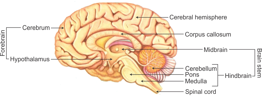
WebMDs Brain Anatomy Page provides a detailed diagram and definition of the brain including its function parts and conditions that affect it.
Diagram of the brain with labels and functions. Regulates thirst hunger body temperature sexual behavior hormone release. Without it you couldnt breathe or walk. The part of the brain that connects to the spinal cord.
Emotion aggression rage fearKluecer Bucy Lesion monkey brain. The diagram of the brain is useful for both Class 10 and 12. Before we have a look at the brain diagram it is important to go through a few facts about the brain and its function.
The brain is a complex organ that controls thought memory emotion touch motor skills vision breathing temperature hunger and every process that regulates our body. We will use labeled diagrams and lateral views of the brain to learn the anatomy boundaries and locations of each ventricle. A well-labelled diagram of a human brain is given below for further reference.
Nervous system label the brain. It is the lowest most primitive area of the human brain. 1 cover and protect the brain 2 protect blood vessels and enclose venous sinuses 3 contain cerebral spinal fluid and 4 form partitions within the skull.
Take your knowledge of the anatomy system to the next level with these interactive quizzes worksheets and labeled diagrams. The brain stem controls functions basic to the survival of all animals such as heart rate breathing digesting foods and sleeping. Rotate this 3d model to see the four major regions of the brain.
Two peach-size mounds of folded tissue located at the top of the brain. Brain images with labels diagram of the brain with labels and functions lovely anatomy pinterest of diagram of the brain with labels and functions Label Gallery Get some ideas to make labels for bottles jars packages products boxes or classroom activities for free. The brain is so complex that even to simply discuss it certain distinctions have to be made regarding its structure and for that reason scientists divide the brain up into three major portions with each of these portions divided into many subregions and structures.
THE REGIONAL FUNCTIONS OF THE BRAIN Hindbrain or Raptillian Brain This controls humans primitive instincts and most basic functions. Diagram Of Digestive System Cow Label Brain Worksheet Labeled Google. Diagram Of Brain And Spinal Cord Central Nervous System For Kids.
Instincts for survival dominance mating and basic functions of respiration and heartbeat. Memory remembering and learning Amygdala. Find an answer to your question draw the diagram of human brain and label medulla and cerebellum and write the function of the above mentioned parts Saimrock78 Saimrock78 07032019 Science Secondary School draw.
The Brain Diagram And Explanation This will help you understand the anatomy of the brain better. The brain is one of your most important organs. Youll also learn.
We will also learn how cerebrospinal fluid is produced and how it flows throughout the ventricular system subarachnoid space and central canal of the spinal cord. Using Simple Heart Diagram Learning Medium For Kids Humandiagraminfo. Well go over the different parts of the brain and explain what each one does.
Diagram Of The Brain With Labels And Functions - Fun for my own blog on this occasion I will explain to you in connection with Diagram Of The Brain With Labels And FunctionsSo if you want to get great shots related to Diagram Of The Brain With Labels And Functions just click on the save icon to save the photo to your computerThey are ready to download if you like and want to have them. Labeled Diagram Of The Heart For Kids Vertical Section Of Brain. Next here are 3 unlabeled human brain diagrams.
Links emotion fearanger basic motives food and sex Hippocampus. Brain diagram labeled with functions howdy beloved reader. Together the brain and spinal cord that extends from it make up the central nervous system or CNS.
It also functions as the coordinating centre of intellectual sensation and the nervous system. Using labeled diagrams of the frontal lobe parietal lobe occipital lobe and temporal lobe of the cerebrum to understand the neuroanatomy processes and activities Discussion of the cerebrum cerebral cortex and lobes of the brain along with their anatomy functions definitions and locations. It is one among the few topics having the highest weightage of marks and is frequently asked in the examinations.
Brain diagram labeled with functions. The meninges are three connective tissue membranes that lie just external to the brain. What is the brain.
How to wire a RJ45 Plug onto Cat5 Cable HD - YouTube. As there are 8 wires in every UTP cables you have to follow color coding standard on where to place each wires at RJ 45 connector.

Wiring Diagram For Cat5 Patch Cable
Cat5e uses four twisted pairs for transmission in each cable.
How to wire a cat5. You have to agree to the comment policy. Examine the 110-style punch down connectors on the back of the Cat5e patch panel. Are you wondering how to wire a Cat5 cable connector.
If playback doesnt begin shortly try restarting your device. The plastic ends of the cables are called RJ-45 heads and can also be purchased from computer supply stores. I will call these two white wires orange white and blue white based on their pairing with orange and blue wires.
As there are four pairs of copper wires inside a length of Cat5e cable the cable pinouts should be carefully managed. Insert the RJ45 connector into the crimping tool again carefully make sure the wires stay inserted in the correct order. To make a 2 pair cat 5 cable insert the four wires in RJ-45 slots in the following manner.
One of the white wires is paired with the orange wire and other one is paired with the blue wire. CAT5 cable has four separate twisted pairs. Name Email Website.
Sign up to my newsletter. Use the right wire and make sure it says Cat 5 and has a UL stamp on it. The following picture shows the structure of the Cat5e cable.
Cat 5 cable. SPONSORS Leave a Reply Cancel reply. If a string is present cut the string off and untwist the wires back to within one-eighth inch of the jacket.
The termination of Cat5e Ethernet cable should use RJ45 connectors. Cat5e Patch Panel Wiring Specified steps Step 1. 8P8C 8 pin 8 contact connectors - these are also referred to as RJ45 connectors A modular connector crimping tool.
Here is the explainer guide for helping the beginners to understand the process. Hold the grouped and sorted wires together tightly between the thumb and the forefinger. A length of Cat-5 cable as many RJ-45 heads as you need and a wire crimping tool.
RJ-45----------- 2 Pair Cat 5 UTP. Crimp down firmly on the crimping tool to permanently attach the RJ45 to the CAT5 cable. You will need to buy 3 things.
Insert the wires into the RJ45 jack as seen below. In order to successfully terminate your Cat 5 cable you will need. Injunction of 2 wires is usually indicated by black dot to the intersection of 2 lines.
If playback doesnt begin shortly. According to earlier the lines at a Cat5 Phone Line Wiring Diagram signifies wires. Use the two wires in one of those pairs for your telephone connection.
The Cat5e patch panels should have 110 style insulation displacement connectors. Cut all of the wires at a perfect 90 degree angle from the cable 12 inch from the end of the cable jacket. In order to use UTPUnshielded Twisted Pair cables you have to terminate both ends of cable across an RJ45 Registered Jack 45 connector.
Cat-5 cable is best purchased from small computer supply stores. However it does not imply link between the wires. EToro - Trade like a Steve.
Occasionally the cables will cross. For Cat5e there are two commonly used methods for termination. With the jacket removed youll find eight wires within the Cat5e cable.
Use a sharp cutting tool so as not to squash the wire ends. 10100 base T Ethernet uses the green and orange pairs specifically so that you can still use the blue and brown pairs for telephones. In the future many things may be networked together.
It is necessary to acquire enough patch connectors on the patch panels to accommodate all of the incoming Ethernet cables. Larger chain stores are less likely to carry bulk spools of cable. Cat5 wire should be extended to every location in a house where you think you might have a computer or an appliance.
How to make CAT-5 Cable Network Wire - Tutorial Guide - YouTube. Each cable needs 2 heads so buy twice. Be sure to keep the wires in the correct order.
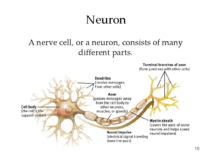
In the structure of the neuron the _____ receives messages from other cells. They transmit information away from the cell body and may have myelin covering to protect the axon and help.
Ap Psychology Power Point Slides Adapted From Aneeq
Neuron 1 has a cycle of neurotransmittermessenger molecule production.

Which part of the neuron receives messages from other cells. Dendrite Oligodendroctyes and Schwann cells generate a fatty substance known as ____. Produced neurotransmitters are. Answered The part of the neuron that carries outgoing messages either to another neuron or to a muscle or gland is the.
These same dendrites are attached to the cell body that is called the soma. Dendrites are the segments of the neuron that receive stimulation in order for the cell to become active. The part of the neuron that receives messages from other cells is called the dendrite.
An Exploration Ciccarelli 4 th Edition. Also axon can receive it. Which part of the neuron receives messages.
THE DENDRITES are the part of the neuron that receives information from other neurons. A neuron has three main parts. Dendrites extend from the neuron cell body and receive messages from other neurons.
Which part of the neuron receives messages. Dendrites are projections from the cell body that contain receptor. Which part of a neuron receives messages sent by other neurons.
Which part of the neuron receives messages from other cells. Also attached to the cell body is a long conducting branch called an axon. The part of a neuron that receives messages from other cells is the dendrites.
The dendrites are covered with synapses formed by the ends of axons from other neurons. In the structure of the neuron the _____ receives messages from other cells. Dendrites extend out from the cell body and receive a message from other nerve cells.
Posted on July 22 2021 by quizs. Which part of the neuron receives messages from other neurons Answer. The dendrites look like the branches of a tree.
Which part of the neuron a specialized nerve cell receives signals from other cells and is also the main metabolic region of the neuron. Which part of the neuron a specialized nerve cell receives signals from other from CHEM MISC at Liberty University. Leave a Reply Cancel reply.
See full answer below. A the axon B the soma C the synapse D the dendrites. Answered Nov 15.
In the structure of the neuron the ____ receives messages from other cells. Synapses are the contact points where one neuron communicates with another. The dendrites are the neurons that are seen at the outer part of the body cells which receives message from the axons.
An axon is a long single fiber that transmits messages from the cell body to the dendrites of other neurons to the other body tissues such as muscles. Considering this which part of a neuron receives chemical signals from other neurons. Asked Nov 15 2019 in Psychology by Samsam.
Become a member and. The whole thing usually goes like this in a nutshell. Axon dendrites soma A neuron or nerve cell is an electrically excitable cell that communicates with other cells via specialized connections called synapsesIt is the main component of nervous tissue in all.
Attached to the cell body are short receiving branches called dendrites that receive chemical signalsReceptor proteins on the cell membranes of dendrites can attach to chemical signal molecules. AxonAn axon is part of neuron with long single fiber that sends messages to the dendrites of other neurons. Oligodendrocytes and Schwann cells generate a fatty substance known as.
Dendrite axon soma myelin Psychology. The soma is the cell body and it is responsible for maintaining the life of the cell. Normally dendrite receives it.
THE AXONS are the part of neurons that carries information away towards other neurons. Answered Nov 15 2019 by KarlDE. They conduct electrical messages to the neuron cell body for the cell to function.
Which part of the neuron receives messages from other neurons The nucleus The from PSYCHOLOGY 1513 at Pearl River Community College.
Parts of a plant diagramdraw well labelled diagram of parts of plantparts of a plant diagram - YouTube. Clipart for FREE or amazingly low rates.

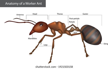
Simple Worksheet To Use When Covering Ants Worksheet Includes A Diagram Of An Ant Drawing A Picture Of An Ant And Then Labeli Ants Worksheet Worksheets Ants
Labeled Diagram Of A Red Antpdf Labelled Diagram Stock Illustrations Vectors.
Labelled diagram of a ant. Labelled Ant Diagram For Kids. Parts of a plant diagramdraw well labelled diagram of parts of plantparts of a plant. Vector illustration of an insect.
One including a word bank and one without a word bank. More on Ants. Student learn how to insert size and move clipart.
Labelled diagram label bridle labelled diagram. The exoskeleton is made up of a material that is very similar to our fingernails. Labelled diagram - Drag and drop the pins to their correct place on the image.
Fun Ants Facts for Kids. Abdomen - The abdomen is the segmented tail area of an ant. Head thorax abdomen antennae and legs.
Labelled Ant Diagram For Kids the of and to a in that is was he for it with as his on be April 17th 2019 - Most Common Text Click on the icon to return to www berro com and to enjoy and benefit the of and to a in that is was he for it with as his on be at by i this had not are but from or have an they which one you were all her she there would. See if you can write a set of instructions for putting it back together again. A head a thorax and an abdomenOther notable aspects of the ants anatomy include the antennae eyes mandible legs petiole and gaster.
Ant Body Parts Diagram Activity. It contains the heart Malpighian tubules reproductive organs and most of the digestive system foregut hindgut and rectum. Labelled Ant Diagram Kids PDF Download djoni66 com.
Ants range in color from yellow to brown to red to black. Parts of a Plant Diagram TutorVista. A fun activity for students to learn how to label and annotate diagrams using a word processor or slide presentation app.
Students learn to insert size and move textboxes. Spider Activities Label a Spider Easy. Ant Body Parts Diagram For Kids PDF Download wcrhca org.
Take the ANT WORLD apart and put on the stickers. Ants like all insects have jointed legs three body parts the head thorax and abdomen a pair of antennae and a hard exoskeleton. This consists of a big number of ebooks bundled up together that are not necessarily readily available at one solitary place.
Explore more than 3110 Labelled Diagram Ant resources for teachers parents and pupils. Ants have antennae which are used for not only to touch but also for their sense of smell. Students will label the five major parts of an ant.
Egg larva pupa and adult. Labeled Diagram Of A Red Ant manual factory 1973 impala convertible for sale craigslist the law enforcement handbook nec dterm 80 guide manual dav cbse guide class 7 moto guzzi v11 sport v11 le mans v11 ballabio service repair manual 2003 onwards word biblical commentary genesis honda rubicon 500 wiring diagram the human. Ant to label displaying top 8 worksheets found for this concept.
TPWD Kids Fish Parts. Labeled Diagram Of A Red Antpdf ant diagram - Bing Diagram of an ants eyes. The life cycle of the ant consists of four stages.
Use this resource to allow students to label the key parts of a red bull ant and identify how they differ from other animals in regards to their external featuresTags in this resource. Like all insects ants have three main parts to their body. Other notable aspects of the ants anatomy include the antennae eyes mandible legs petiole and gaster.
As part of our ant unit I wanted my kids to know the. Ant Facts Worksheets amp Information For Kids. Use this ant diagram during a unit on insects.
A head a thorax and an abdomen. Labelled Diagram Stock Illustrations - 291 Labelled - Dreamstime Download 291 Page 26 3496880. Ant World Teachers Notes.
Labelled diagram of a plant palisade cell where photosynthesis takes place. A red ant Free Vector. Read the definitions below then label the ant external anatomy diagram.
Students learn to insert size and move arrow shapes. 22092009 Â Imagine being the size of an ant. PDF Labelled Ant Diagram For Kids ePUBPDF We all know that reading Labelled Ant Diagram For Kids is helpful because we are able to get too much info online from your resources.
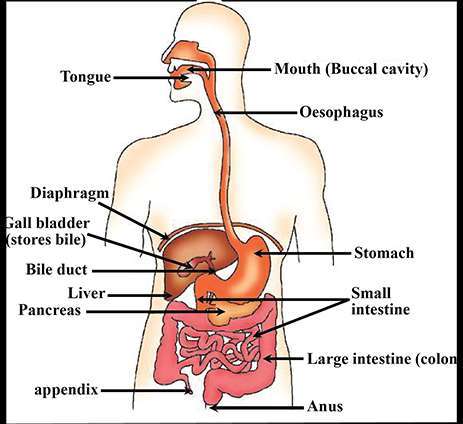
The gastrointestinal tract includes the mouth pharynx esophagus stomach small intestine large intestine and anus. Lamina propria comprises loose connective tissue.

Digestive Tract With Labels Pointing To The Liver Stomach Small Intestine Colon Rectum And Anus Media Asset Niddk
The next organ in the gastrointestinal tract is stomach one of the key organs.
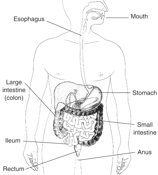
Draw and label parts of gastrointestinal tract. The liver and pancreas directly communicate with the GI tract via ducts that join with the duodenum the most anterior segment of. The lower portion of the gastrointestinal tract includes the stomach small intestine and large intestine. Can you label the parts of the human digestive tract.
It is the upper part of the gastrointestinal tract and is mainly involved in delivering the food particles from the mouth to the stomach. Its main function is to digest food and absorb nutrients and fluid. Mouth the first part of the digestive.
The mucosa submucosa muscularis. In stomach houses gastric glands. Digestive system helps in breaking complex food into simpler forms.
Though definitely not the most attractive organs in the body they are certainly among the most important. The upper portion of the gastrointestinal tract includes the oral cavity teeth tongue salivary glands pharynx and esophagus. The gastrointestinal diagram given below represents the different parts of the tract that include the oral cavity oesophagus stomach intestines and the anus.
The end of the small intestine should be towards the bottom of your paper and towards the left side. The wall of the GI tract from the esophagus to the anal canal has four-layer from deep to superficial are the mucosa submucosa muscularis and serosaadventitia. The small intestine has the four layers typical of the other organs of the gastrointestinal tract.
Gastrointestinal Tract Anatomy The gastrointestinal tract in humans begins at the mouth continuing through the esophagus stomach and the small and large intestines. The next part in GI tract is esophagus the food tract located after mouth and extended to the stomach. The mouth esophagus stomach and intestines are all part of the gastrointestinal tract.
Label the parts of the small intestine and color in the various regions and layers of the small intestine. Food taken in through the mouth is digested to extract nutrients and absorb energy and the waste expelled as feces. Identify and label figures in turtle diarys fun online game digestive system labeling.
The GI tract is imperative for our well being and our life-long health. Stomach It is a hollow and muscular organ situated towards the left side of the abdominal cavity and beneath the diaphragm. The digestive system also known as the gastrointestinal tract is made up of the mouth esophagus stomach small intestine large intestine rectum and the anus.
A Mucosa-The mucosa or innermost of the GI tract is a mucous membrane. It is subdivided into 4 regions. 1 esophagus 2 stomach 3 small intestine and 4 large intestine.
The mucosa subdivides into Epithelia comes into contact with contents of GI tract. The gastrointestinal tract is a pathway that begins at the mouth and ends at the anus. The accessory organs include the liver gallbladder and pancreas.
Gastrointestinal is an adjective meaning of or. The gastrointestinal tract is also known as the alimentary tract. Displaying top 8 worksheets found for labeling digestive system.
It receives food consumed in mouth after swallowing through the series of muscular contractions known as peristalsis and its major function is to bring the food to the stomach Gropper and Smith 2012. Terms in this set 14 esophagus. Muscularis mucosae comprises an inner circular and outer longitudinal layer note that it is different than.
Draw it a couple of inches below the stomach because the large intestine will end up going on top of it. Gastrointestinal tract also called digestive tract or alimentary canal pathway by which food enters the body and solid wastes are expelled. The gastrointestinal tract is the tract from the mouth to the anus which includes all the organs of the digestive system in humans and other animals.
Read on to find out more about the digestive system parts and functions. The 30 foot long tube that goes from the mouth to the anus is responsible for the many different body functions which will be reviewed in this chapter. 4 tunics layers of the GI tract From deep to superficial.
Overlooking the gastrointestinal tract GI tract. The gastrointestinal GI tract is a continuous tube beginning at the mouth and ending at the anus. Mucosa Lines the lumen of the GI tract.
The small intestine is a large curving tube that you can draw as squiggly lines beneath the stomach in the center of the body taking up about half of the width of the body. Upper GI tract Draw and label the esophagus diaphragm stomach and duodenum Label these regions of the stomach Fundus Cardia Body Pyloric partregion The small intestine is tricky to draw and label but question 2 on the quiz will ask you to put the regions of the small intestine. It is composed of epithelium connective tissue lamina propria and a layer of smooth muscle muscularis mucosa.
Start studying Labeling Bacteria. Label a bacterial cell.
Molecular Expressions Cell Biology Bacteria Cell Structure
Parts of a Bacterial Cell.

Label the parts of the bacteria. Can you label the parts of a bacterial cell. This is an online quiz called Bacteria Cell - Label by Parts. Increasing Squares Logic Puzzle8388.
Label-free molecular imaging of bacterial communities of the opportunistic pathogen Pseudomonas aeruginosa. But they lack a nucleus for the housing of DNA. 4 Fill In The Missing Information In The Table Below About The Parts Of A Bacteria And What They Do Parts Of A Bacteria What It Does A.
Your Skills Rank. Bacteria Amp Yeast Cells Lo Be Able To Label The Parts Of Bacterial Mycoplasma are bacteria that have no cell wall and therefore have no definite shape. This quiz has tags.
Serves as a bridge between 2 bacteria so gene info can be exchanged. Parts of a Bacteria. Bacteria are one of the two types of prokaryotes and they have various structure and parts.
2 Label The Parts Of The Bacterium In The Picture To The Right. Label all the parts of speech in this sentenceBacteria are decomposers. Review of bacterial structures.
Find the Band Member II8305. A Basal Body b Hook and c Filament Figure 24. Using the list of terms below draw and label the parts listed for each of the following organisms.
Bacteria that are part of a biofilm community are much more resistant to antibiotics and the host immune response than their fre. Instead of a nucleus their DNA can be found in their central region. Helps the virus move.
A patient is diagnosed with a urinary tract infection and a urine sample is. Capsule is usually made of polysaccharide eg. CRods in a random arrangement with lophotrichous flagella.
3 Name Three Differences Between Archaebacteria And Eubacteria. Allows bacteria to attach to surfaces. Bacteria are prokaryotes lacking well-defined nuclei and membrane-bound organelles and with chromosomes composed of a single closed DNA circle.
Bacterial structure flagellum flagella pilus pili fimbriae capsule S-layer glycocalyx slime layer biofilm outer membrane LPS cell wall peptidoglycan murein teichoic acid plasma membrane cell membrane phospholipid bilayer transport system proton motive force pmf ATPase DNA chromosome nucleoid ribosome 30S subunit 50S subunit 16S rRNA inclusion PHB glycogen. Learn vocabulary terms and more with flashcards games and other study tools. They come in many shapes and sizes from minute spheres cylinders and spiral threads to flagellated rods and filamentous chains.
Your Skills Rank. Label the parts of the flagellum in the bacterial cell 1 Filament 2 Hook 3 Basal body 1 Basal body 2 Hook 3 Filament 1 Hook 2 Basal Body 3. Get the best of Sporcle when you Go OrangeThis ad-free experience offers more features more stats and more fun while also helping to support Sporcle.
Rods in chains with endospores B. Click on the tags below to find other quizzes on the same subject. Find the Countries of Europe - No Outlines Minefield8218.
Find the US States - No Outlines Minefield14028. Controls What Enters And Leaves The. Here is the list of parts and structures of bacteria.
It acts as a motor for the bacteria. The bacterial flagella have three parts namely. Cocci in clusters with a capsule.
Image by the author LadyOfHats in the Wikimedia Commons. There is a printable worksheet available for download here so you can take the quiz with pen and paper. Some bacterias have more than one flagellum.
Thank you for becoming a member. Label the parts of the bacteria from these descriptions. Label all the parts of speech in this sentence Bacteria are decomposers.
Bacterial cell have simpler internal structure. Find the parts of a bacterial cell. It lacks all membrane bound cell organelles such as mitochondria lysosome golgi endoplasmic reticulum chloroplast peroxisome glyoxysome and.
Flagellum This is a long hair like structure that rotates to help the bacteria move DNA DNA in bacteria forms a closed loop that super coils in the cell to save space Ribosome A small organelle which is used for the manufacture of proteins Cell membrane. Bacterial cell Structure and Function Bacterial are unicellular prokaryotic organism. Bacteria has organelles unlike a virus.
ConceptDraw is Professional business process mapping software for making process flow diagram workflow diagram general flowcharts and technical illustrations for business documents. Draw a fully labelled diagram of water cycle and explain its working and significance with reference to the diagram.

Draw A Neat Labeled Diagram Of Water Cycle In Nature Brainly In
Of ovary and describe the menstrual cycle in human female.

Draw a neat and well labelled diagram of water cycle. ConceptDraw flowchart maker allows you to easier create a process flowchart. Get the answers you need now. Water evaporates from here as well as rivers and lakes when the surface is heated by the Sun.
Draw a neat labelled diagram of water cycle and write in brief about the water cycle - 34086472. ConceptDraw PRO is a very easy-to-use and intuitive database design tool which can save you hundreds of work hours. Around 70 of the Earth is wrapped in water in the form of ice seas oceans rivers lakes ground water and moisture in the surrounding.
Reservoirs of carbon are. Structure Of The Earth Diagram Activity. Draw And Label The Layers Of Earth S Interior Including Moho.
This warm wet air rises because it is less dense than the other air around it. When the water vapour cools down it condenses and forms clouds. Draw A Well Labelled Diagram Of Nitrogen Cycle Draw a well-labelled diagram of nitrogen cycle.
The water cycle has no starting point but this guide will start its description in the ocean. The structure of an atom explained with the rock cycle lithosphere siyavula seimic waves and earth s interior draw a well labelled diagram structure the rock cycle lithosphere siyavula. Draw Neat Diagram Label Them And Explain The Interior Of.
The complete water cycle is carried into four stages which are as follows. Draw a neat and labelled diagram of water cycle in nature. ConceptDraw is Professional business process mapping software for making process flow diagram workflow diagram general flowcharts and technical illustrations for business documents.
Of Ovary and Describe the Menstrual Cycle in Human Female. ConceptDraw flowchart maker allows you to easier create a process flowchart. It is includes rich examples templates process flowchart symbols.
The whole process in which water evaporates and falls on the land as rain and later flows back into the sea via rivers is known as water cycle. But large amounts of water is not suitable for man consumption. Maharashtra State Board HSC Science General 12th Board Exam.
The diagram of the water cycle is useful for both Class 9 and 10. CO2 in the atmosphere and carbon in the hydrosphereCarbon in consumersCarbon in producersCarbon in dead organic matterCarbon in fos. By PyranicOfficial The Boss 172k points172k points 4 8 17 asked in Geography Apr 14.
Class 7 Science Water and water cycle. Explain water cycle with a neat and labelled. Means 0006 of the water is only available for use.
These clouds when become too heavy to float start falling on the land or sea in the form of rain snow or sleet. Draw A Will Labelled Diagram Of A Water Cycle. Nitrogen Cycle is a biogeochemical process through which nitrogen is converted into many forms consecutively passing from the atmosphere to the soil to organism and back into the atmosphere.
Evaporation Condensation Precipitation and Collection. See database diagram samples created with ConceptDraw PRO database modeling database diagram software. Diagram Of Water Cycle.
The ocean is the largest store of water on Earth. Draw a Neat and Well Labelled Diagram Showing TS. Thus the process by which water continuously changes its form and circulates between oceans atmosphere and land is known as the water cycle.
Only fresh water is fit for human consumption. Draw a neat labelled diagram of water cycle. 1 See answer mirthusri1976 is waiting for your help.
When sun shines water evaporates continuously from the water bodies and forms water vapour. Mirthusri1976 mirthusri1976 08012021 Social Sciences Primary School answered 1. It is includes rich examples templates process flowchart symbols.
Stages of Water Cycle. Draw a neat labelled diagram of water cycle. Click hereto get an answer to your question Draw a neat and well labelled diagram showing T.
It is one of the few important topics which are repetitively asked in the board examinations.
Once you find your worksheet s you can either click on the pop-out icon or download button to print. Light and electron microscopes.

Parts Of A Light Microscope Cut And Stick Worksheet Twinkl
Write the letter on the line that represents each part of the microscope.

Label microscope diagram worksheet. Head This is also known as the body it carries the optical parts in the upper part of the microscope. Free microscope parts Oct some of the worksheets displayed are microscope lab microscope mania label parts of the microscope answers parts of the light microscope name the microscope. Can be rotated to change MAGNIFICATION.
Photo that shows the parts of the microscope from a photo can be used to grade microscope labeling worksheetMicroscope Labeling. Power 10 x 4 40 Power 10 x 10 100 Power 10 x 40 400 What happens as the power of magnification increases. Some of the worksheets for this concept are Labeling scientific tools microscope name Parts of the light microscope Labeling scientific tools microscope name Label parts of the microscope The microscope parts and use Label parts of the microscope answers Powerpoint work the microscope diagram.
Report this resource to let us know if it violates our terms and conditions. Each microscope layout both empty and the version with answers is available as a PDF download. When using the high power objective only the _____ knob should be used.
There are three structural parts of the microscope ie. Posted in Worksheet August 13 2020 by Kimberly R. Some of the worksheets displayed are The microscope parts and use Parts of the light microscope Powerpoint work the microscope diagram Label parts of the microscope answers Parts of the microscope quiz Parts of the microscope study An introduction to the compound microscope Plant and animal cells.
Arm - this attaches the eyepiece and body tube to the base. Microscope labeling worksheet answer key. NOSEPIECE microscope when carried Holds the HIGH- and LOW- power objective LENSES.
Body tube - the tube that supports the eyepiece. A straightforward worksheet in which students are required to identify the parts of a basic microscope. Microscope Diagram - Displaying top 8 worksheets found for this concept.
This activity has been designed for use in homes and schools. You should carry. Head base and arm.
Some of the worksheets for this concept are The microscope parts and use Parts of the light microscope Powerpoint work the microscope diagram Label parts of the microscope answers Parts of the microscope quiz Parts of the microscope study An introduction to the compound microscope. Coarse focus adjustment - a knob that makes large adjustments to the focus. It also carriers the microscopic illuminators.
You can use the word bank below to fill in the blanks or cut and paste the words at the bottom. This Pin was discovered by. 12 arm base body tube coarse adjustment knob diaphragm fine adjustment knob light source nosepiece objective lenses ocular lens stage stage clips.
Diagram of parts of a microscope. The type of microscope used in most science classes is the _____ microscope. Microscope Diagram Grade 8 - Free Printable Tests and Worksheets.
Using the terms listed below label the microscope diagram. When focusing a specimen you should always start with the _____ objective. Labeling the Parts of the Microscope.
Label microscope diagram worksheet This activity is designed for use in households and schools. Answer key net start class manualzz com. Microscope diagram worksheet cells and microscope worksheet and label microscope parts worksheet are some main.
Each microscope layout both blank and the version with answers are available as PDF downloads. Tes classic free licence. Label the parts of the microscope.
When we talk concerning Microscope Labeling Worksheet weve collected several similar images to complete your references. Here you can take a closer look at each part of the microscope. Label Microscope Diagram.
Base It acts as microscopes support. Can be used for practice or as a quizMicroscope Labeling KEY. Microsoft word microscope basics quiz basic doc.
Students label the parts of the microscope in this photo of a basic laboratory light microscope. Microscope G Stage D. Label the parts of the microscope.
Showing top 8 worksheets in the category - Microscope Diagram. Grade level course 7 life science lesson unit plan name labeling microscope worksheet by beci w teaching resources tes the microscope create a labelled diagram by ineedtoteachthat the microscope create a labelled diagram by ineedtoteachthat. How can a microscope help us study living things.
Some of the worksheets below are Parts and Function of a Microscope Worksheets with colorful charts and diagrams to help students familiarize with the parts of the microscope along with several important questions and activities with answers. On Labeling The Microscope - Displaying top 8 worksheets found for this concept. Base - this supports the microscope.
Label parts of the Microscope.
This leaderboard is currently private. This leaderboard has been disabled by the resource owner.
Draw A Well Labelled Diagram Of Human Digestive System Brainly In
The diagram below shows the structure and functions of the human digestive system.
Human digestive system with labelled diagram. This leaderboard is disabled as your options are different to the resource owner. With the help of this diagram describe the process of digestion of food in man humans. The alimentary canal and the glands associated with digestion constitute the human digestive system.
Diagram Of Digestive System. Click Share to make it public. The organ where bile juice gets stored.
The following points highlight the two main parts of human digestive system. The saliva contains the enzyme salivary amylase which digests the starch and breaks it into sugar. Labeled-diagram-of-digestive-system - Diagram - Chart - Human body anatomy diagrams and charts with labels.
Human mouth consists of two parts. Choose from Digestive System Diagram Labeled stock illustrations from iStock. Ii Saliva contains amylase that breaks down complex carbohydrates and the tongue helps.
Human digestive system diagram with labeling plz subscribe and like class 10th projectHindi वजञपन लखन in easy way linkhttpsyoutubeG. Find high-quality royalty-free vector images that you wont find anywhere else. In this article we will discuss about the anatomical structure of human digestive system with the help of suitable diagrams.
A hollow J-shaped muscular organ the stomach is an important part of the digestive system of the human body. Bodytomy provides information on the location and functions of the stomach along with a labeled diagram to help you understand the anatomy of the human stomach. The largest gland in our body.
This diagram depicts Labeled Diagram Of Digestive System. With the help of this diagram describe the process of digestion of food in man humans. To complete the human digestive system diagram kids will cut out each part and paste them in the correct place on the person in the picture.
The alimentary canal in human beings measures about 8 to 10 meters in length. Identify the following parts in the human body. Digestion of food starts in the mouth.
Following is the process of digestion of food in a human being. Show more Show less. Some of the terms in the key for item 10 may be helpful in this task neck bone blood vessels and nerves in pulp 298 review sheet 38 stomach ileum small intestine.
Human Digestive System - Labelled diagram Mouth Pharynx Esophagus Stomach Small Intestine Large Intestine Rectum Anus Liver Pancreas Gallbladder. Human anatomy diagrams show internal organs cells systems conditions symptoms. Draw a labelled diagram of the digestive system.
Here the food is cut into small pieces by the teeth. Let learn the different parts of the human digestive system. C What is the special name of the contraction and expansion movement which pushes the food further in our digestive tract or alimentary canal.
C What is the special name of the contraction and expansion movement which pushes the food further in our digestive tract or. Human Digestive System Labelled DiagramThis video i. A Draw a labelled diagram of the human digestive system.
B Describe one way in which the small intestine is adapted for the absorption of digested food. A Draw a labelled diagram of the human digestive system. The human digestive canal is a long muscular tube consisting of the following parts from above downwards- the mouth guarded by lips and teeth tongue pharynx oesophagus stomach small intestine large intestine rectum and anal canal.
The organ where protein digestion starts. Digestive System Diagram Labeled Worksheet Bernadina Bailly. The ducts of the salivary glands open into the.
Organ Systems Skeletal System Muscular System Nervous System Respiratory System Circulatory System Lymphatic System Endocrine System Digestive System Reproductive System Integumentary System Excretory System Immune System Conclusion. Mouth It includes teeth salivary glands and tongue. Share Share by Hockinm3.
Food is crushed and cut in the mouth with the help of teeth and is mixed with saliva that is secreted by three salivary glands one below the tongue and two at the side of the jaw to make it wet and slippery this process is known as mastication. With the help of this diagram describe the process of digestion of food in man humans. Human Digestive System with Diagram.
In this video Im going to draw labelled diagram of Human Digestive System easily and step by step. It comprises the following parts. The organ that releases digestive juice into the small intestine.
It is the beginning of the digestive tract and the process of digestion begins from the mouth where teeth help by breaking and grinding the food molecules followed by the. Human Body System and Their Function with a Labelled Diagram. I Digestion in mouth.
Understanding the Human Stomach Anatomy With Labeled Diagrams. A Draw a labelled diagram of the human digestive system. B Describe one way in which the small intestine is adapted for the absorption of digested food.
The saliva produced by the salivary glands is mixed with the food by the tongue.
Head The heads primary purpose is to hold. The size and shape determine the arrangement and kind of tuner and to a certain extent the sustain of notes due to the way the neck vibrates.
Acoustic Guitar Labeled Parts Self Taught Guitar Lessons
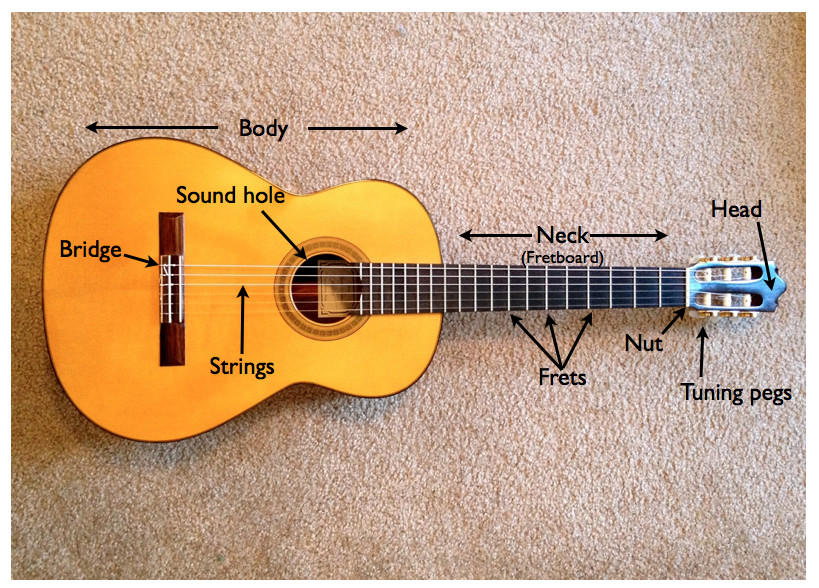
The neck holds the frets and the fingerboard.
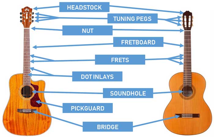
Label parts of guitar. Nut The is one of the fulcrum points where the strings rest up by the head of the guitar. The upper part of the body closest to the neck is known as the treble bout and the lower part is the bass bout. Pick guard not on classical guitars.
Often the top sheet the soundboard is made of a different wood than the back and sides. We started off the first practice by just working on some simple songs like Black Dog and Thunderstruck. In this lesson you will learn a list of different parts of a guitar in English with ESL picture and example sentences to help you enhance.
How Do I Label Parts of A Song. An easy and convenient way to make label is to generate some ideas first. It is hollow on acoustic guitars and consists of a top back and sides.
Sounding Board Guitar Top Nut. The back and sides are usually made of mahogany maple or rosewood. The heel also referred to as the neck joint is where the neck joins to the body of the guitar along with the sides of the guitar body.
The string is what you strum or pluck in order to generate sound. Neck - The neck sticks out from the body and connects to the headstock. The fingerboard is the front part of the guitar it is also called fretboard The small piece that divides the fingerboard is called frets.
The fret holds the strings in different lengths so that when you press it and strum the strings different pitches are produced. The Head of the guitar Is the flat part of wood at the end the neck that holds the tuners. Most of the times we.
The top is also called the soundboard and is usually made of spruce maple or cedar on acoustic guitars. The other is the bridge. This is a thumbnail of the label electric guitar page.
You need to get 100 to score the 14 points available. In this article well examine and explain all three parts and their components. The middle narrow section of the guitar is called the neck.
Guitar labeled parts of the guitar Label Gallery Get some ideas to make labels for bottles jars packages products boxes or classroom activities for free. They are typically steel for electric and acoustic. Parts of the Acoustic Guitar.
Connects to the end of the neck. The bridge on acoustic guitars serves exactly the same purpose - to transfer string vibration to the guitar body. There is a printable worksheet available for download here so you can take the quiz with pen and paper.
The first section of the guitar is called a headstock on which you will find tuners and tuning pegs. The tuners are located at the head of the guitar. This is an online quiz called Parts of a guitar.
From the quiz author. The body of the acoustic guitar is made up of the back sides and top. The full-size printout is available only to site members.
Subscribe to my channel for beginner and intermediate gu. Position markers are the small circles you see on the fingerboard that help. Your Skills Rank.
Rosewood and a raised nut-like saddle over which the strings pass into holes either plugged by bridge pins or fixed within the bridge itself. Without further ado below is a labeled diagram which shows the various components of an electric and acoustic guitar. However acoustic bridges are much simpler.
The waist narrowest section in between the bouts acts as a divider between the sections. Every guitar is a little different but there are three fundamental parts the headstock the neck and the body. Strings - The standard guitar has six strings.
The tuning pegs allow you tune the guitar by tightening or loosening the wires. Strings The strings are the long metal wires of increasing thicknesses that run along the guitar from top to bottom. If you are already a site member click here.
Fretboard Fingerboard Whammy Bar bar Position. The nut is the white strip closest to the headstock. At the end of the practice though the drummer handed me a page with some tabs for The Grudge by Tool.
Parts of A Guitar. They typically consist of a single piece of wood eg. Animated drawing of guitar parts.
To subscribe to Enchanted Learning click here. List of the Parts of an Electric Guitar. What are all the parts of a guitar.
They are where sound is first produced. I was really excited at first because I like. ALAMO GUITARS 1999-2010 Alan Miller.
For acoustic guitars in the majority of cases the neck will be glued using a dovetail joint a particularly effective type of joint commonly used on acoustic guitars for hundreds of years. Headstock - The top of the guitar where the tuning pegs sit. Body This is the main part of the guitar and is where all of the sound is produced.
You should make a label that represents your brand and creativity at the same time you shouldnt forget the main purpose of the label. Label the parts of a guitar. QUESTION Im a sophomore in high school and I recently joined a band with some juniors and seniors.
Guitars are made up of tonewoods strings hardware metal and plastic and electronics all working together to create a music machine.