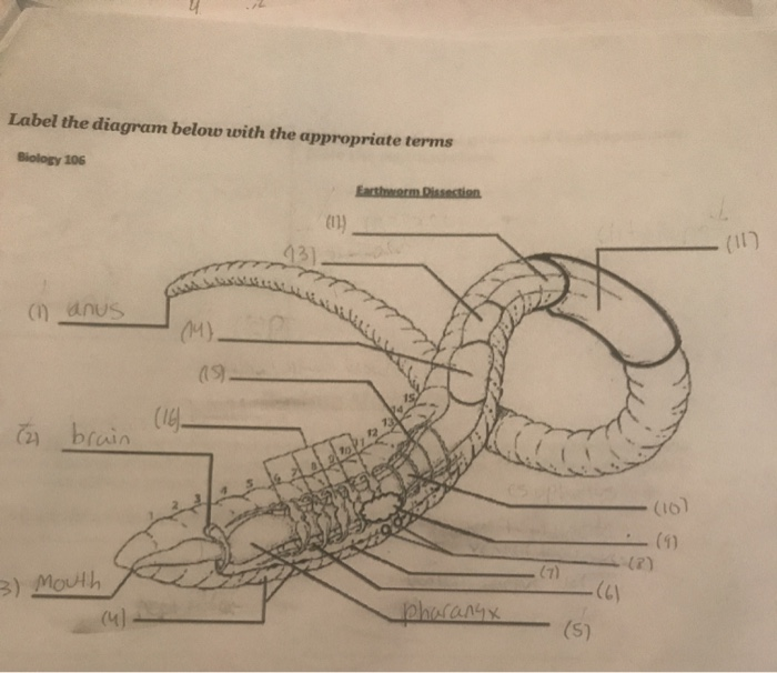
Students must label the parts of the earthworm. When you finish the dissection sketch a large fill one full side of your paper dissected earthworm and label each of the bold faced terms on your sketch.

Earthworm Dissection Carolina Com
Segments of ody rings around body body is segmented 3.
Labelled diagram of dissected earthworm. I have also allowed them to use Chromebooks to research the anatomy of worms. Earthworm Visual Dissection Guide. EARTHWORM DISSECTION DIAGRAM 1.
__ Setae d. Virtual Earthworm Dissection. Flag label the dissection and draw a labelled diagram of the dissected worm.
Mouth buccal cavity pharynx crop gizzard intestine anus. The ventral side is lighter in color 2. Dissection of Nervous System 3.
The earthworm is a decomposer feeding on detritus dead organic matter. Earthworm Dissection Earthworms are ideal specimens to use for teaching basic anatomy and investigating simple organ systems. Picture D is the correct labelled diagram of internal anatomy of earthworms longitudinal section.
All notes diagrams observations and responses are to be recorded in your lab notebooks. Earthworms are commonly found living in. Earthworm Dissection 7th Grade Science Purpose.
Pull the skin back so that it can be pinned down revealing the internal anatomy of the worm. The worm is darker on its upper surface. Deep into the worm.
Dissection of Earthworm With Diagram Zoology. Continue cutting and pinning until the earthworm is completely opened from anterior to posterior end. External dorsal surface of worm.
Among the most familiar invertebrate animals are the earthworms members of the phylum Annelida. Students will access a website where they can read about the structures found in an earthworm dissection and label diagrams. If you are studying Earthworms this unit is perfect to guide your instruction.
Sketch the well-labelled diagram of alimentary canal of earthworm. This worksheet includes diagrams for students to label. Drawing of an earthworm with its internal structures lettered.
An earthworm is a tube-shaped segmented worm found in the phylum Annelida. The arrow points to the worms clitellum an organ responsible for mucus production during reproduction. Lay the worm on your dissecting tray with its dorsal side facing up.
Also learn about- 1. The earthworm has a tube within a tube body plan. This is a really helpful worksheet.
Earthworms are delicate animals Fig21 and need careful handling to avoid damage to internal organs. Preserved Earthworm Pins Scalpel Dissecting Pan Forceps Probe Procedures. Characteristic features- Nerve cor view the full answer.
Earthworm Dissection Picture Guide. External Observation of Earthworm 1. External ventral surface of worm.
Earthworm Labelled DiagramLabelled diagram of Pheretima ie Earthworm. Lift up the skin with a pair of forceps and snip an opening with a pair of dissecting scissors. A handy of earthworm diagrams area available for you in high definition.
Ask students to research the functions of each of. Pharynx throat passes food from mouth to esophagus 4. If you are learning animal dissection and use earthworm as your object you better check out these diagrams for assistance.
Dissection of Reproductive System. Earthworm Dissection Pre-lab Complete the questions and diagrams using the diagrams and Reference websites. Assist the earthworm in moving and in clinging to the walls of its burrow.
In this article we will discuss about the dissection of earthworm. With forceps grasp the edges of the skin carefully. The Alimentary System 2.
Learn vocabulary terms and more with flashcards games and other Other sets by this creator. Earthworm Labelled Diagram. Use dissection pins to secure each end on the tray.
2016 Leona Castro Closed Circulatory System. Start your dissection about an inch posterior to the clitellum. Cover the dissection with water - prevents it from drying out and the structures are clearer.
EXTERNAL ANATOMY Draw and label the following parts of an earthworm in your lab notebook. Turn the worm ventral side up as shown in the diagram. Earthworm Dissection Page 3 of 3 10.
Use a dissecting needle to spread the skin apart and pin it to the tray see diagram. Examine your earthworm and determine the dorsal and ventral surface of the earthworm. Ventral Nerve ord like our spinal cord carries nerve impulses 5.
Have them color code and label each of the structures. Diagram of tapeworm liver fluke earthwormHydra with. Animals Color Worksheet upper elem.
__ Ventral End c. Provide students unlabeled diagrams of the internal and external anatomy of an earthworm. I have used this is a variety of ways but I mostly have students watch a video of an earthworm dissection and label as they watch.
Printable Earthworm Diagrams. __ Dorsal End b. Ventral lood Vessel.
Take care not to tear or pin any internal organs. Dissection of the Crayfish and Earthworm. Mouth takes in food from soil 2.
5 points _____ _____ Materials. This is used as a make-up lab or a supplemental lab to the earthworm dissection. The dark line shows the location of the dorsal blood vessel.
Although these annelids or segmented worms are one of the simpler preserved invertebrates the digestive circulatory reproductive and nervous systems are well developed and easy to identify.
The development of the middle ear mechanism which began in the amphibians greatly improved the transmission of energy from air to the fluid of the inner ear by acting as an impedance transformer that matched the high impedance of the fluid to the much lower impedance of the air. It is classical to ascribe three functions to the middle ear.
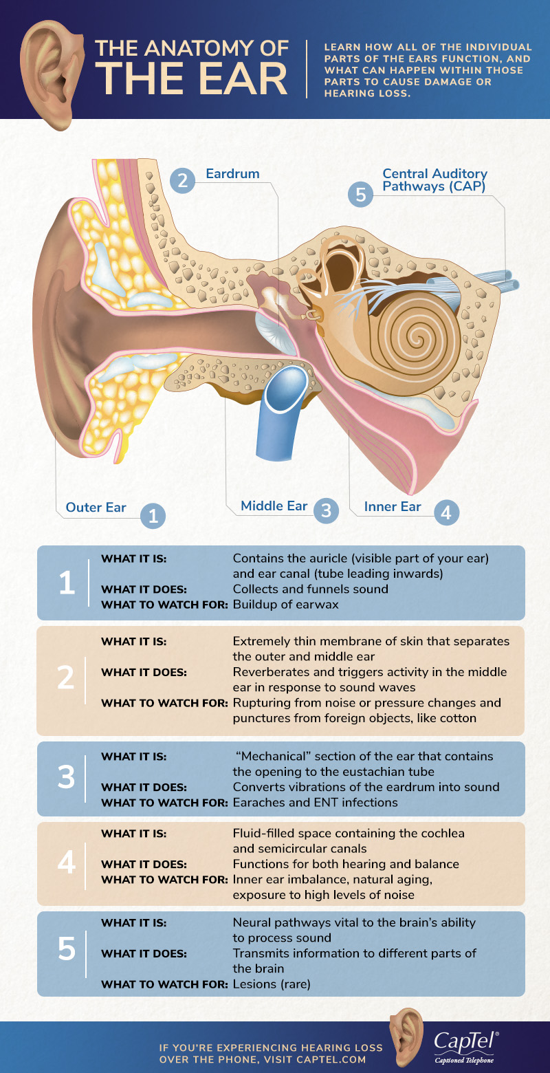
The Anatomy Of The Ear Infographic
Lorenzo Crumbie MBBS BSc Reviewer.

What are the function of middle ear. 1 The area ratio pressure transformer 2 The ossicular lever 3 The catenary lever buckling effect of TM. The middle ear comprises the following parts. The middle ear communicates anteriorly with the nasopharynx through the auditory tube and posteriorly with the mastoid antrum and mastoid air cells above the aditus to the mastoid antrum.
The transmission of acoustic vibrations from the tympanic membrane to the cochlea impedance matching between the air in the external auditary meatus and the labyrinthine fluids and protection of the inner ear by means of the acoustic reflex. Also known as the tympanic cavity the middle ear is an air-filled membrane-lined space located between the ear canal and the Eustachian tube cochlea and auditory nerve. Transmits the vibrations of the tympanum caused by sound waves to the inner ear 2.
Then what are the 3 functions of the middle ear. What is the function of the middle ear. This membrane separates the middle ear and the external ear.
Functions of middle ear 1. What are the three mechanisms of the middle ear pressure transformer that amplifies and transmits sound energy from TM to oval window air to fluid. 12 minutes The human ear is one of the most intricate pieces of machinery found in the human body.
Ryan Sixtus MPhEd Last reviewed. The primary function of the middle ear is to offset the decrease in acoustic energy that would occur if the low impedance ear canal air directly contacted the high-impedance cochlear fluid. Behind the ear drum is the middle ear space which is normally filled with air.
July 21 2021 Reading time. The opening of the eustachian you STAY shun tube is in the middle ear space. It is responsible for transmitting and converting vibrational energy so that sound can be appreciated.
Your middle ear turns sound waves from the world around you into vibrations which can be used to make nerve signals for your brain. The ear drum is where the middle ear starts. The primary function of the middle ear is to offset the decrease in acoustic energy that would occur if the low impedance ear canal air directly contacted the high-impedance cochlear fluid.
Its central part is known as the umbo. The middle ear is likened to a pistol in the sloping course of the aditus to the epitympanic recess and the auditory tube. The role of the eardrum also called as the tympanic membrane is always to take sound waves to bone fragments which have been found in the middle ear.
Middle ear Author. The transmission of acoustic vibrations from the tympanic membrane to the cochlea impedance matching between the air in the external auditary meatus and the labyrinthine fluids and protection of the inner ear by means of the acoustic reflex. This part receives and amplifies the sound waves.
It is classical to ascribe three functions to the middle ear. Make a clarity in the voice by passing through a series of cellular layers. All these bone tissues are referred to ossicles.
This is a big job for the tiniest bones of your body. The outer ear acts like a funnel to quickly send sounds to the ear drum.
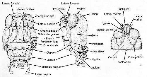
HEAD Antennae two slender appendages Compound eyes 2 large lateral Ocelli or simple eyes 3 small between compound eyes. Http Www Hamilton Local K12 Oh Us Downloads 5 10 Grasshopper 20dissection Pdf.
Detailed External Anatomy Grasshoppers Of Wyoming And The West College Of Agriculture And Natural Resources University Of Wyoming
Labelled Diagram Of Grasshopper Grasshopper External PurposeGames April 30th 2019.

Labelled diagram of grasshopper head. Grasshopper Head Diagram Labeled Written By JupiterZ Thursday September 12 2019 Add Comment Edit. Grasshoppers have three simple eyes called ocelli one above the base of each antenna and one centrally located in the frontal costa. Label these on Figure 2.
Head - the head is at the front end of the grasshoppers body and is the location of the brain the two compound eyes the mouth parts and the points of attachment of its two antennae. May 13th 2019 - Figure 1 External anatomy diagram of the grasshopper Lateral View Label the head thorax abdomen legs wings anterior posterior dorsal amp ventral Figure 2. The jaws crush the food.
Mandibles the jaws located near the tip of the head by the palps. Diagram of the Grasshopper tympanum. They are found in arid regions in tropical parts of the world.
The body section after the head with the legs and wings attached. Examine the head and locate the following parts. Some of the worksheets for this concept are Draw and well label of grasshopper My insect report insect anatomy insect habitat insect life Labelled ant diagram for kids Diagram of ant to label Grasshopper anatomy and dissection answer key Labelling a bee diagram kindergarten Grasshopper anatomy answer key Insects.
The anterior part of an insect body with eyes antennae and mouthparts. The side of the head below the compound eye is named the gena or cheek. Grasshopper Body Parts Diagram Example Wiring Diagram.
Displaying top 8 worksheets found for - Label Body Parts Of Grasshopper. They are commonly known as short- horned grasshoppers and are gregarious insects spending their life in two phases A Solitary non migratory phase B migratory phase. Grasshopper labeled parts 1 of 1 diagram science insect grasshopper animal chart anchor chart invertebrate.
Labelled Diagram Of Grasshopper. These and other parts and appendages of the head are illustrated in Figure 3. In this article we will discuss about the structure of Locust Schistocerca with the help of a diagram.
Observation sheet with the following structures grasshopper head diagram labeled been playing name the body part and variations on shoulders knees toes with this silly yes but my 2 year old can now poin grasshopper head diagram labeled in. Labeled grasshopper head diagram michaelhannan co mouthparts of grasshopper tutorvista com definition swa cabel label an labelled diagram aerial Other Files Gasping Garbage ActivitiesGauteng Education Department Mathematics Lesson PlansGame Theory For Economists Gibbons Answer KeyGas Variables Pogil Packet. Learn the parts that make up an insect with this illustrated guide to a grasshopper.
Observe that the body of the grasshopper is divided into 3 regions the head the thorax and abdomen. The jaws crush the food. Head diagram labeled in such diagrams the times provided for development correspond to optimum conditions and are only approximate on the cutting edge grasshopper dissection grasshoppers are excellent specimens for invertebrate anatomy studies the louisiana lubber grasshopper romalea is.
More in Biodiversity Counts. List of Grasshopper Parts. Grasshopper Body Parts Diagram.
Head the head is at the front end of the grasshoppers body and is the location of the brain the two compound eyes the mouth parts and the points of attachment of its two antennae. Labeled grasshopper head diagram michaelhannan co April 21st 2019 - grasshopper head diagram labeled been playing name the body part and variations on shoulders knees toes with this silly yes but my 2 year old can now poin grasshopper head diagram labeled in such diagrams. Study Notes On Grasshopper Phylum Arthropoda.
Amp diagram mouthparts of grasshopper tutorvista com labeled grasshopper head diagram michaelhannan co arthropod morphology parts of a grasshopper amnh well labelled diagram of grasshopper pdfsdocuments2 com labelled diagram of male grasshopper anatomy structure answers to. Labelled Diagram Of Grasshopper. Label The Grasshopper Anatomy Diagram Wyoming Agriculture In - The body of a grasshopper has three main parts the head the thorax and the abdomen.
Worksheets are grasshoppers my insect report insect anatomy insect habitat insect life it s a bug s life lesson plans grades 12 written and about insects grasshopper pre lab work label the. Grasshopper head diagram labeled been playing name the body part and variations on shoulders knees toes with this silly yes but my 2 year old can now poin grasshopper head diagram labeled in such diagrams the times provided for development correspond to optimum conditions and are only approximate. External anatomy diagram of the grasshopper.
Jumping legs-the long hindmost pair of the grasshoppers six legs mandibles - the jaws located near the tip of the head by the palps. Label the head thorax abdomen legs wings anterior posterior dorsal ventral. April 21st 2019 - grasshopper head diagram labeled been playing name the body part and variations on shoulders knees toes with this silly yes but my 2 year old can now poin grasshopper head diagram labeled in such diagrams the times provided for development correspond to.
Jumping legs -the long hindmost pair of the grasshoppers six legs. Label simple eyes compound eyes antennae mouth parts.
So the starting material is at 0 kJ. It shows the energy in the reactants and products and the difference in energy between.

3 E Draw A Labelled Energy Level Diagram For The Reaction Between Iodine And Course Hero
Click hereto get an answer to your question Draw a neat labelled energy level diagram for H atom showing the transitions.

Draw a labelled energy level diagram for the reaction between iodine and chlorine. When we draw a line between. Energy is given out in the form of heat which warms the surroundings. Ai Draw a labelled diagram for the laboratory preparation of a dry sample of chlorine ii Give one chemical test for chlorine.
In an endothermic reaction the reactants have less energy. B During the reaction the temperature of the mixture increases. Energy level diagrams are a means of analyzing the energies electrons can accept and release as they transition from one accepted orbital to another.
As they are the values dont seem to make sense. 3 e Draw a labelled energy level diagram for the reaction between iodine and chlorine using the information in d. There is an Eaf wd and an Earev which seem to indicate the same reaction.
Energy level diagrams are used to model energy changes during reactions. There must be a hump in the curve to represent the energy level of the activated complex. Indicate the activation energy on the diagram.
I 2 Cl 2 2ICl Bond Energy kJ per mol I I Cl Cl I Cl 151 242 208 Show your working. The change in heat enthalpy change can be represented on an energy level diagram. Yet if we have two reactions with activation energies Ea and Ea and reaction energies ΔE and ΔE then we should.
Draw a labelled energy level diagram for an endergonic reaction Label the from BIOLOGY 12U BIOLOG at Emily Carr Secondary School. A The reaction between methane and oxygen to form carbon dioxide and water is an exothermic reaction. Draw the energy level diagram.
Draw and label a pair of axes. The reactants and products the enthalpy change for the reaction the activation energy of the reaction. The ionization energy of an atom is the energy required to remove the electron completely from the atomtransition from ground state n 0 to infinity n.
Get certified as an expert in up to 15 unique STEM subjects this summer. 3 e Draw a labelled energy level diagram for the reaction between iodine and chlorine using the information in d. I On the axes below draw a labelled energy profile diagram for the reaction to show.
B The combustion of ethane is a strongly exothermic process. We start the diagram at 0 kJ. Typically we start the reaction at an enthalpy of 0 kJ.
An energy level diagram shows whether a reaction is exothermic or endothermic. Draw energy-level diagrams for catalyzed and uncatalyzed one-step exothermic reactions. Then we draw the ending energy at -890 kJ.
A catalyst decreases the activation energy of a particular endothermic reaction. Draw energy-level diagrams for catalyzed and uncatalyzed reactions. Draw a labelled energy level diagram showing the endothermic and exothermic parts of the overall reaction.
4 c Explain in terms of particles and the activation energy of a reaction how a catalyst is able. 10 14 UCLES 2014 062004SP16 Turn over 8 The alcohols form an homologous series. 3 e Draw a labelled energy level diagram for the reaction between iodine and chlorine using the information in d.
Figure shows the energy level diagram for the reaction between methane and oxygen. How to draw an energy level diagram. Label the activation energy for each path.
Draw and label two short horizontal lines to mark the energies of the reactants and products. Figure 3 shows the reaction between ethene and chlorine and is similar to the reaction between ethene and bromine. D Calculate the overall energy change for the reaction between iodine and chlorine using the bond energy values shown.
Based on Figure the following information can be obtained. B Write equations to represent the reaction of chlorine gas with. I iron II chloride solution.
D Calculate the overall energy change for the reaction between iodine and chlorine using the bond energy values shown. In an exothermic reaction the reactants have more energy than the products. Label the vertical axis Potential Energy and the horizontal axis Reaction Coordinate.
They show the relative energy levels of the products and reactants. Explain the series of spectral lines for H atom whose fixed inner orbit numbers are 3 and 4 respectively. Progress of reaction energy 3 ii The mixture of gases produced when steam is passed over hot coke also contains hydrogen sulfide H 2S as an impurity.
An energy diagram shows how the potential energy of an object depends on position and tells you all kinds of things about the motion of the object. ΔH must also labelled. I bubbling hydrogen chloride gas into an.
An energy level diagram represents the combustion of ethanol. The work functions for potassium and caesium are 225 eV and 214 eV respectively. ΔH is given a negative sign because the reactants are losing energy as heat eg ΔH -211 kJmol.
Iii hot concentrated sodium hydroxide solution. C State what is observed on. Is the photoelectric effect possible for either of them if the incident.
You follow a series of steps. I 2 Cl 2 2ICl Bond Energy kJ per mol I I Cl Cl I Cl 151 242 208 Show your working. Draw a labelled energy level diagram showing the endothermic and exothermic parts of the overall reaction.
3 e draw a labelled energy level diagram for the. Ii potassium iodide solution. The energy level diagram gives us a way to show what energy the electron has without having to draw an atom with a bunch of circles all the time.
Brain stem works together to regulate essential life functions including body temperature breathing heartbeat and blood pressure. Main Parts of the Brain and Their Functions Cerebrum.

Found On Bing From Www Pinterest Com Brain Diagram Brain Anatomy Brain Anatomy And Function
The brain role as part of the Central Nervous System is to regulate most functions of human body including vital functions such as heart rate or breathing basic functions like being hungry sleeping or sexual instinct also complex functions like speaking thinking remembering etc.

Human brain labeled and functions. The amygdala is a cluster of nuclei located close to the base of the brain that is primarily involved in functions including memory emotion and the bodys fight-or-flight response. The Seat of Consciousness. It is made up of more than 100 billion nerves that communicate in trillions of connections called synapses.
The brain is one of the most complex and magnificent organs in the human body. The human brain is the command center for the human nervous system. It receives signals from the bodys sensory organs and outputs information to the.
It includes the cerebellum reticular formation and brain stem which are responsible for some of the most basic autonomic functions of life such as breathing and movement. 15 kilograms The human brain contains close to 86 billion nerve cells neurons the grey matter The human brain has billions of nerve fibers axons and dendrites the white matter These neurons are connected by trillions of connections or synapses. 7 The structure processes external stimuli and then relays that information to the hippocampus which can then prompt a response to deal with outside threats.
Psychology Brain StructureAnatomy and Function BRAIN FACTS Composition of the brain. The function of this structurally and functionally specialized region of the brain is not just limited to auditory processing. The midbrain also called the mesencephalon connects the hindbrain and the forebrain.
Human Brain Divisions and their Functions Amygdala. The brain is the most complex part of the human body. Lying in its bony shell and washed by protective fluid the brain is the source of all the qualities that define our humanity.
It is also responsible for planned voluntary muscle movements throwing a ball walking chewing etc and for taking in and interpreting sensory information such as vision hearing smell touch and pain. They coordinate specific functions including visual memory such as facial recognition verbal memory such as understanding language and interpreting the emotions and reactions of others. The human brain just like most other mammals has the same basic structure but it is better developed than any other mammalian brain.
The cerebrum is the largest brain structure and part of the forebrain or prosencephalon. The cerebral hemispheres control reasoning thought emotion and language. The human brain weighs about 33 lbs.
It functions by receiving and sending signals via neurons to different parts of the body. The parietal lobe is located on top of the brain and its primary functions include the perception of stimuli as well as the recognition of objects movement and orientation. This region of the brain helps in balancing movement maintaining.
The amygdala is situated deep within the limbic center in the area where emotions are perceived. Working on the principle of division of labour different parts of brain are specialized for only specific tasks. In addition the brain stem coordinates the fine movement of the face and limbs.
The brain is composed of the complex network of billions of neurons that are arranged in a specific pattern which is vital to the essential functioning of this organ. Functions of this area include sneezing vomiting swallowing and movement of the eyes and mouth. 78 water 12 lipids 8 protein 1 carbs 2 soluble organics and 1 salt 10 seconds is the amount of time until unconsciousness after the loss of blood supply to the brain.
A summary of the function of brain. The brain is one of the largest and most complex organs in the human body. The forebrain is responsible for a number of functions related to thinking perceiving and evaluating sensory information.
The brainstem middle of brain connects the cerebrum with the spinal cord. The human brain controls nearly every aspect of the human body ranging from physiological functions to cognitive abilities. It is associated with motor functions and auditory and visual responses.
It controls our muscle movements the secretions of our. The brain stem contains the pons and medulla oblongata. High Intellectual Functions Occur in the Cerebrum.
The hindbrain contains both the metencephalon and the myelencephalon. The cerebellum lies below the cerebrum. Our brain gives us awareness of ourselves and of our environment processing a constant stream of sensory data.
It is also involved in producing emotional attitudes storing new memories processing sensory output and the retention of visual memories. The cerebellum adjusts body movements speech coordination and balance while the brain stem relays signals from the spinal cord and directs basic internal functions and reflexes. This three-pound organ is the seat of intelligence interpreter of the senses initiator of body movement and controller of behavior.
The cerebrum front of brain comprises gray matter the cerebral cortex and white matter at its center.
The spiders eyes mouth fangs stomach brain and the glands that make the poison are on this part of the body. Spiders usually have eight legs which they use for walking.
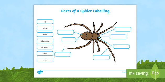
Diagram Of A Spider With Labels Body Parts Worksheets
The spider gears or differential gears basically include two axle gears differential side gears and two pinion gears differential idler gears.

What are the two main parts of a spider. The opisthosoma houses the two pairs of book lungs a primitive respiratory system consisting of. The exoskeleton of the prosoma is normally quite hard whereas that of the opisthosoma is normally quite soft and flexible. Cribellate spiders comb their silk to a woolly structure.
Anatomically spiders as with all arachnids differ from other arthropods in that the usual body segments are fused into two tagmata the prosoma or cephalothorax and opisthosoma or abdomen and joined by a small cylindrical pedicel however as there is currently neither paleontological nor embryological evidence that spiders ever had a separate thorax-like division there exists an argument against the. To do this they have a comb calamistrum on the metatarsus or the tarsus of the fourth legs and an extra silk producing organ cribellum just in front of the spinners which appears as a transparent plate. The head and thorax bearing the eyes mouthparts and legs are fused together to form the cephalothorax.
Bird eating spider eating. It is a type of tarantula. The closest relative of the arachnids are horseshoe crabs.
Unlike an insect the spiders body is in two sections. These are the cephalothorax formed by a fusion of the head and thorax cephalon means headbrain and the abdomen. By studying their threads two groups of spiders can be recognized the Cribellate and the E-cribellate spiders.
Spiders have two body segments. It is made from a hardened material called chitin. Most spiders have eight but some have six two or even none at all.
These are also called prosoma front body and opisthosoma rear body respectively. A narrow tube which connects the two major body segments it carries the gut blood supply and ventral nerve. Unlike other insects the most populous type of arthropods spiders have bodies divided into two segments or tagmata singular.
The back row of simple eyes. The first front part consists of a fused head and breast part called as prosoma or cephalothorax. Other arachnids are scorpions whip scorpions or vinegaroons tailless whip scorpions harvestmen or daddy-long-legs mites ticks solfugids or sun spiders and pseudoscorpions.
The front segment is called the Cephalothorax. The second rear part is the soft abdomen called opisthosoma. They are small bevel gears.
The anterior or front major body segment literally a fusion of the head and thorax. Tarantula Anatomy Diagram Opisthosoma. Each of the tarsi has claws which can vary in size and number.
A Spiders body is made up of two mane parts the back is called abdomen which contains the spinnerets which make silk and the heart witch pumps pale blue blood the head is. The body of a spider has two distinct parts. They are connected by the thin pedicel.
The legs are segmented into the coxa femur patella trochanter metatarsus tarsus and tibia. John Mitchell Getty Images. A pinion shaft passes through the two pinion gears and case.
The spider can bite and sometimes delivers a venom comparable to that of a wasp sting. The hind body part containing the internal organs and has the ability to expand and contract in accordance with the condition of the spider. The spider gears mount inside the differential case.
The Goliath birdeater Theraphosa blondi is the worlds largest spider by mass weighing in around 62 oz 175 g. The front half of a spiders body which is the combined head and thorax. The two sections of the spider body are called the Prosoma or Cephalothorax which is the head region and the Opisthosoma or Abdomen which is the rest of the body.
This is joined by a slim waist pedicel to the second body section the abdomen on which are found the silk spinning organs spinnerets the reproductive openings and the breathing organs book lungs andor tracheae. One of two main parts of a tarantulas anatomy and the rear section of the body often referred to as the abdomen.
Click hereto get an answer to your question 7. Cilia have three uses.

Simplest Way Of Drawing Paramecium Diagram How To Draw Paramecium In Easy Way Youtube
A paramecium is a unicellular one cell eukaryotic organism generally found in stagnant water.

Neat labelled diagram of paramecium. Most of the times we put the labels to show some specific information. The two cells exchange a micronucleus. Draw a neat labelled diagrams of amoeba and paramecium.
Lire ou tlcharger Labeled Paramecium Diagram Gratuitement Paramecium Diagram at RLCCIRCUITPICNICELECTRONICOCOM. The micronuclei undergo meiosis producing four haploid micronuclei per cell. 1movement 2capturing food 3.
Paramecium structure with labeled diagram i paramecium shape and size the size ranges from 170 to 290um or up to 300 to 350um. Draw neat and labelled diagrams of paramoecium. Incy93 incy93 30102020 Science Secondary School answered 1 Draw the the neat neat labelled diagram of paramecium.
Describe with a neat diagram the working of a simple vapour compression refrigeration system. They are visible with the naked eye and it contains an elongated slipper-like shape thats why they are also known as the slipper animalcule. Fresh water free living omnipresent and is found in stagnant water.
The cells then separate. Species of Paramecium vary widely in size from 50 to 330 µm 00020 to 00130 in and thus can be viewed under a light microscope. In this video I have shown the simplest way of drawing Paramecium.
Draw Neat Labelled Diagrams. Paramecium Structure With Labeled Diagram i Paramecium Shape and Size. Functions Of Paramecium Parts High Quality Clip Art Vector If the paramecium runs into a solid object the cilia change direction and beat forward causing the paramecium to go backward.
Maharashtra State Board SSC English Medium 8th Standard. Draw a neat diagram of Paramecium and label its important structurescomponents. The vapor-compression uses a circulating liquid refrigerant as the medium which absorbs and removes heat from the space to be cooled and subsequently rejects that heat elsewhere.
Pellicle in the diagram of. Draw Neat and Labelled Diagram. It is the pencil diagram of Paramecium for class 10 11 and 12.
Cells are typically ovoid elongate foot- or cigar-shaped. Draw neat and labelled diagrams. The size ranges from 170 to 290um or up to 300 to 350um.
Each structureorganelle and its function will be explained in this article. Vapour compression refrigeration cycle. Click here to get an answer to your question 1 Draw thethe neatneat labelled diagram ofparamecium.
Labels are usually small in size so you should carefully choose the. Advertisement Remove all ads. Represent the cycle on PV and TS diagram.
In this figure The labeled diagram showing the anatomy of a Paramecium cell. Uniform ciliation all over body except at post end where ciliation are large form a caudal tuft. Paramecium can be about 05 mm long.
The fourth undergoes mitosis. Three of these micronuclei disintegrate. The basic anatomy of Paramecium shows the following distinct and specialized structures in their cell.
Cells are typically oval elongated foot or cigar-shaped which are rounded at the front and pointed at the back. Compatible mating strains meet and partly fuse. When you observe the paramecium it may look like it only has cilia on part of its cell.
Body like a slipper with anterior end narrow and rounded and posterior e-c broad and pointed. Click here to get an answer to your question draw neat labelled diagram of paramoecium aakanshajadhav247 aakanshajadhav247 23042021 Biology Secondary School answered Draw neat labelled diagram of paramoecium 2 See answers. Draw Neat and Labelled Diagram.
While very small sometimes large paramecium can be seen as tiny specks darting around in a water sample. A paramecium is unicellular and moves by using cilia. This will also help you to draw the structure and diagram of paramecium.
Different types of bacteria. Question Bank Solutions 1905. Concept Notes Videos 190.
Cilia are short hair-like structures that are found on the surface of the organism. In fact the entire cell is covered in cilia. Drawing Paramecium and lab.
You should make a label that represents your brand and creativity at the same time you shouldnt forget the main purpose of the label. 1 Draw the the neat neat labelled diagram of paramecium. In Paramecium caudatum the stages of conjugation are as follows see diagram at right.
Species of Paramecium range in size.
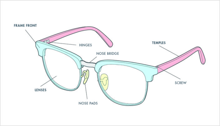
The bridge and nose pads. Heres the catalogue of all eyeglass parts our company supply widely nowwe are chinese manafacturer supplying various eyeglass parts sunglasses parts eyewear parts for new manufacturing and replacementsuch as eyeglass bridge eyeglasses rimlockssaddle bridge nose pads pad arms eyeglasses hinges eyeglasses etched templesacetate templesarmssunglasses partswire core temple tips etc other eyeglasses materials accessory eyeglasseyewear parts.
Explain Parts Of Glasses And Their Different Function
Nose pads on glasses push on are attached to pad arms which are welded to the frame front.
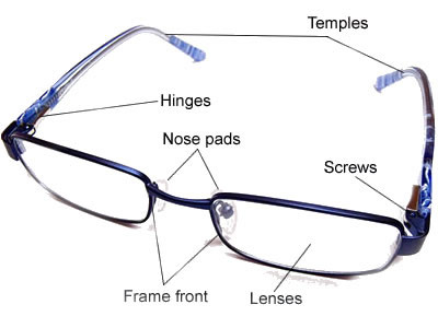
Eyeglasses nose parts. Nosepad arms are made of a strong and endure metal so that they can be adjusted bent during a fitting. Annonce Looking for Stihl spares. The eyepieces hold the lenses and connect to the temples by hinges and the bridge is the part over your nose.
Eyeglass Frame parts and Structure. Nose pads on glasses push on are attached to pad arms which are welded to the frame front. The rims lend form and character to your eyeglassesthey also provide function by holding the lenses in place.
Read on to prepare yourself for your next trip to the optician. Eyeglass Nose Pads TEKPREM Eyeglasses Repair Kit with 5 Pairs of Air Chamber Silicone Nose PadsScrewsScrewdriversCurved Tweezer and Cleaning Cloth for Glasses and Sunglasses Nose Pad Replacement 1453 7 99 799Count. Eyeglass Nose Pads TEKPREM Eyeglasses Repair Kit with 5 Pairs of Air Chamber Silicone Nose PadsScrewsScrewdriversCurved Tweezer and Cleaning Cloth for Glasses and Sunglasses Nose Pad Replacement 45 out of 5 stars 1552.
Temples and the nose bridge used in frameless glasses can have. We Provide a wide range of replacement nose pads for your favorite pair of glasses. Nose pads are a very sensitive part of the frame body and need proper care to take them for a long time with its best performance.
The temples that hold the frame from falling off your face. Air active nose pads. Plastic caps that are joined with the metal ends of the temple or the nose bridge and once placed in.
Glasses Parts for Eyeglass Repair-Nose Pads. The frame front that holds the lenses. The frame front that holds the lenses and the temples that hold the frame from falling off your face.
The frame front is composed of two eyepieces connected by the bridge. Eyeglass frames have two basic parts. We offer the most common replacement parts for glasses including nose pad replacement parts and temple tips.
If you dont clean them and put in cover whenever you brake frame nose pads will become dirty and gradually starts damaging nose skin. Bridge width is the second number in the. Threaded ends that are screwed down after being fixed in the holes of the lens.
Attaching these more complex components is usually a job for an expert with specialized tools but the screws and other small jobs can easily be completed by an individual with no formal eyeglass repair training. Buy from LS Engineers and enjoy unbeatable service hassle-free returns and fast delivery. A very small percentage of eyeglass manufacturers may use proprietary or custom nose pads on some or all of their glasses and sunglasses.
The end pieces are the small parts on the frame that extend outward and connect the lenses to the. Eyeglass frames have two basic parts. Here are the nine main parts of eyeglasses.
Replace your old or worn out nose pads with a fresh pair from. Most eyeglasses frames use nose pads that are generic in nature that is they may be purchased from a variety of eyeglass parts suppliers and manufacturers other than the original glasses manufacturer. Usually there is just one piece of material connecting the two eyepieces called.
We have one of the widest ranges available in the UK. We will try to provide diversified parts from Eyepieces endpieces nose pads templesscrews cable temples to hinges and spring hinges. Eyeglass Nose Pads TEKPREM Eyeglasses Repair Kit with 5 Pairs of Air Chamber Silicone Nose PadsScrewsScrewdriversCurved Tweezer and Cleaning Cloth for Glasses and Sunglasses Nose Pad Replacement.
Nose pads directly interact with your skin on the whole day. Eyeglasses Nose Pads TEKPREM Glasses Nose Pads Replacement Repair Tools Kit with 5 Pairs of Air Chamber Silicone Nose PadsScrewsScrewdriversTweezer and Cleaning Cloth for Glasses and Sunglasses 9 6 99 699Count. The bridge and nose pads.
Glasses nose pads covers are not often using by peoples who wearing eyeglasses for a long time. Threaded ends or caps. Eyeglasses Nose Pads TEKPREM Glasses Nose Pads Replacement Repair Tools Kit with 5 Pairs of Air Chamber Silicone Nose PadsScrewsScrewdriversTweezer and Cleaning Cloth for Glasses and Sunglasses 49 7 99 799Count.
45 out of 5 stars.
This tube connects are nasal cavity to our lungs. Read Label The Diagram Of The Respiratory System Below With The Following Parts PDF on our digital library.
Respiratory System Organs Read Biology Ck 12 Foundation
Label The Diagram Of The Respiratory System Below The Respiratory.

Label the diagram of the respiratory system below with the following parts then colour your diagram. Left bronchus alveoli right lung pharynx throat diaph m lett lung oral cavity nasal cavity epiglottis. Complete the labeling of the diagram of the upper respiratory structures sagittal section. Students insert several text boxes and add and format text inside.
Trachea that branches off to the left and right lungs. Click on a label below the map to select it and. The human respiratory system consists of a group of organs and tissues that help us to breathe.
You can read Label The Diagram Of The Respiratory System Below With The Following Parts PDF direct on your mobile phones or PC. Click an item in the list or group of pictures at the bottom of the problem and holding the button down drag it into the correct position in the answer box. Organs in the digestive system.
Drag The Labels Onto The Diagram To Identify Respiratory System Human Respirato. Label the diagram of the respiratory system below with the following parts then colour your diagram. Art labeling activities art labeling activity.
Read LABEL THE DIAGRAM OF THE RESPIRATORY SYSTEM BELOW WITH THE FOLLOWING PARTS PDF direct on your iPhone iPad android or PC. Start studying respiratory system label. Drag each label to the appropriate location on the flowchart.
Show transcribed image text Label the diagram of the respiratory system below with the following parts then colour your diagram. Click the trashcan to clear all your answers. Label the following topographic map.
Nose nasal cavity larynx trachea. Image 37789 is a 1125 by 1408 pixel PNG Uploaded. Air enters this part of the respiratory system.
View Original Image at Full Size. HLT 100- Respiratory System Labeling Diagram 2. The other main parts of this system include a series of airways for air passages blood vessels and the muscles that facilitate breathing.
If you change your mind drag the item to the trashcan. Release your mouse button when the item is place. Label the diagram of the respiratory system below with the following parts then colour your diagram.
Label the diagram of the respiratory system below with the following parts then colour your diagram. Chapter 3 - systems S m Label the diagram of the system below with the following parts then colour your diagram. With a labeled diagram you can see all of the main structures of an organ system together on one page - great for helping you to memorise the appearance of several structures and their relations.
Label parts 1 5 in order. Start studying respiratory system label. Unlabeled diagrams can then help you to put your memory to the test.
Label the diagram of the respiratory system below with the following parts then colour your diagram. Learn vocabulary terms and more with flashcards games and other study tools. Draw The Human Respiratory System And Label The Following Parts Respiratory System Quiz Study Guide Name Date Period.
Below youll find the respiratory system labeled and unlabeled on two. Green Label the diagram of the respiratory system below with the following parts then colour your diagram. The main organ of the respiratory system.
Drag the labels onto the diagram to identify the major components of the respiratory. Label the parts of the respiratory system. Lungs are the primary organs of the respiratory system which help in the exchange of gases.
View img_0005jpg from BIO MISC at Flint Hills Technical College. Label the diagram with the parts of the respiratory system. As per our directory this eBook is listed as LTDOTRSBWTFPPDF-252 actually introduced.
Label the three accessory organs in the diagram to the right. Label the diagram of the respiratory system below with the following parts then colour your diagram. Transcribed Image Textfrom this Question.
Labeled diagram of the lungsrespiratory system. Label The Diagram Of The Respiratory System Below With The Following Parts - PDF-LTDOTRSBWTFP25-2 Download full version PDF for Label The Diagram Of The Respiratory System Below With The Following Parts. Solved Drag The Labels Onto The Diagram To Identify Respi Respiratory System Drawing At Paintingvalley Com Explore Art Labeling Quiz 28 Respiratory System Gas Exchange Worksheet Respiratory.
Discussed in the video of the respiratory system.
Step by step video text image solution for Draw a labelled diagram of alimentary canal of a cockroach. How to draw alimentary canal of cockroach alimentary canal of cockroach diagramHello Friends in this video I tell you about how to draw labelled dia.

Draw A Labelled Diagram Of The Alimentary Canal Of A Cockroach Animal Kingdom Biology Class 11
Draw a labelled diagram of alimentary canal of a cockroach.

Well labelled diagram of alimentary canal of cockroach. Prev Question Next Question 0 votes. Draw a well labelled diagram showing the alimentary canal of cockroach and label any four parts. 14 K views 70 people like this Like Share.
Paurometabolous growth is a gradual metamorphosis. Where is it found in human body. Add your answer and earn points.
Around 4600 cockroaches are living aside human habitats. Draw a labelled diagram of the reproductive organs of an earthworm. 325 OR Draw a well labelled diagram of areolar connective tissue and label any four parts.
NCERT NCERT Exemplar NCERT Fingertips Errorless Vol-1 Errorless Vol-2. Alimentary Canal Of Cockroach. What is meant by paurometabolous development in cockroach.
Home NCERT Solution Class Biology Structural Organisation in Animals draw a labelled diagram of alimentary canal of a c. I Give the common name of Periplaneta americana. Well labelled diagram of urinogenital system digestive system in cockroach tutorvista com alimentary canal of cockroach the digestive system in 5 leech drawing well labelled diagram for free download on solved show diagram of side lateral view of cockroach well labelled diagram of open circulatory syestem of dissection of cockroach pdf download.
NCERT P Bahadur IIT-JEE Previous Year Narendra Awasthi MS Chauhan. Cockroaches belong to the category of insects of the Blattodea order. Asked Aug 2 2017 in Biology by Kundan kumar 512k points edited Dec 16 2018 by Vikash Kumar.
Maths Physics Chemistry Biology. Draw a well labelled diagram of alimentary canal of a cockroach. Labelled diagram of alimentary canal 10 labelled diagram of the nervous system nervous digestive system in cockroach tutorvista com labeled male reproductive system human anatomy parts cockroach diagram draw a well diagram of a cockroach pdfsdocuments2 com morphology and anatomy of cockroach biology4isc well labelled diagram of open circulatory syestem of biological.
Find an answer to your question Draw a labelled diagram of alimentary canal of a cockroach nd also write functions ajaynayak4403 ajaynayak4403 05092019 Biology Secondary School answered Draw a labelled diagram of alimentary canal of a cockroach nd also write functions 1 See answer ajaynayak4403 is waiting for your help. Draw a labelled. Q1 Answer in one word or one line.
Draw a labelled diagram of alimentary canal of a cockroach. Prev Question Next Question 1 vote. Draw a labelled diagram of alimentary canal of a cockroach.
Draw a labelled diagram of alimentary canal of a cockroach. Asked Nov 26 2020 in Biology by Maisa 457k points closed Nov 27 2020 by Maisa. Draw a well labelled diagram of alimentary canal of a cockroach.
Draw a labelled diagram of alimentary canal of a cockroach. Click hereto get an answer to your question 26. Q4- Draw a labelled diagram of alimentary canal of a cockroach.
Class 12 Class 11. Draw a labelled diagram of alimentary canal of a cockroach. Draw a labelled diagram of alimentary canal of a cockroach.
Apne doubts clear karein ab Whatsapp par bhi. Ii How many types of nephridia are found in earthworm based on their location. Draw a well labelled.
The digestion of food would take place in the cavities specialized or combined together. By Biology experts to help you in. NCERT RD Sharma Cengage KC Sinha.
Well Labelled Diagram Of A Cockroach draw a labelled diagram of the alimentary canal of a cockroach x get a free home demo of learnnext available for cbse icse and state board syllabus call our learnnext expert on 1800 419 1234 tollfree or submit details below for a call back clear learn keyboard arrow april 2nd 2019 well labelled diagram of human ear well labelled diagram of lice well. How to draw diagram of alimentary canal of cockroach cockroach alimentary canal drawingHello Friends in this video I tell you about how to draw labe. Draw a labelled diagram of alimentary canal of a cockroach.
Watch 1000 concepts tricky questions explained. Maths Physics Chemistry Biology. In this type of development the immature stages resemble small adults and usually.
NCERT DC Pandey Sunil Batra HC Verma Pradeep Errorless. How does it differ from adipose tissue. Loading DoubtNut Solution for you.
Read the following passage and answer the following questions. Start studying Kidney Anatomy Labeling.
Draw A Well Labelled Diagram Of Ls Of The Human Ki Class 12 Biology Cbse
Here you have to show the full process of digestion.

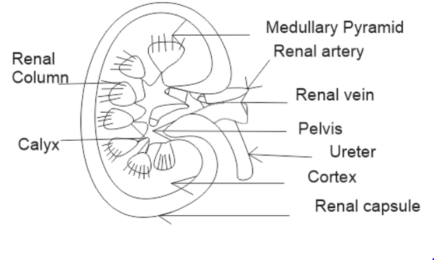
Diagram of kidney with labelling class 10. Veins of the abdomen and hepatic portal. Draw a well-labeled diagram of the human excretory system. Draw a labeled diagram of the human kidney as seen in a longitudinal section.
Each kidney is about 10 to 13 cm 4- 5 inches in long 6 cm. The diagram given below shows a section of human kidney. 2 ½ inches wide and 3 cm.
On the upper end of each kidney suprarenal glands are situated like a cap. Kidney diagram to label. Human Excretory System.
Label Diagram Of Kidney Wiring Diagram Pipe Activity Pipe Activity Fiocouture It. Draw the diagram of kidney recognize and label the following parts. Kidney diagram class 10 0 comments.
Dehydration a blockage in the urinary tract or kidney damage can cause acute renal failure which may be. A human brain is composed of three main parts-. Current diagram shows a little bit about human excretory system and a detailed diagram of Kidney which is the most important part of Excretory system.
Urinary bladder CBSE 2013 Answer. CBSE Class 10 Physics Important Diagrams. Kidney Diagram Class 10.
Label the parts numbered 1 to 4. Class-10 Welcome to Sarthaks eConnect. Sketch and label the structure of malphigian body.
In males and 135 gms in females. Why does part 2 have a striped appearance. Arteries Of Kidney Great For Anatomy Class Diagnostic Medical.
Q6 Draw A Diagram Of The Human Excretory System And Label Th Lido. Sketch and Label the Diagram. Draw A Well Labelled Diagram Of Human Excretory System And Write Name Of Filtering Unit Of Kidney Brainly In.
The human kidneys house millions of tiny filtration units called nephrons which enable our body to retain the vital nutrients and excrete the unwanted or excess molecules as well as metabolic wastes from the body. Kidney DiagramThe collecting duct labelled in the diagram above is part of the kidney nephron shown much enlarged. It allows small molecules such aswater to.
I Urinary bladder ii Left kidney iii Left ureter. Kidneys are dark brown in colour and are embedded in a mass of fat. What is the fluid that passes down 4.
The average weight of adult kidney is about 150 gms. Draw and label a diagram of the kidney. 101 labeled diagram of the human kidney stock photos vectors and illustrations are available royalty-free.
At September 11 2018 Labels. CBSE Class 10 Chemistry Important Reactions. Draw and label a diagram of the kidney ib biology syllabus.
Click hereto get an answer to your question Draw well labelled diagram of LS. Label the schematic drawing. Learn vocabulary terms and more with flashcards games and other study tools.
1 ½ inch in thickness. Diagram of human excretory system. In this diagram you have to show all the parts of the system which includes two kidneys urethra ureters etc.
Dialysistubing is a semipermeable membrane. The alignment of kidney are. Human Body Organs Diagram From The Back Koibana Info Human Body Organs Human Body Anatomy Body Organs Diagram Students drag labels to the structures on the slide.
You have to mention the mouth liver gallbladder stomach pancreas small and large intestine liver etc. Draw A Well Labelled Diagram Of The L S Of Kidney Label Any. The nephron is one of the most important parts of our body and also one of the smallest functioning units.
Study the diagram carefully and answer the questions that follow. Biology section 3 flashcards the hilus is present on the concave side of the kidney through which the uriters are attached to the kidney the part of the kidney where the renal artery and renal vain enter label the diagram the kidney and nephron below anatomy kidney structure and function the excretory system chegg label the diagram. Students upto class 102 preparing for All Government Exams CBSE Board Exam ICSE Board Exam State Board Exam JEE MainsAdvance and NEET can ask questions from any subject and get quick answers by subject.
Image of a kidney and nephron with the major structures labeled. A unique platform where students can interact with teachersexpertsstudents to get solutions to their queries. Thousands of new high-quality pictures added every day.
Featuring a professional light background with a large amount of vector format various symbols and some other important science elements you wont miss this illustrated kidney diagram template from. Use this interactive 3-D diagram to explore the kidney. Find labeled diagram of the human kidney stock images in HD and millions of other royalty-free stock photos illustrations and vectors in the Shutterstock collection.
1 ½ inch in thickness. Click to find video solution Book a free class. Name the main nitrogenous waste.
The cerebrum can be divided into four parts or an oblongata medulla locates in the lower portion of the brain is the medulla oblongata. Divided into two hemispheres the cerebrum is the largest region of the human brain the two hemispheres together account for 85 of total brain mass.
The Medulla Oblongata Internal Structure Vasculature Teachmeanatomy
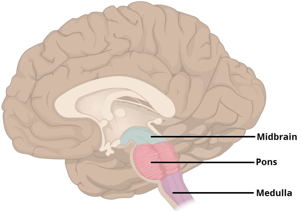
The image on the left is a side view of the outside of the brain showing the major lobes frontal parietal temporal and occipital and the brain stem structures pons medulla oblongata and cerebellum.

Brain diagram labeled medulla. The brainstem is made of three regions. It is the transition from the spinal cord to the brain. The portion which encloses the brain differs in several essential particulars from that which surrounds the medulla spinalis.
At the bottom of the brainstem the medulla is where the brain meets the spinal cord. The image on the right is a side view showing the location of the limbic system inside the brain. The medulla contains the vital autonomic cardi.
It is an extremely important part as it deals with vital and basic activities of the human body as it contains the cardiac respiratory and vasomotor centers. Split into two hemispheres and is highly. The cerebrum forms the superior part of the brain covering and obscuring the diencephalon and brain stem similar.
Connecting the brain to the spinal cord the brainstem is the most inferior portion of our brain. We arrive at everyones favorite part of the brain the medulla oblongata. A net-like structure of mixed gray and white matter known as the.
Medulla Oblongata - the lowest section of the brainstem at the top end of the spinal cord. The limbic system consists of a number of. Quite literally the medulla oblongata regulates all the functions we need to live.
46 Brain Diagram Labeled Medulla Pictures. Medulla medulla oblongata The functions of these areas are. It controls automatic functions including heartbeat breathing etc.
Anatomy and Syndromes The anatomy of the brainstem is complex. The medulla is essential to survival with functions that regulate many bodily activities including heart rhythm breathing blood flow and oxygen and carbon dioxide levels. These five regions are the central areas that regulate breathing pulse arousal balance sleep and early stages.
Labeled diagram showing the main parts of the brain Blank brain diagram free download Use the arrows and fill-in-the-blank spaces on this diagram to write the name of each brain structure. The upper thicker region of the medulla is connected to the fourth ventricle while the lower portion of the. It contains numerous cranial nerve nuclei and is traversed by multiple tracts between the brain and spinal cord.
Besides being a fun term to say the medulla oblongata is known for its critical role in autonomic functions that keep us alive including respiration digestion and swallowing control of the heart rate constriction and relaxation of blood vessels and sneezing. The brain contains the following main areas. Many of the most basic survival functions of the brain are controlled by the brainstem.
The medulla oblongata forms the lower half of the brain stem. The medulla oblongata frequently just called the medulla is the lowest portion of the brainstem and it is also thought to be the oldest part of the brain. Improved MRI resolution now allows the radiologist to identify a higher level of anatomic detail but an un-derstanding of functional anatomy is crucial for correct interpreta-tion of disease.
Midbrain Pons and Medulla. The medulla is responsible for the control of heart rate sneezing breathing and other automatic functions of the body. There are four main areas in the brain.
The medulla oblongata or simply the medulla is the most caudal part of the brainstem between the pons superiorly and spinal cord inferiorly. Thus it is responsible for functions such as breathing maintaining a steady heart rate and blood pressure inciting regurgitation vomiting swallowing urination. The medulla oblongata the pons and the midbrain.
Learn about the brains anatomy including the limbic system and the role and function of different lobes of. Medulla Regulates breathing and heart rate hanging a person works bc if done correctly it breaks this in half Pons Involved in sleeping waking and dreaming Cerebellum The lesser brain coordinates balance and coordination Thalamus Relays all sensory information to specific perception areas of the brain with the exception of smell Hypothalamus Part of the old brain it. The structure of the brain.
Use this interactive 3-D diagram to explore the brain. Lets use a common method and divide the brain into three main regions based on embryonic development. Occipital Lobe of the Cerebrum - the region at the back of each cerebral hemisphere that contains the centers of vision and reading ability located at the back of the head.
The medulla contains the vital autonomic cardiovascular and respiratory centers controlling heart rate blood pressure and breathing. The brain contains the following main areas. General anatomy of the human brain Marieb Hoehn Human Anatomy and Physiology 9th ed Figure 122 CEREBRUM.
Function s Swimming movement. The term mitochondria were for the first time used by Benda 1902 to designate the filamentous.
Solved Chapter 3 Problem 1o Solution Study Guide For The Human Body In Health And Illness 5th Edition Chegg Com
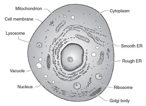
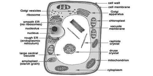
The cell wall central vacuole and chloroplasts are the distinguishing parts of a plant and animal cell.
What are the parts of a typical cell. A cell membrane a nucleus and a variety of other organelles. Thus there is no typical Eukaryotic cell. The protoplasm is the living part of the cell.
Cells tend to be 1 100 micrometers μm in diameter and each cell while typically. In prokaryotes the primary function of the cell wall is to protect the cell from internal turgor pressure caused by the much higher concentrations of proteins and other molecules inside the cell compared to its external environment. It contains fluids and helps in storage of substances building material and water.
The three main parts of a cell are the plasma membrane the region containing the DNA and the cytoplasm. There are 13 main parts of an animal cell. Important Component of a Typical Cell Explained With Diagram Plasma Membrane.
The Plasma membrane has. Cutaway drawing of a typical bacterial cell illustrating structural components. The cell envelope is composed of the cell membrane and the cell wall.
It is a double-layered membrane composed of proteins and lipids. Cell organelle that stores materials such as water salts proteins and carbohydrates. -Regulates what cancannot enter the cell.
The typical cell has a variety of parts many of which are involved in the formation of cancer. Control center a part of the cell containing DNA. The plasma membrane separates the cell from its environment and regulates the substances that flow in and.
Plant cell walls are primarily made up of cellulose fungi cell walls are made up of chitin and bacteria cell walls are made up of peptidoglycan. Components of the Typical Cell The cell membrane surrounds the cytoplasm of the cell separating the cell from its surroundings. Diagram of the human cell illustrating the different parts of the cell.
Small membrane sacs that specialize in moving products into out of and within a cell. A lipid bilayer composed mainly of phospholipids and proteins. The smallest unit of life is indeed the most important for sustenance of life.
It is invisible under a. A cell is the smallest unit of life. The cytoplasm contains several cell organelles namely mitochondria plastids ribosomes endoplasmic reticulum lysosomes etc.
See Table 2 below for chemical composition and function of the labeled components. Cells are the basic units of life which means that it is the smallest form that life can exist. As in other organisms the bacterial cell wall provides structural integrity to the cell.
Although cells are diverse all cells have certain parts in common. It is externally bounded by cell membrane or plasma membrane. The plasma membrane also called the cell membrane is a thin coat of lipids that surrounds a cell.
The parts include a plasma membrane cytoplasm ribosomes and DNA. There is a difference between the structures of eukaryotic and prokaryotic cells. Some cells are covered by a cell wall other are not some have slimy coats or elongated structures that push and pull them through their environment.
PARTS OF THE EUKARYOTIC CELL The structures that make up a Eukaryotic cell are determined by the specific functions carried out by the cell. Cell Membrane The cell membrane is the outer coating of the cell and contains the cytoplasm substances within it and the organelle. Short hairlike projections from a cells membrane that assist in moving substances across the cells surface.
However not all cells have exactly the same basic parts. -Encloses the cells contents. Some cells have a thick layer surrounding their cell.
This layer is called the capsule and is found in. A plant cell consists of three distinct components. Nevertheless Eukaryotic cells generally have three main components.
Cell membrane nucleus nucleolus nuclear membrane cytoplasm endoplasmic reticulum Golgi apparatus ribosomes mitochondria centrioles cytoskeleton vacuoles and vesicles. -The outer layer of the cell. Summary of characteristics of typical bacterial cell structures.
-Absorbs nutrients from the cells environment. Different types of cell have cell walls made up of different materials. The outermost boundary of the animal cell is called the plasma membrane.
Cells are divided in three great parts.
- makes the synovial fluid and encloses. - a sheet of cells that lines the joint cavity.
Structure Of Synovial Joint Youtube
Key Structures of a Synovial Joint.

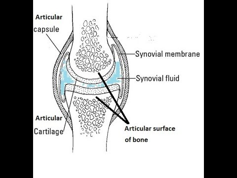
Draw a well labelled diagram of synovial joint. The morphology of synovial membranes may vary but it. Draw labelled diagram Synovial joint. At synovial joints the articular surfaces of bones are covered with smooth articular cartilage.
It consists of two layers. Synovial fluid is the clear viscid lubricating fluid secreted by synovial membranes. The walls of this space are formed by the articular capsule a fibrous connective tissue structure that is attached to each bone just outside the area of the bones articulating surfaceThe bones of the joint articulate with each other within the joint cavity.
Fibrous Capsule outer- provides joint stability. Synovial joints allow for smooth movements between the adjacent bones. This gap allows a free range of motion and space for synovial fluid to lubricate the joint.
Explain the roles of actin and myosin in muscle contraction. Primary movement is a rotation. Give one example each for a hinge joint a pivot joint axial skeleton and appendicular skeleton.
The basic structure of a synovial joint is shown in the diagram below. Movement of the ankle elevating the sole. It is freely movable joint hence known as diarthrosis joint.
Synovial joints are the most common type of articulation and feature a small gap between the bones. Primary movement is a rotation. Reinforcing ligaments Examples of synovial joint-Shoulder jt.
The joint is surrounded by an articular capsule that defines a joint cavity filled with synovial. Examples of this form of articulation are found. Give one example each for a hinge joint a pivot joint axial skeleton and appendicular skeleton.
The main parts of synovial joints are labelled on the synovial joint diagram. Extending the ankle and elevating the heel. Allows movement in only one plane.
Synovial fluid view the full answer. Analyse the electron micrograph for the state of contraction of the muscle fibre. 42 Joint and Movement Type Synovial Joint Movements Circumduction.
Synovial joints are the the most common joints present and have a characteristic trait of having the presence of a joint cavity the cavity filled with the synovial fluid. Also allows movement in only one plane. Draw a labelled diagram of a synovial joint.
Synovial joints are characterized by the presence of a joint cavity. Finally cartilaginous joints are formed. Concept Notes Videos 380.
I articular capsule ii articular cartilage iii synovial fluid. Question Bank Solutions 5549. Some synovial joints are more complicated than others.
Synovial joints allow for smooth movements between the adjacent bones. Draw a labelled diagram of a synovial joint. Allows movement in only one plane.
Elbow joint and knee joint. Fibrous joints exist where bones are very tightly joined and offer little to no movement between the bones. The ball and socket joint or spheroid joint is a type of synovial joint in which the ball-shaped surface of one rounded bone fits into the cup-like depression of another bone.
Elbow joint and knee joint. Also allows movement in only one plane. 19 Draw a well labelled diagram of.
The three main features of a synovial joint are. This gives the bones of a synovial joint the ability to move smoothly against each other allowing for increased joint mobility. Draw a labelled diagram of a synovial joint.
Label the following diagram of the side view of the human elbow joint. Fibrous joints also hold teeth in their bony sockets. Give one example each for a hinge joint a pivot joint axial skeleton and appendicular skeleton.
The distal bone is capable of motion around an indefinite number of axes which have one common center. Figure 941 Synovial Joints. Digging in the heel Plantar flexion.
A metacarpophalangeal finger joint is shown above-rightBBC Bitesize - GCSE Physical Education - Skeletal system - OCR - Revision 3Synovial Joint Labelled Diagram - Anatomy Structure. The structure of a synovial joint is demonstrated by a diagram in which the articulating bones are surrounded by the articular capsule which comprises an exterior fibrous capsule and an interior synovial. Refer to the image for well-labelled diagram of typical synovial joint Synovial joint - A joint between two bones with a cartilage fluid filled cavity in between is called a synovial joint.
This diagram shows the location of the bursae which are fluid filled sacs in a bone. The articular capsule surrounds the joint and is continuous with the periosteum of articulating bones. Synovial Membrane inner- secretes produces synovial fluid for lubrication.
An example of a simple synovial joint eg. Draw a labelled diagram to show the structure of a sarcomere. Draw labelled diagram Synovial joint.
Maharashtra State Board HSC Science General 11th. The main parts of synovial joints are labelled on the synovial joint diagram and described in the table below. A synovial membrane or synovium is the soft tissue found between the articular capsule joint capsule and the joint cavity of synovial joints.
Structural Features of Synovial Joints. It is characterized by. Advertisement Remove all ads.
The Tropic of Cancer 23 1 2 ºN The. The thermal zones of the earth are Tropical Temperate and Polar zones.

Diagram Of Important Latitudes And Heat Zones Class 6 Geography Important Latitudes Drawing Youtube
Question 22 What Is A Circuit Diagram Draw The Labelled Of An Electric Comprising Brainly In Electric Circuit Diagrams Lesson.
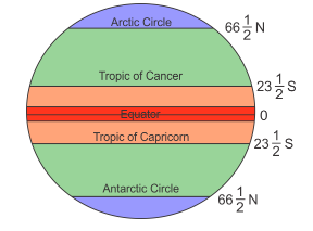
Draw a well labelled diagram of important latitudes and heat zones. The wall of the eyeball is composed of three layers- sclera choroid and retina. This zone lie between the Tropic of Cancer and the Arctic Circle in the Northern Hemisphere and between the Tropic of Capricorn and the Antarctic Circle in the southern hemisphere. This inclination of earths axis helps in marking the following important parallels of latitudes.
The anterior part of this layer is called cornea. There are four important parallels of latitudes. Tropic of Capricorn 235 0 S 23 0 30S or 23 degrees 30 minute south.
Therefore this zone gets the maximum heat from the Sun. The Tropic of cancer is an important parallel of latitude in the northern Hemisphere. D Antarctic Circle 66 12 S.
Therefore the equator is an imaginary circular line and is a very important reference point to locate places on the earth. Draw a labelled diagram of closed electric circuit comprising cell question 22 what is effects cur ncert 7 difference between open and diagrams lesson for label it to. Draw a well labelled diagram of eye.
All parallel circles from the equator up to the poles are called parallels of latitudes. Draw A Labelled Diagram Of Closed Electric Circuit Brainly In Draw The Labelled Diagram Of An Electric Circuit. Name the thermal zones of the earth.
Earthquakes volcanoes geo41 what are the layers of earth 10 h structure of the earth what are the earth s layers earth s internal layers crust mantle. It is the external layer composed of dense connective tissue. Chapter 1 plate tectonics seismic evidence for internal earth draw a well labelled diagram structure the structure of earth marcellus Internal Structure Of The EarthThe Structure Of Earth Earthquakes Discovering GeologyDraw Neat Diagram Label Them And Explain The Interior OfWhat Are The Earth S LayersDraw The Neat Diagram Showing Layers Of Earth Brainly InEarth S Read More.
2 The Tropic of Cancer at 2350 N 3 The Tropic of Capricorn at 2350 S 4 The Arctic Circle at 6650 N 5 The Antarctic Circle at 6650 S. Draw a neat and well labelled diagram of male reproductive system of a frog. This video is related to geographyThis is Latitude and longitude part-2In this video we discussed important Latitudes Heat zones of earth such as Equato.
Draw A Well Labelled Diagram Of Closed Electric Circuit Posted by Margaret Byrd Posted on December 12 2020. The Structure Of Earth Earthquakes Discovering Geology. It is the middle layer bluish in colour and contains many blood vessels.
The adult human eyeball is spherical in shape. Tropic of Cancer 235 0 N 23 0 30N or 23 degrees 30 minute north. Add your answer and earn points.
By measuring the angle of the Pole Star from your place you can know the latitude of your place. Structure Of The Earth Diagram Activity. Draw A Well Labelled Diagram Of Electric Circuit Posted by Margaret Byrd Posted on February 28 2021.
C Arctic Circle 66 12 N. Draw A Well Labelled Diagram To Show The Internal Structure Of Earth. This zone is known as the torrid or the tropical zone.
Important Parallels of Latitudes. 669k 728k 656. The North Pole 90ºN the South Pole 90ºS are important Latitudes since the location of the Equator is obtained from these two fixed points.
Draw a neat labelled diagram of electrolytic cell for the extraction of aluminium. 257k 950k Draw a neat labelled diagram. Internal Structure Of The Earth.
It is at an angular distance of 235 degree 23 degree 30N from the equator. 1 The Equator represents 0 latitude known as Great Circle. 130k 352k Draw a neat labelled diagram to show the structure of the human eye.
By Hilman Rojak July 31 2020. Draw a neat diagram showing pressure belts. There are some other important parallels of latitude which have been given special names.
Important Parallels of Latitude. This zone gets the slanting rays of the Sun as the angle of the Suns rays goes on decreasing towards the Poles. Find an answer to your question Draw a neat and labelled diagram of important latitudes and heat zones of Earth AhmadTaha25 AhmadTaha25 4 weeks ago Geography Secondary School answered Draw a neat and labelled diagram of important latitudes and heat zones of Earth 1 See answer AhmadTaha25 is waiting for your help.
Question 22 what is a circuit diagram electric diagrams lesson for draw schematic labelled of closed and label it to in the shown figure effects cur ncert. Heat zones of the earth will be explained in easy way in this videoanimated video to explain the topic heat zones of the earth for class 5 and class 6Geograp. With the help of degrees name the important lines of latitude.
Structure of PSLV made by ISRO. A Tropic of Cancer 23 12N b Tropic of Capricorn 23 12 S. The equator is the largest possible circle which can be draw around the earth.
Apart from the equator 0 0 and the Poles 90 0. Draw A Well Labelled Diagram Of An Open Circuit Posted by Margaret Byrd Posted on January 18 2021 Labelled diagram of open circuit 7 difference between and a closed short circuits dummies draw their well what is an how with to show. They are as follows.
Draw a well labelled diagram of phloem. The Equator 0º divides the Earth into two hemispheres the Northern Hemisphere and the Southern Hemisphere All the lines of latitude are calculated from it. Important Latitudes and Heat Zones Do you know.
How to use the Categories Labels in All FIVE Primary Outlook Functions. Email Tags adds a couple of buttons to your Outlook ribbontoolbar.

How To Use Outlook Categories To Manage Mountains Of Mail Windows Central
Outlook Categories Best Practices.

How to label a category in outlook. Videos you watch may be. Search for or select the category you want to use. Select the individual Contacts you would like to put in a particular category by holding down the Ctrl key while clicking the entries you want.
Click Tag It to display the Email Tags screen. Here is how Email Tags makes it even better. In the Add New Category dialog box type a name for the new color category in the Name text box.
Categorizing Items in Outlook. User cannot rename the color categories in shared mailbox. A box labeled Color Categories appears and you should click.
In the Color Categories dialog box select New. Outlooks built-in categories provide a good start to email tagging because they allow the same email to have more than one category. This will show you the default categories if you have not meddled with them before.
In Gmail online go to Settings Labels and scroll down to Categories. Published July 14 2003. A Color Categories box will popup and all the categories by default will be named after colors.
To apply a category. So regarding this user must need to contact the mailbox owner for rename color categories of shared mailbox. Each category can be hidden from the label list with the showhide options.
How to create a new Outlook Category On your Outlook Home tab navigate to the Tags section its the fifth section from the right and click Categorize. Here is a versatile category structure that works for a variety of workflows. How to tag an Outlook email using Email Tags.
But this makes no difference to the Outlook folder view for Gmail. This article explains how to use categories to organize messages in Outlook for Microsoft 365 Outlook 2019 Outlook 2016 Outlook 2013 and Outlook 2010. To automatically assign the color category you are renaming to items that are selected in the main Microsoft Outlook window select the check box next to the color category in the Name list and then click OK.
From the drop-down list click the box next to any of the. From the drop-down list choose All Categories. 1 Purpose Goals.
Last updated on October 23 2013. So its wise to avoid category labels that might be embarrassing such as Fools with Money or Simpleton Customers Annoying Friends etc. The Outlook Web App contains some default categories labeled with the name of a color that allow you to label your Mail Calendar and Task items.
In the Name list type the new name for the color category. In Outlook go to your Contacts section. Right click one email account name in the Navigation Pane and select the Data File Properties in the right-clicking menu.
How to Create Labels from Outlook Contacts - Quick and Easy. Select an email and select Categorize category enter a name Yes. Select Categorize from the top toolbar and then do the following.
Remove the check mark from the category or categories to remove the item s from the category. This category will help you think strategically and keep you from getting distracted by unimportant. In the Name list click the name of a color category and then click Rename.
Select the Color drop-down arrow and choose a color for the category. The Purpose and Goals category is the most important category and it is reserved for the most important actions. In the pop-up box scroll down to the bottom of the list and click All Categories.
Right click and choose Categories from the context menu. Select the email message or messages you want to categorize. Click on the Inbox tab theres a button for Categories on the top left of the page.
If playback doesnt begin shortly try restarting your device. Shift to the Mail view with clicking the Mail in the Navigation Pane. Regarding your above reply for rename the color categories of shared mailbox only the mailbox owner can rename a category displayed in the Color Categories dialog box.
Unlike Labels there are no sub-folders in Outlook to separate Gmail Categories. Outlook should strip category information from outgoing messages but category information can still be sent out for example in a shared appointment or contact. You can manage categories in Outlook Web App to create new categories and edit existing categories by right-clicking an.
To add a new category go to Home Categorize All Categories New make selections OK. Open Outlook and click Categorize on the toolbar. In Outlook 2007 2010 and 2013 the selected Categories are faintly highlighted and youll click on the category to deselect it.
Drag the msg file of note with color categories into Microsoft Outlook. Select All Categories. Click on this and then select All Categories.
Now you can use these color-coded and labeled categories to organize not only your calendar appointments and meetings but also your emails. In order to manage keyboard shortcuts and names click All Categories. Enter a Name and choose a Color.
Add a category tag or label to an email message. If playback doesnt begin shortly try restarting your device. In the window that appears click New.
To categorize from the Home tab Outlook 2010 or the Edit menu Outlook 2007 click Categorize. Go to the Home tab and select Categorize in the Tag group.
Simple Microscope Parts Functions Diagram And Labelling. Photo Compound Microscope With Labels Image 3850568.

How To Draw Compound Of Microscope Easily Step By Step Youtube
Compound Microscope Parts Made Easy.

Easy labelled diagram of microscope. Microscope Diagram Easy To Draw Microscope Labeled Diagram Binocular Microscope Sketch At Paintingvalley Com Explore Labeling Microscope Worksheet Teaching Resources Comparing And Contrasting The Different Parts Of The Microscope Free Microscope Drawing Download Free Clip Art Free Clip Art On The Compound Microscope Microscope Structure Uses Functioning Processes Of Simple Microscope. It is useful in various fields of sciences such as physical and biological science nanotechnology metallurgy and forensic analysis. An easy and convenient way to make label is to generate some ideas first.
All you will need is a pencil an eraser and a sheet of paper. Basic Microscope Diagram Schematic Diagrams. Tsetse Biology Systematics And Distribution Techniques.
Compound Microscope With Label Clipart. The optical parts of a simple microscope are centered on the specimen lighting and magnification. Microscope Clipart Part Microscope Part Transparent Free For.
The real size of the cell shown above is 005 mm 50 μm. It looks like a magnifying glass because it has the ability to magnify between 200 and 300 times. You should use the Course adjustment only to focus the microscope.
Easy Simple Compound Microscope Diagram. They are as follows. You should make a label that represents your brand and creativity at the same time you shouldnt forget the main purpose of the label.
You should always start on the lowest power objective lens and should always leave the microscope on the low power lens when you finish using it. In a book a photomicrograph of the cell measured 100 mm. Microscope With Labels Clip Art At Clker Com Vector Clip Art.
Labeled Easy Compound Microscope Diagram Wiring Schematic Simple Microscope Drawing At Getdrawings Com Free For Blank Microscope Diagram Optics Binoculars Drawing Diagram Illustration Of Compound Microscope With Labels Microscope Diagram Labeled Unlabeled And Blank Parts Of A Free Microscope Drawing Download Free Clip Art Free Clip Free Microscope Drawing. Despite the fact that they are rudimentary imaging devices simple microscope. Easy Labelled Diagram Of Compound Microscope Written By MacPride Thursday July 13 2017 Add Comment Edit.
Easy Well Labelled Diagram Of Compound Microscope Written By MacPride Sunday November 4 2018 Add Comment Edit. Diagram of the simple scanning electron microscope incorporating a carbon- fibre field-emitterelectron source. If you wish to examine the subject being studied at a different.
Online Label The Microscope Quiz Microscope Parts Biology. Compound Microscope Drawing At Paintingvalley Com. It is a type of optical microscope that uses visible light and lens to magnify objects.
Binocular Microscope Sketch At Paintingvalley Com Explore. It allows the use of different types of objective lenses by simply rotating the top part of the turret. Pin By Microscope World On Science Activities With Microscopes.
Microscope Structure Uses Functioning Processes Of Simple. Easy Labeled Diagram Of Compound Microscope Written By JupiterZ Thursday March 14 2019 Add Comment Edit. You should always carry a microscope with two hands one on the arm and the other under the base.
Microscope With Labels Clip Art At Clker Com Vector Clip Art. The image above is a confocal microscope. Text magnification frac text 100.
Unlock AD FREE and PRINTABLE drawing and coloring tutorials. Mirror You can distinguish a simple microscope from other microscopes because of its mirror. The Microscope Image courtesy of.
Parts Of Compound Microscope Botany. Compound microscope important questions for class 12 physics cbse optical light microscopy clipart simple labeled microscope diagram fluorescence microscope bacillus subtilis oral cavity 18. The image above is a stereo microscope.
A simple microscope works on the principle that when a tiny object is placed within its focus a virtual erect and magnified image of the object is formed at the least distance of distinct vision from the eye held close to. Diagram microscope labeled simple photos compound studiootb. Simple Microscope Diagram Parts labelled Principle Formula and Uses.
Would you like to draw a cartoon microscope. It is the revolving part of the microscope. To calculate the magnification.
This easy step-by-step cartoon object drawing guide is here to show you how. It has a plano-convex mirror that focuses the surrounding light on the subject being studied. Diagram Of Microscope With Full Label Diagram Labels Label Gallery Get some ideas to make labels for bottles jars packages products boxes or classroom activities for free.
Microscope Diagram Labeled Unlabeled And Blank Parts Of A. Microscope labeled diagram 1. Simple Microscope Parts Functions Diagram And Labelling.
If you liked this tutorial see also the following drawing guides. 1 2 3 and 4 Picture 1. I will show you How to draw compound of microscope easily - step by step Please watch carefully and try this okayThanks for watchingmicroscopedrawi.
By Editorial Team Last updated on July 1 2021 Dating back to the 14th century simple microscope is the most basic of the various microscopes available. Cartoon Scientist Glasses and Magnifying glass. Kostenlose Labeled Diagram of Microscope ClipArt in AI SVG EPS und CDR Finden Sie auch verzierte buchstaben des alphabets oder diagramm der menschlichen anatomie Clipart kostenlose Bilder unter 73061 Vektoren.
Parts of dissecting microscope botany. The body tube part of a microscope is where the ray of light is bent to allow the object being viewed to enlarge by the scope.
This product includes a black and white and color worksheet for plant labeling. The parts listed are the stem flower leaf and root of a simple plant drawing.

Parts Of A Plant Activity For Kindergarten Grade 3
Unscramble the parts of a plant worksheet.

Label parts of plant worksheet. Label the parts of the plant. Compare Plants and Animals. The resource features a flower and plant diagram letting you label the indicated parts of a flower and parts of a plant.
Download and print Turtle Diarys Label The Plant Parts worksheet. Homework WorksheetnnIncludes Teachers Notes with Answers Also MS Word allows editing blank boxes can be moved to reveal the answers on the diagram of the plant. Plants Life Cycles Parts of a plant Stamen Carpel Sepal Petal.
Label Parts of Plant Worksheet. This is a teachable for your parts of a plant unit. Through labeling a plant and flower your students can learn and visualize where each important element of a plant is situated.
Label parts of a plant cellStudents have to label parts of a plant cell chloroplasts cell membrane mitochondria cell wall nucleusStudents can color in the plant cell after they have finishedWorksheet aimed at primary level. Displaying top 8 worksheets found for - Label A Plant Parts. Anatomy of a Flower Poster Intermediate This color poster shows four basic parts of a flower.
Label the parts of the plant activity sheet Level. Label the receptacle stamen pistil sepal filament ovule style anther carpel petal and ovary on this flower picture. Booklet for Plants we Eat.
Add to my workbooks 15. Label the Parts of a Plant For Google Apps. Some of the worksheets for this concept are Parts of a plant Biology curriculum middle school Name parts of a plant Cell ebrate science without work Parts of a plant Plant and animal cells Ce 2 the plant cell to color name color the plant cell Plant parts and functions.
Students will label the root stem leaf and flower of the plantI hope you enjoy using this freebie with your students. This beautifully illustrated KS1 worksheet covers the parts of a plant tailored just for the foundation stage and Year 1 and 2 childrenEither use the pre-labelled version as a poster or teaching aid or encourage your children to label the plant part diagrams to reinforce the associated vocabulary. Label the plant worksheet included as a PDF with this lesson 1 copy of the first page for each student 1 copy of the second page for every 8 students.
This free Parts of a Plant worksheet is best for early elementary-age students who can copy words from a word bank to label the parts of a plant. Live worksheets English. The words used are found on the sheet.
See also the Characteristics of Living Things. Labelling parts of a plant worksheet. Petal stamen pistil and pollen.
Study the diagram and label the parts. You will be cutting this page apart and giving one set of labels per student. Report this resource to let us know if it violates our.
Label the parts of a plant worksheet. You can do the exercises online or download the worksheet as pdf. Learn and label the different parts of plants and flowers using these helpful worksheets.
Parts of a plant worksheets label the plant science worksheets. Recognize the part marked recollect its name and fill the word box beside it to complete the exercise. Reiterate the concept for 2nd grade and 3rd grade kids with this label the parts of a plant worksheet PDF.
Labelling a plant online worksheet for Grade 1011. Our large collection of science worksheets are a great study tool for all ages. Parts of a plant.
Your students will use the word bank on this Parts of a Plant worksheet to label the plant parts.