Write in numbers or pictures in each of the boxes labeled with a sign. In this video I show you how to use and play a paper Fortune Teller Cootie Catcher.

Teaching With Cootie Catchers Lessons4now
Once you created your origami you are ready to play your game.

How do you label a cootie catcher. You may rely on it. You can add designs or use colorful markers to make the outside of your cootie catcher pop. When you flatten out the catcher the inside tabs should be labeled on the outside with a number.
For an added challenge have students come up with the content to include. Flip your square over so you now see the inside tabs. Cootie Catcher Fortune Teller Fold two corners together and crease firmly.
The player will choose a number and you will open the fortune teller message by using your fingers inside the cootie catcher pockets. Finally fold the paper in. Open and close the Cootie Catcher while counting out the number picked on step 3.
Write the numbers 1-8 in each triangle and add a picture of each emotion on the inside. Cootie Catcher Fortune Teller Fold two corners together and crease firmly. When youve stopped counting look inside and let your friend choose again.
Under the inside flaps you have to write a fortune or prediction. Whats another name for cootie catcher. Folding instructions for blank cootie catcher template.
On top of those flaps you need to write a number. Make it simple by labeling the outside with colors and numbers on the inside. In each of the boxes labeled with text write your fortunes in the direction that the tiny sample text is written in the corners of the template so that it makes sense when you open it up.
Turn your square over so the flaps are facing up. Fold each corner point into the center. Under the inside flaps you have to write a fortune or prediction.
This is the editable cootie catcher template for reading and speaking games. When you are holding the catcher each of the four corner tabs should be labeled with a color. Flip it over and fold all four of these corners into the center.
Flip it over and fold all four of these corners into the center. How to Make a Cootie Catcher. Red has three letters so stop after three moves and ask your friend to choose again.
Then turn the paper over. What do I write in a cootie catcher. A fortune teller also called a cootie catcher chatterbox salt cellar whirlybird or paku-paku is a form of origami used in childrens games.
Place the square facedown and fold each of the four corners in so the points meet in the center. Look for the number on the square selected open and close the Cootie Catcher the right number of times. Want to try it out.
Make them pick one. Stop at the last letter to reveal some of the numbers on the flaps. Spell out the color opening and closing the cootie catcher along with the letters.
Fold the corners toward the center again. 1-- Find a friend and ask him or her to pick a colour on top flaps 2-- Moving the flaps in and out and side to side in time with the lettersspellout the name of the colour 3--Stop on the last letter with the cootie catcher open to reveal the numbers ask your friend to pick a numberCount out the number and open the the flaps in and out and side to side like step 2When your done counting your first number get. Who invented Cootie catchers.
Ask a friend to choose one and then move the cootie catcher with your fingers so it snaps open first one way and then the other imagine a mouth talking. On top of those flaps you need to write a number. See how to make a Paper Fortune TellerCootie Catcher step-by-step ins.
Turn the paper over so the flaps are now facedown. Parts of the fortune teller are labelled with colors or numbers that serve as options for a player to choose from and on the inside are eight flaps each concealing a. Count the numbers open close side to side then choose again.
To use your cootie catcher you will stick your thumb and two fingers in the pockets and open the origami to reveal a fortune teller. On each square write the name of a color. Open up and down and side to side as you count the right amount they picked.
Create a Cootie Catcher that asks about the character actions in The Westing Game Puffin Modern Classics. Typically there are three main sections of the cootie catcher that need to be labeled. You can change the texts and pictures accordingly.
Heres a FREE set of 4 themed Cootie Catchers for personal andor classroom use waiting for you to add the content your students need. The printable cootie catcher is blank and you can make it any way you want. Fold each corner point into the center.
As you do this spell out what they have chosen for example if they choose red spell out R-E-D. When you label a completed cootie catcher numbers go on the outside most flaps the ones your fingers go under colors go on the next layer of flaps and questions go on the very inside of the cootie catcher underneath the inside flaps. Add your fortunes and information to the cootie catcher.
Cut out your cootie catcher and color it. Here are some answers you might write under the flaps. Place the square faceup and fold and unfold the square in diagonals from corner to corner so you end up with x shaped creases.
What do you put inside a cootie catcher.
The Cell The Biology Primer. Find Human Cell Diagram stock images in HD and millions of other royalty-free stock photos illustrations and vectors in the Shutterstock collection.

Draw A Neat Diagram Of A Typical Animal Cell And Label The Following Organelles 1 The Organelle Called Science Cell Structure And Functions 10429465 Meritnation Com
Diagram of the human cell illustrating the different parts of the cell.

Labelled diagram of typical cell. Comparison of Prokaryotic Cells and Eukaryotic Cells and 2. In prokaryotic cell there is naked DNA without histone protein but in Eukaryotic cell there is DNA with histone protein. However it typically exists in three forms ie.
LPS teichoic acid etc surrounding the bacterium like a shell and lies external to the cytoplasmic membrane. It is a tough and rigid structure of peptidoglycan with accessory specific materials eg. You can also put your logo at the top or bottom corner of the label.
Structure and Functions With Diagram Let us make an in-depth study of the structure and functions of cell. Stock vector labelled diagrams of typical animal and plant cells with editable layers 222613513 in cell diagram simple the structure and contents of a typical animal cell every has membrane cytoplasm nucleus but not all cells have rat liver cell a scheme of the typical cell membrane structure with lipid bilayer integral proteins. Cell is a compartment where all the activities of life takes place.
Targeting Strategies for Multifunctional Nanoparticles in. A typical bacterial cell. Structure of a typical animal cell click to enlarge.
Prokaryotic cells article Khan Academy. Label Gallery Get some ideas to make labels for bottles jars packages products boxes or classroom activities for free. The Golgi Apparatus is the cell organelle mostly present in eukaryotic cells which is responsible for the packaging of macromolecules into vesicles so that they can be sent out to their site of action.
A typical bacterial cell - Labelled diagram free-floating DNA plasmid circular ring of DNA slime capsule stops the cell from drying out flagellum helps the bacterium move through liquids cell membrane and cell wall the label was too big to tell these apart. The lipid molecules on the outer and inner part lipid bilayer. Which biological activities will be hindered and why.
Most of the times we put the labels to show some specific information. Illustration of Labelled diagrams of typical animal and plant cells with editable layers. On the contrary plant cells lack centrioles and intermediate filaments which are present in animal cells.
Examining a diagram of the plant cell will help make the differences clearer. Labelled diagram of a plant cell under microscope posted on march 18 2011 by admin onion cells stained with methylene blue look at the images of onion cells as they would be seen under a microscope draw each magnification label appear high picture plant and animal cell diagrams. It is a double-layered membrane composed of proteins and lipids.
Labelled Diagram Of A Typical Animal Cell. In prokaryotic cell DNA is circular but in Eukaryotic cell DNA is double helix. Vector art clipart and stock vectors.
A Draw a fully labelled diagram of a typical receptor tyrosine cell surface recepto b Name the other classes of enzyme coupled receptors and describe their mechanism of action. Thousands of new high-quality pictures added every day. Labeled diagram of a typical plant cell.
Cisternae vesicles and tubules. Typical Animal Cell Diagram Labeled Best Of Typical Animal And Plant Animal Cell Coloring Page Lovely 25 Best Typical Plant Cell Ideas On Click The Animal Cell Diagram To Enlarge It Wiring Diagram Blog Eukaryotic Cells Biology I Typical Animal Cell Plant And Animal Cells S Cool The Revision Website Animal Cell Anatomy Diagram Structure With All Parts Nucleus Cells Alive Plant Cell. Typical Plant Cell Diagram Labeled Battery electricity Wikipedia.
1in prokaryotic cell there is primitive nucleus but in Eukaryotic cell there is well developed nucleus. After reading this article you will learn about. Room Full of Crazy TV Tropes.
Western Wood Products Association. A Draw an annotated diagram of a typical student microscope b Explain the. Unit 2 Cells Biology Junction.
The single circular double-stranded chromosome is the bacterial genome. An easy and convenient way to make label is to generate some ideas first. Cell Membrane The cell membrane is the outer coating of the cell and contains the cytoplasm substances within it and the organelle.
Labels are usually small in size so you should carefully choose the font of the texts to make sure it is readable. C Taxol from the Yew tree stabilises microtubules. Hello Guys In this Video Im going to teach you guys How to Draw Structure of a cell Labeled DiagramSubscribe to the Channel and also share this video.
Other structures include cytoplasmic membrane mesosomes. The structure of the Golgi Complex is pleomorphic. Labelled diagram of a typical animal cell.
INLINE PICTURES VERSION W1TP. Intro to eukaryotic cells article Khan Academy. It gives shape to the cell.
It is 10-25 nm in thickness. Structure and Components of a Human Cell. Human Cell Diagram Stock Vector Royalty Free 400272997.
It is brown to pale-grey in colour and measures 215-3 cm x 12-15 cm. This is the well labelled diagram of liver fluke.

How To Draw Liverfluke Fasciola Hepatica Easy Way Step By Step Youtube
In these organs they produce pathological lesions leading to parasitic diseases.
Labelled diagram of a liver fluke. Acoelomate a labeled diagram of a fluke labelled diagram of liver draw it a generalized life cycle of the liver fluke albendazole oral liver flukes troccap fluke flatworm britannica com liver flukes xanadufarms free download here pdfsdocuments2 com a well label diagram of a liver inner. The anterior body part is broader than the posterior part which is blunt in outline. Infections in humans usually occur after eating contaminated raw or undercooked freshwater fish or watercress.
Diagram Of Liver Fluke. Liver flukes are a group of about fifteen different species of parasites that share a number of characteristics during their life cycle and result in a similar etiology when they infect humans. Liver fluke disease is a chronic parasitic disease of the bile ducts.
The liver is a very hardworking organ that filters more than a liter of blood per minute. Lungworm and liver fluke to threaten livestock this autumn. Liver flukes are an important cause of acute and chronic disease in grazing sheep and cattle.
Add your answer and earn points. Liver fluke disease fasciolosis is caused by the trematode parasite Fasciola hepatica. Disease can result from the migration of large numbers of immature flukes through the liver or from the presence of adult flukes in the bile ducts or both.
Your risk of infection increases if you travel to parts of the world where the. Labeled Diagram Of Liver Fluke. Suggested in case of lung flukes.
The mouth of liver fluke is anterior and terminal surrounded by. Some selected sexual organs concerning with the passages of the spermatozoa and the eggs were obseved in detail. How to draw a liver fluke in exam is.
Draw A Neat Labelled Diagram Of Liver Fluke State The Phylum To Which It Belongs And Brainly In Fluke eggs which are passed in the faeces control of liver fluke disease should be an important part of a farm health plan drawn up with the witholding period information is carried on the product label and datasheet and advice should be. The conclusion of this study is that the Laurers canal may be the copulatory organ of the female reproductive system as Miyazaki et al. Fasciola hepatica also known as the common liver fluke or sheep liver fluke is a parasitic trematode fluke or flatworm a type of helminth of the class Trematoda phylum Platyhelminthes.
It may reach a size of 3 cm in length and 15 cm in breadth. They are principally parasites of the liver of various mammals including humans. Liver fluke can infect all grazing animals and man but mainly affects sheep and cattle.
640x480 well labeled diagram of liver fluke labelled diagram of liverLiver fluke in sheep also known as. A well-labeled pencil sketch diagram of liver fluke - 21306762 olubimioye olubimioye 22082020 Biology Secondary School answered A well-labeled pencil sketch diagram of liver fluke 1 See answer olubimioye is waiting for your help. Liver cell cardiac cell nerve cell skin cell.
640x480 well labeled diagram of liver fluke labelled diagram of liver. This is the well labelled diagram of liver fluke. Liver Fluke Labelled Diagram çšå¾çæ œççæžœ Chart Map Map Screenshot.
Body of liver fluke is soft flattened leaf-like with a triangular head lobe Fig. Draw A Neat Labelled Diagram Of Liver Fluke. Liver fluke is a collective name of a polyphyletic group of parasitic trematodes under the phylum Platyhelminthes.
In this article we will discuss about the external morphology of liver flukes. It is dorsoventrally flattened oval in shape like a leaf and faint brownish in colour. Free ncert and other textbook solutions.
From wikipedia the free encyclopedia. A related parasite fasciola. They are principally parasites of the liver of various mammals including humans.
For example the liver makes and processes many body fats. Draw a neat labelled diagram of Liver fluke State the phylum to which it belongs and its - 34158509. Fluke infection is estimated to cost the uk agriculture industry about 300 million a year.
Combination with levamisole have a specific. It infects the livers of various mammals including humans and is transmitted by sheep and cattle to humans the world over. A labeled diagram of a fluke labelled diagram of.
The body is covered with a cuticle the greater portion of which bears minute spines. While most infected persons do not show any symptoms infections that last a long. Capable of moving along the blood circulation they can occur also in bile ducts gallbladder and liver parenchyma.
Diagram of Liver Fluke How To Draw Liver Fluke Labelled Diagram Biology Diagram - YouTube. A labeled diagram of a fluke liver fluke anatomy gallery human anatomy learning. Diagram Of Liver Fluke With Label.
Diagram Of Liver Fluke With Label. 231 draw and label a diagram of the ultrastructure of a liver cell as an example of an animal cell. Beranda Diagram Of Liver With Labelling.
Liver fluke is a collective name of a polyphyletic group of parasitic trematodes under the phylum platyhelminthes. Anatomy of the human pancreas explained with labeled diagrams. Structure of Liver Fluke.
In this article we will discuss about the external morphology of liver flukes. Vector illustration in flat style isolated over white background.
Browse 378318 the human body stock photos and images available or search for the human body anatomy body or the human body photos to find more great stock photos and pictures. Each of these muscles is a discrete organ constructed of skeletal muscle tissue blood vessels tendons and nerves.
Labeled Muscles Of The Human Body Anterior View 3d Rendering Stock Photo Download Image Now Istock

The muscular system is responsible for the movement of the human body.

Pictures of the human body muscles. Human body muscle system the muscles of the human body that work the skeletal system that are under voluntary control and that are concerned with movement posture and balance. Find high-quality stock photos that you wont find anywhere else. Thousands of new high-quality pictures added every day.
For mobile and web. For general help questions and suggestions try our dedicated support forums. Search from Human Body Muscles stock photos pictures and royalty-free images from iStock.
Contains such icons as bodybuilding heartbeat swimming cycling running diet. In these organs muscles serve to move substances throughout the body. Find human muscle anatomy stock images in HD and millions of other royalty-free stock photos illustrations and vectors in the Shutterstock collection.
- muscles body stock illustrations. Find high-quality stock photos that you wont find anywhere else. Find Download the most popular Human Body Muscle Photos on Freepik Free for commercial use High Quality Images Over 9 Million Stock Photos.
Attached to the bones of the skeletal system are about 700 named muscles that make up roughly half of a persons body weight. - human body muscles stock illustrations. Superficial and deep posterior muscles of upper body Anterior and posterior muscles of the upper arm Anterior and posterior muscles of the lower arm Anterior and posterior muscles of upper leg Posterior muscles of lower leg Anterior muscles of lower leg Respiratory muscles.
Outer superficial muscles between ribs slopes downward and an. Smooth muscle tissue - human muscles anatomy stock illustrations. Search from Human Body Muscles Labeled stock photos pictures and royalty-free images from iStock.
Affordable and search from millions of royalty free images photos and vectors. We hope your visit has been a productive one. For mobile and web.
Human back muscles - human muscles anatomy stock illustrations. Newest results human muscle. There are around 650 skeletal muscles within the typical human body.
Contraction leads to flattening of the muscle leading to inhal. Thousands of new high-quality pictures. Browse 219601 human muscle stock photos and images available or search for human muscle anatomy or human muscle tissue to find more great stock photos and pictures.
Fitness and workout line icons. - human muscles anatomy stock pictures royalty-free photos images. Unit 8 upper body muscles descriptions and pictures.
Muscle tissue is also found inside of the heart digestive organs and blood vessels. Flexes neck to look down unilaterally. 3d render depicting the anatomy of a human muscular system.
The human body - human muscles anatomy stock illustrations. Nevertheless the exact number is difficult to define. Browse 87557 human body muscles stock photos and images available or search for anatomy or human anatomy to find more great stock photos and pictures.
Select from premium Human Body Muscles of the highest quality. Find muscle anatomy stock images in HD and millions of other royalty-free stock photos illustrations and vectors in the Shutterstock collection. This is a table of skeletal muscles of the human anatomy.
Human heart and vascular system - the human body stock pictures royalty-free photos images. Choose from Human Body Muscles Pictures stock illustrations from iStock. Download human body anatomy muscles stock photos.
In this image you will find frontalis orbicularis oculi zygomaticus masseter orbicularis oris sternocleidomasteoid deltoid pectoralis major biceps brachii iliopsoas adductor longus. Contains such icons as bodybuilding heartbeat swimming cycling running diet. If youre having any problems or would like to give some feedback wed love to hear from you.
If you need to contact the Course-NotesOrg web. Find high-quality royalty-free vector images that you wont find anywhere else. Fitness and workout line icons.
Almost every muscle constitutes one part of a pair of identical bilateral muscles found on both sides resulting in approximately 320 pairs of muscles as presented in this article. Browse 94444 muscles body stock photos and images available or search for oxygen or the human body to find more great stock photos and pictures. Find the perfect Human Body Muscles stock photos and editorial news pictures from Getty Images.
Describe the structure and give the name of four cell organelles. The tabernacle consisted of a tent-like structure the tabernacle proper covered by rug-like coverings for a roof and an external courtyard 150 feet by 75 feet.

Draw A Labelled Diagram Of The Basic Body Plan Of Chordates Brainly In
Click hereto get an answer to your question Draw a labelled diagram of the structure of mature dicot embryo.

Draw a labelled diagram of the basic body plan of chordates. 167953095 stock photos online. New users enjoy 60 OFF. What are the 5 major body plan characteristics of the Phylum Chordata Draw a from BIO 102 at Santa Barbara City College.
Draw a labelled diagram of the experimental set-up to study the latent heat of vaporisation of water. Soft iron core PrincipleTransformer is based on the principle of electromagnetic mutual inductionWhen the current flowing through the primary coil changes an emf is induced in the secondary coil due to the change in magnetic flux linked with the primary coil. The total surface area of the cerebral cortex is about 2500 cm2 and when stretched it will cover the area of a night table.
The fence was made of linen hangings held by pillars. It contains 8 proteins 1 carbohydrates 2 soluble organics and 1 insoluble salts. Draw a neat labelled diagram showing the LS of a typical flower.
Quizlet flashcards activities and games help you improve your grades. A flower is a seed-bearing part of a plant consisting of reproductive organs stamens and carpels that are typically surrounded by a brightly coloured corolla petals and a green calyx sepalsFlowers are attractive and appear in different colours and shapes to. All vertebrates are built along the basic chordate body plan.
Draw a labelled diagram of the basic body plan of chordates. A stiff rod running through the length of the animal vertebral column with a hollow tube of nervous tissue the spinal cord above it and the gastrointestinal tract below. I Labelled diagram of a step-down transformer.
Basic chordate body plan study guide by Venabiezer includes 8 questions covering vocabulary terms and more. The brain is composed of 77 to 78 water and 10 to 12 lipids. The worksheet has two pages.
The whole compound was surrounded by a high fence about 7 feet in height. Drawing Anatomy Diagram Anatomy Of Human Body. Download 782 Fertilization Diagram Stock Illustrations Vectors Clipart for FREE or amazingly low rates.
Asked May 21 2020 in Structure of Living Organisms by Rukmani 511k points. Your lungs human body human body systems match column a with. This course covers the NCERT syllabus for Chordates.
It has a diagram of the human body to label and a diagram of the human face to label on the same page. The first page of. The diagram below shows the experiment set up to study the latent heat of vaporisation of water.
I have created a simple body and face worksheet for the study of basic body parts. This course is all about Phylum Chordata and their detailed classification their general features including examples on a systematic basis. Ii Turn ratio in terms of voltage isn iii For an ideal transformer according to.
Draw a labelled diagram of an animal cell. Diagrams of the Tabernacle and Basic Layout. Advertisement Remove all ads.
Of the spinal cord of man. Wiring Diagram 10 Major Muscles In Human Body A Draw A Labelled Diagram Of The Human Digestive System Skeletal System Function And Components Amazon Com Internal Organs Of The Human Body Anatomical The Human Organ Systems Human A. Correct answer to the question.
Click hereto get an answer to your question Draw a labelled diagram of the TS. Atent heat of vaporisation.
Eyeglasses frames have several components and the names of the different frame parts are useful to learn. Eyeglasses are where fashion meets function but your eyeglasses cant encompass either of these qualities if they are sliding down your face every three minutes.

Grandmother Wearing Eyeglasses Silver Haired Grandma Pack Of Body Parts Emotions And Things Build Your Personal Design Of Cartoon Character Vecto Stock Vector Image Art Alamy
Ihr Warenkorb ist leer.
Eyeglasses body parts. Httpsgentlmneyeglasses-IICheck out part I of our series here. Eyeglass frames have two basic parts. 45 out of 5 stars.
Check your local glasses store or you can search online to find the parts you need. Framesfashion carries a wide selection of eyeglasses temple. 2020 popular 1 trends in Tools Consumer Electronics Apparel Accessories with Eyeglasses Frames Parts and 1.
Glasses of mirrors Body parts - Glasses also known as eyeglasses or spectacles are vision eyewear consisting of glass or hard plastic lenses mounted in a fr. Product details Add to quote. Body parts and glasses Cacciamali body part glasses Cacciamali body parts.
If your glasses are too tight too big or dont sit on your nose correctly this diagram can help. Choose from a variety of sizes and styles for your precious broken eyeglasse and also great look on your favorite pair of glasses. Beliebte Heiße Suche Ranking-schlüssel wörter-Trends in 2021 in Kleidungaccessoires Werkzeug Heimwerkerbedarf Heim und Garten mit eyeglasses parts nose und Heiße Suche Ranking-schlüssel wörter.
1 of 2. If you cant find replacement parts from the manufacturer company do not worry. After just a few simple steps you will be prepared to order the perfect pair of glasses.
Where to find eyeglasses temple replacement. And it can also classified. ADATTATORE FANALI HELLA PARAURTI 100 E.
Knowing the parts of your glasses will help you find a pair that not only improves your eyesight but that will fit well and look great. The frame front that holds the lenses and the temples that hold the frame from falling off your face. BODY PARTS AND GLASSES for bus and coach on sale original and after market FRA is leader on providing parts and accessories for bus.
Skip to main contentus. The leading manufacturers or firms in the eyeglasses market provide eyeglass replacement parts for their glasses. Shop the top 25 most popular 1 at the best prices.
Upgrade Version Eyeglass Repair Kit1500 Pcs More Complete Glasses Screws Kit and Nose Pads with 6 Pcs Screwdrivers and 3 Pcs Tools for Glasses Eyeglasses and Sunglasses Repair. Product details Add to quote. Search parts bus for De Simon.
Ask for a quote. Shop the top 25 most popular 1 at the best prices. The frame front is composed of two eyepieces connected by the bridge.
The eyepieces hold the lenses and connect to the temples by hinges and the bridge is the part. Eyeglasses glasses temple have three types based on tooth type. While there are a few face measuring apps available nothing is more accurate than taking the measurements yourself.
Hello Select your address All. Premium-eyeglasses frames parts mit kostenlosem weltweiten Versand auf AliExpress. Where to find replacement parts.
2020 popular 1 trends in Tools Apparel Accessories Home Appliances Watches with Parts for Eyeglasses and 1. Flat tooth Convex tooth and Dent tooth. Check out our glasses for parts selection for the very best in unique or custom handmade pieces from our shops.
There are parts for every frame even if they are not from the original manufacturer. Take a look at our detailed glasses diagram below and read more about each frame part below the main image. Click here for our full guide on finding the right pair for your face shape.
The part of the laptop with the letters is called the keyboard. Special keys computer Keys.
How To Use A Computer Keyboard Digital Unite
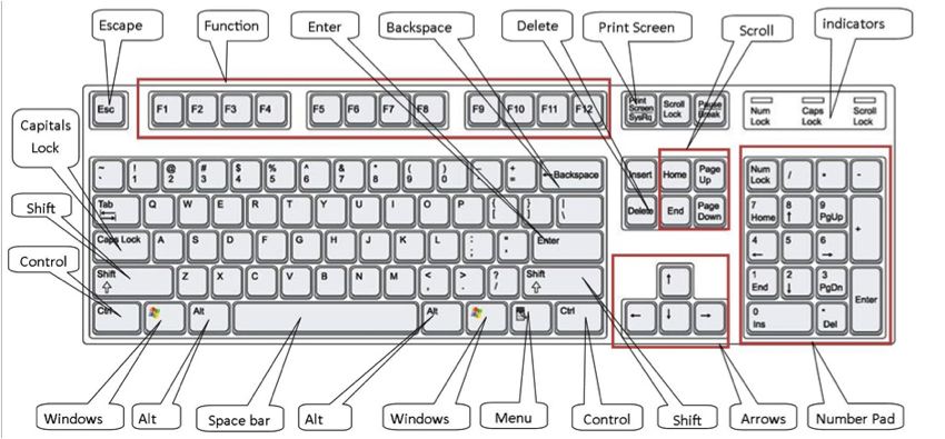
A membrane keyboard is a computer keyboard without separate keys.

What are the different parts of a computer keyboard. An example could be a command or a character that can be used in a document. Apple Mac computers have a command key. 4 Membrane Keyboard.
The shape of the keyboard is rectangular and the buttons are arranged horizontal contain about 108 Keys. 5 PARTS OF A KEYBOARD. The keyboard is designed to input the data by typing letters symbols numbers ABC123email protected.
Home basic computer parts motherboard processor memory monitor hard drives CDDVD drives keyboard mouse computer cards power supply computer case Resources. The keyboard is one of the most important parts of computer. Parts Of A Computer Keyboard.
Computer Keyboard Parts and Functions. Robotron Z1013 using the membrane Keyboard. Thats why we say keyboard.
Typing a key from the keyboard sends a small portion of data to tell the computer which key was pressed. That works out to. For information on keyboard shortcut key combinations eg CtrlS or AltF4 see our computer keyboard shortcut keys page.
There are three different types of PC keyboards are the original PC keyboard with 84 keys the AT keyboard with also 84 keys and enhanced keyboard with 101 keys. The basic parts of a desktop computer are the computer case monitor keyboard mouse and power cord. The thicker body of the keyboard holding the mechanical or silicone dome switches are also very distinct.
There are two main different types of keyboards. The parts of a typical computer keyboard includes. Though computer keyboard comes in several types and variations the basic elements are the same in all keyboards.
MAJOR FUNCTIONS OF THE KEYBOARDS- these are the 5 major functions these are the Function keys All F keys Alphanumeric keys 9 Special keys Cursor keys Arrow and the numeric keypad. Membrane keyboards are a unique computer keyboard that uses pressure pads instead of individualseparate keys. Regarding this what are the parts of keyboard and its function.
Basic Parts of a Computer. Other sets of keys common to almost all keyboards are entering and editing keys eg Enter Delete Insert modifier keys eg Control Shift navigation keys eg arrows for up down. Mechanical and membrane types.
These keyboards are the opposite of mechanical keyboards. These parts include the alphanumeric keypad the numeric keypad the arrow keys the control keys and the function keys. The keyboard is used for writing work on the computer.
A keyboard is one of the ways to communicate with a computer. People also ask what are parts of computer keyboard. Watch the video below to learn about the basic parts of a computer.
PC keyboards also have a Menu key that looks like a cursor pointing to a menu. The keys found on a Computer keyboard can be categorize as Alphanumeric keys which contains numbers and letters Punctuation keys that has comma period semicolon and other keys and Special keys with function. In front of the keyboard is a touchpad which you can touch to move your cursor the arrow on your computer screen.
Most computer keyboards are easily recognizable because of the spaces between each individual key. A computer key that you press together with another key so that the other key does something different from what it usually does. Unlike traditional ones membrane keyboards have symbols and characters printed on a flexible flat surface also called the membrane.
There are five main parts to most desktop keyboards. A standard keyboard has 108 keys plus 4 shell parts top bottom 2 support legs an interior circuit board an attachment cable a rubberized key mount for each key 46 rubber feet on the bottom and possibly a usb port soldered to the circuit board. Each part plays an important role whenever you use a computer.
The computer can use this information in many ways. Click to explore further. A board is a flat surface and this board is covered with buttons called keys.
PC keyboards have a Windows key that looks like a four-pane wavy window. With this type there are different parts or a major part that we need to know including the different keys its of this major function. Cursor- Control Keys Navigation key Numeric Keys.
Functions of Every Key - Keys on the Computer Keyboard and their Functions.
They are actually a form of larvae from the common mealworm beetle Aka the flour bettle. Mealworms that birds reptiles and other animals love arent really worms.
Why These Mealworms Don T Miss A Meal Bug Squad Anr Blogs
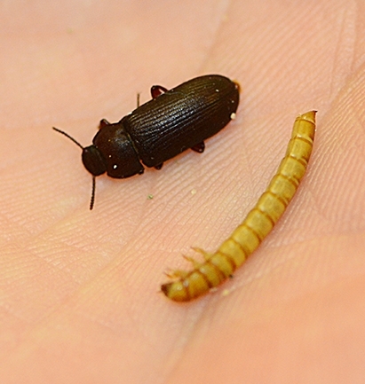
Mealworms do not bite and are not even capable of biting a human.

What is a mealworm beetle. Mealworms are the brown worm-like larvae of darkling beetles. After 2-3 months you can start using the. As far as mealworm nutrition facts are concerned they are rich sources of proteins and contain low fats.
Facts about mealworm beetles The female mealworm beetle lays up to 500 eggs. They remain as pupae for about one to three weeks before emerging as adult darkling beetles ready to. Mealworms are not worms they are the larval stage of the darkling beetle Tenebrio molitor.
It will go from white to brown to black. Mealworms are the larvae of darkling beetles. There are over 20000 different types of darkling beetles and mealworms come from the species Tenebrio molitor.
Mealworm is a basic food for birds and some pets in captivity. The darkling beetle is a holometabolic insect meaning it has four life-stages. The larvae are often sold in pet stores as live food for lizards and other small carnivorous pets.
Egg larvae pupae and imago adult. They are the larvae of darkling beetles. A mealworms life cycle goes eggLarvaepupaeadult beetle.
They remain as pupae for about one to three weeks before emerging as adult darkling beetles ready to eat and reproduce. Darkling because of its colour once the mealworm has finished turning into an adult beetle. Mealworm to Darkling Beetle in under two minutes - YouTube.
Mealworms are holometabolic insects AKA insects that develop in four distinct stages. Mealworm beetles Tenebrio molitor are widespread throughout Britain. The mealworm life.
Once they hatch mealworms molt repeatedly over the course of several months until they are about 137 of an inch long and are ready to pupate. Once they hatch mealworms molt repeatedly over the course of several months until they are about 137 of an inch long and are ready to pupate. A darkling beetle experiences complete metamorphosis which means that it has four distinct stages of life.
Mealworms can be found throughout most of the world where they prefer warm dark and damp places. They are the second of four stages of life and exist to eat and grow until they have enough energy stored to begin transformation into pupae and then beetles. Besides serving as a live feed for pets mealworms are also consumed by humans.
Each one of these life stages is distinct from one another making the transformation of egg to adult a complete metamorphosis. Hence it is commonly sold as a live food in pet stores. The mealworm is a darkling beetle in larva form where it remains for 90 to 114 days before turning into a pupa.
The amount of time it takes the insects to go through these stages depends on the temperature of their environment and availability of food. The mealworm beetles will lay eggs and eventually die continuing the cycle. Egg larva pupa and adult.
Darkling beetle is one of the names that the adult insect is known by. Mealworms are the larval stage of the darkling beetle. Mealworms are the larvae of the darkling beetle also known as Tenebrio molitor.
Other insects that are holometabolic include butterflies moths bees and wasps. Generally the life cycle of a mealworm can take anywhere between four months and a year. This looks like an adult Yellow Mealworm beetle Tenebrio molitor.
Egg larva pupa and adult. Mealworms are the larval stage of the darkling beetle. Darkling beetles mealworms are one of over 20000 species of beetles in the Tenebrionidae ten-eb-ri-on-i-dea family Dellinger and Day 2018.
Although the mealworm looks a bit like a worm it has six small jointe. The adult beetles fly and are attracted to light. Mealworms are not actually worms Even though that what we call them.
The larvae will also infest stored foods like cereals and grains. If playback doesnt. Mealworm to Darkling Beetle in under two minutes.
Add another layer of bran or oats whenever the bedding starts to get low. Mealworm beetles go through four distinct stages of development. Mealworms are one of the easiest insects to maintain and a common feeder for pet bearded dragons.
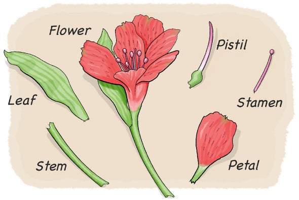
Diagnostic Characters of Rosaceae Floral Formula and Floral Diagra. Learners will 1 identify the different parts of a flower and understand their function.
Dissect A Flower Scientific American
Parts of a Flower Diagram All the Flower Parts are Labeled Plant Parts and Their Function Including Diagrams.
Labelled diagram of rose flower. You will sometimes see the term quartered especially in reference to old garden roses. This fain lb has a great economic importance for mankind. Filament - the filament is the part of the flower that holds the anther and part of the stamen the male reproductive organs of the plant.
Looking at a diagram of a flower you can easily see the individual parts of the flower. In green rose the petals are leaf-like in structure and green in colour. Parts of a hibiscus flower complete flower fiower part sepal plant ovary flower anatomy diagram of flower parts of a flower flower diagram parts of the flower.
Refer children to the flower diagram chart. Floral formula and floral diagram Rose plant Economic Importance. Apr 26 2016 - Most plants in the same family have similar characteristics.
How to Identify Six Plant Families Using Their Flowers Parts of a flower Flower structure Diagram of a flower. Each member of corolla is called a petal. Rose family General characters floral formula floral diagram economic importance and common species January 31 2019 Angiosperm families Botany Rosaceae Rose family Rosaceae Rose family In this article we will discuss about.
The androecium is the male reproductive whorl of the flower. In cultivated roses many stamens gradually change into petals. Explain that each flower is unique with its own special beauty.
Answer verified by Toppr. It has great importance in temperate cold region. In Canna the stamens and the style become petaloid.
While flowers are composed of the same Location. See flower parts diagram stock video clips. What is the overhead for each department.
A rose with more than 25 petals in three or more rows is called double. Fresh flowers 1000 square feet. The five hairy red spots shown below is a close-up view of the tip or stigma female part of the flower.
Download a powerpoint showing labelled and unlabelled versions of these diagrams both parts of a plant and parts of a flower from the link on the right. Classroom with tables for children to work in small groups Objectives. The function of anther is to produce pollen grains that contain the male gametes.
This family is ranked third in the flowering families for commercial importance in thetemperate zone. 87 KB Rosa floral diagramsvg 670 768. It is the first or outermost protective whorl.
The pinkish-white hibiscus flower seen below is from one of my hibiscus plants. Fine china 700 square feet. The pistil and stamen in the middle of the flower are surrounded by brightly colored petals.
11 -14 KS3 14 -16 KS4 Post 16 Plant growth health and reproduction. Rosa floral diagramjpg 719 747. It is the second or attractive whorl present inner to calyx.
Draw a diagram showing longitudinal section of a flower and label on it. The stigma is located at the tip of the pistil. Individual member of calyx is called a sepal which is generally green.
Individual members of the whorl are referred to as stamens. This is the part of the flower thats sticky and collects pollen. Many fruits are cbtained from the plants of this family.
Pottery 300 square feet. Oxford Floral allocated overhead of 7815 by floor space. The water lily shows a gradual transition from sepals to petals and from petals to stamens.
Gifts 800 square feet. Youll recognize the pistil in a plant diagram because it looks like a small knob that protrudes from the flower. Both male and female organs are found on the same flower.
Each stamen consists of a stalk called the filament and sac like structure called the anther. 1201 flower parts diagram stock photos vectors and illustrations are available royalty-free. A rose with 13 to about 25 petals in two or three rows is said to be semidouble.
Try these curated collections. Diagram Label the Flower anther - the anther is the tip of a flowers stamen the male reproductive organs of the plant - it contains the pollen. A very full flower having more than 45 to 50 petals in numerous rows is known as very double.
20 KB Rose flower bud longitudinal cut section spanish labels ovario estilo estigma estambre pétalo sépalo receptáculo pedicelosvg 640 560. I have labelled it to illustrate the different part of a typical flower. Stigma ovary anther filament.
It contains the heart Malpighian tubules reproductive organs and most of the digestive system foregut hindgut and rectum. Egg larva pupa and adult.

Ant Printout Enchanted Learning Software
The eggs are very small soft and oval-shaped.

Labelled diagram of ant. And congrats on being most clicked this week. As you color the diagram showing the basic anatomy of the ant discuss and label the major parts. Ant To Label - Displaying top 8 worksheets found for this concept.
What a great printable and lesson. April 18 2014 at 509 pm. What are the mandibles used for.
Unfertilized eggs produce male ants. Where do you find claws. And each structure has its own special function.
To what part are the legs connected. A well labelled diagram of a spider ant labeled Label Gallery Get some ideas to make labels for bottles jars packages products boxes or classroom activities for free. Read below to quickly identify what type of ant may be on your property decide if you have ants or termites and learn the many different species of ants.
It is protected by an exoskeleton. Ants begin as eggs. Like all insects an ants body is divided into three main parts.
Apr 8 2021 - Use this ant diagram during a unit on insects. Color label and discuss. They can lift 10 times their own weight.
Imagine being the size of an ant. They are exceptionally strong for their size. Ant Body Parts Diagram Activity.
Some of the worksheets for this concept are My insect report insect anatomy insect habitat insect life Labelled ant diagram for kids Diagram of ant to label 2 ant bodies Insect diagram for children Draw and well label of grasshopper Ask a biologist Match and snap. Abdomen - The abdomen is the segmented tail area of an ant. Head thorax abdomen antennae and legs.
Students will label the five major parts of an ant. It can take a few weeks or as long as a year for the life cycle to be completed. Ants have many body parts that are normally hard to see without a magnifying glass or microscope.
Students will label the five major parts of an ant. Thanks for sharing it on Science Sunday. Ants life cycle includes four stages.
Be careful - a face-to-face encounter with an ant would be scary and potentially life-threatening. What a great ant anatomy diagram and with the cut and paste aspect even preearly writers can do this. This product is now both a printable AND a TpT EASEL activity for y.
One including a word bank and one without a word bankUPDATE. The life cycle of the ant consists of four stages. Ants range in size from about 008 inch 2 mm to up to about 1 inch 25 mm long.
Students learn to insert size and move textboxes. Egg larva pupa and adult. One including a word bank and one without a word bankUPDATE.
Some of the worksheets for this concept are Insects It s a bug s life lesson plans grades 12 written and My insect report insect anatomy insect habitat insect life The miracle of ant therapy master class work Ant anatomy activity Labelled ant diagram for kids epub Label the insect Insect study. At the bottom of this guide is a breakdown of the various. Read the definitions below then label the ant external anatomy diagram.
Use this resource to allow students to label the key parts of a red bull ant and identify how they differ from other animals in regards to their external featuresTags in this resource. The queen ant can lay many eggs at a time. Fertilized eggs produce female ants queens workers or soldiers.
An easy and convenient way to make label is to generate some ideas first. Head thorax abdomen antennae and legs. The head the thorax and the abdomen.
Student learn how to insert size and move clipart. Ant eggs are oval shaped and tiny they are on the order of 1. But if you avoided being eaten you could learn a lot about ant anatomy from a close-up view.
Thank you for sharing at Sharing Saturday. Ant Body Parts - Displaying top 8 worksheets found for this concept. The queen ant finds a good place to lay her eggs.
Labelled diagram - Drag and drop the pins to their correct place on the image. Use this ant diagram during a unit on insects. What might be found inside the head.
Ants have a hard waterproof exoskeleton which is made of a material called chitin. A fun activity for students to learn how to label and annotate diagrams using a word processor or slide presentation app. This product is now both a.
Some methods work better for certain ant species than other methods and not every ant killer product is labeled for all ant species.
Serious injuries such as stress fractures can cause your feet to. Monday 04 January 2021 0000.

Is This Serious Why Does The Top Of My Foot Hurt
Do not worry if youre not sure what the problem is.

Why does the inside of my feet hurt when i walk. Why does the side of my foot hurt when I walk. Calluses are generally painless and relatively easy to eliminate. Its different from exercise-related soreness because it occurs only during movement and stops after short periods of rest says Dr.
Foot Pain While Sleeping. Pain on the inside of the foot near the big toe may be caused by a bunion a bony growth located at the base of the big toe joint. Ordinarily your ankle joint supports your entire body weight.
Mortons Neuroma As weve explained in previous blog posts Mortons neuroma is a condition categorized by the abnormal thickening of the tissue around the nerves that connect to your toes. It accounts for more than 70 percent of all ankle injuries. It may also occur after an ankle sprain.
Choose which area of your foot hurts most to read about treatments when to get medical help and possible causes of foot pain. Pain along the inside of the foot may be due to inflammation of a tendon posterior tibialis that attaches to the bone that is the keystone of the arch navicular. Metatarsalgia refers to pain located in the area of the ball of the foot.
Why Does the Inside of My Foot Hurt. Because we use our feet so much occasional aches and pains are common especially after walking for long periods. Gout is a type of painful inflammatory arthritis caused by too much uric acid in the blood Neville explains.
Pain along the inside arch of the foot. The inside of the foot refers to the inner edge of the foot along the side of the big toe. 20 At first symptoms may only be occasional but they may become more common or even constant as the condition develops.
Overuse is a common cause of foot pain but an underlying injury or medical. So if you have mild or severe inside foot pain it could be due to an ankle sprain. Foot gout is a type of inflammatory arthritis causing sudden intense pain swelling and redness typically in the big toe.
Symptoms typically flare up at night. A person who runs excessively or places their foot abnormally may develop peroneal tendonitis. Your Achilles tendon which attaches to your heel bone at the back of your foot can become irritated and inflamed when its overused says.
Follow the advice on this page and see a. Poor footwear selections tough surfaces and improper form can all contribute to foot pain. Why is there a sharp pain in my foot when I walk.
Peroneal tendonitis This condition causes the peroneal tendons to swell or become inflamed resulting in pain on the lateral side of the foot and the heel. When the bottoms of your feet hurt during exercise it makes getting through a sweat session that much tougher. Pain in this area can be indicative of various foot conditions.
Calluses are areas of skin that have been thickened and toughened by repetitive friction between the skin of your foot and an exterior surface like the inside of a badly fitting pair of shoes. This condition develops as a result of the loss of support provided by the ligaments that connect the metatarsal bones the five bones that make up your forefoot. They usually form on areas that take your weight when you are walking or running.
Pain in the bottom of your foot is often caused by exercising too much or wearing shoes that are too tight. Your symptoms might also give you an idea of whats causing your pain. Coronavirus COVID-19 Resource Center.
Inflammation of the tendon across the front of the ankle causes top of foot pain typically when walking down stairs or on slopes. Muscle soreness caused by exercise lasts for hours or days after a workout and can still hurt whether youre moving or standing still. Mortons neuroma causes a sharp stabbing pain when you walk or put pressure on your foot.
Scott Neville DPM a podiatrist in Mooresville Indiana says that for some people pain and swelling in the foot can be a sign of gout. Ankle sprain is a common cause of pain on the inside of the foot. The thickened tissue compresses.
Its caused by thickening of a nerve between your toes usually between the third and fourth toes possibly due to injury. If your feet hurt at night or while you have them elevated youll want to speak to a doctor to rule out one of these three conditions. If your foot pain from working out persists even after making changes consult your doctor to examine the underlying cause.
Click Share to make it public. Draw neat and well labelled diagram of Human respiratory system.
A Draw A Labelled Diagram Of The Respiratory System Of Human Beings With Diaphragm At The End Of Expiration Sarthaks Econnect Largest Online Education Community
This leaderboard has been disabled by the resource owner.
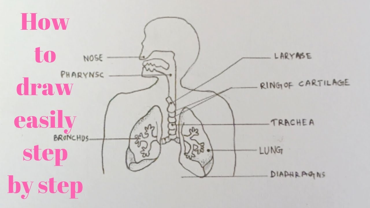
Human respiratory system with labelled diagram. View Original Image at Full Size. The respiratory system blank printable. The human respiratory system consists of a pair of lungs and a series of air passages leading to the lungs.
B List four conditions required for efficient gas exchange in an organism. Changes in the volume and pressure in the lungs aid in pulmonary ventilation. Transport of oxygen and carbon dioxide occurs with the help of respiratory pigment called hemoglobin.
Haemoglobin though is purple coloured but oxyhemoglobin is of bright red colour. Write postulates the cell theory. The air is exhaled back through the same pathway.
Features of the Human Respiratory System. Diagram of human respiratory system with labels. Many more lungs and other anatomical models available online via this link.
In this video we will study human respiratory system and its different parts which help in the process of brea. The respiratory system helps in breathing known as pulmonary ventilation. This leaderboard is currently private.
Take a look at the labeled diagram of the respiratory system above. Brainly User Brainly User New questions in Biology. Labeled Diagram Of The Respiratory System Of A Human was posted in June 7 2017 at 529 am.
This HD Wallpaper Labeled Diagram Of The Respiratory System Of A Human. Lungs are the respiratory organs in humans and other animals. For educational purposes only.
Follow me to draw. In this video Im going to draw the labelled diagram of Human Respiratory System. Put this list together in the correct order by writing their numbers below.
How to draw diagram of human Respiratory system easily - step by step - YouTube. Haemoglobin the iron-containing respiratory pigment is a red coloured pigment of blood which has a very high affinity for oxygen. The air inhaled through the nose moves through the pharynx larynx and trachea into the lungs.
Given below is a labeled diagram of the human lungs followed by a brief account of the different parts of the lungs and their functions. The route of air - on its proper way from the outside into the lungs and out again. As you can see there are several structures to learn.
All the main parts of the respiratory system labeled. Who proposed it hey friends help me with this only G_i_r_l. What is the benefits of neem tree.
Air enters the nose through the nostrils. Description from Labeled Diagram Of The Respiratory System Of A Human pictures wallpaper. Oxygen is transported from lungs to the body cells in the form of oxyhemoglobin.
The human respiratory system functions are mentioned below. Labeled diagram of the lungsrespiratory system. Learn how to draw diagram of human respiratory system using a very easy to understand method.
Labeled Diagram Of The Respiratory System Of A Human download this wallpaper for free in HD resolution. Choose from Respiratory System Labeled Diagram stock illustrations from iStock. Asked Oct 12 2019 in Biology by Suchita 664k points a Draw a labelled diagram of the respiratory system of human beings with diaphragm at the end of expiration.
Show more Show less. The Respiratory System diagram easily and step by s. This video shows the structure of lungs.
Find high-quality royalty-free vector images that you wont find anywhere else. Image 37789 is a 1125 by 1408 pixel PNG Uploaded. Use the mouse or tap the screen to label this diagram of the complete human respiratory system combining the head and thorax chest regions.
Share Share by Sumanbalasingh2. How to draw diagram of human Respiratory system easily - step by step. The structure of the lungs is created in such a way that it helps the exchange of gases.
Human Respiratory System Diagram If you carefully observe the respiratory system diagram you will be able to see the various organs involved in its functioning. The entire respiratory tract passage consists of the nose pharynx larynx trachea bronchi and bronchioles. 2 See answers TheRuhanikaDhawan TheRuhanikaDhawan Neat and well labelled diagram of Human respiratory system.
The gas exchange process is performed by the lungs and respiratory system. When air passes through the nose it is warmed moistened and filtered. Spend a few minutes reviewing the name and location of each one then try testing your knowledge by filling in your own diagram of the respiratory system unlabeled using the PDF download below.
This leaderboard is disabled as your options are different to the resource owner. Human respiratory system - Labelled diagram Nasal cavity Pharynx Trachea Lungs Bronchi Bronchioles Alveoli Diaphragm.
This activity has been designed for use in homes and schools. Microscopes R Us Pack Microscopic Biology Lesson Plans Biology Lessons High power objective 6.
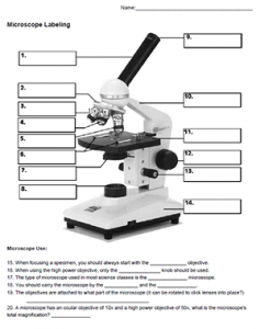
Microscope Labeling
Click to Download.

Label microscope parts worksheet with answers. Microscope parts and use worksheet answer key along with labeling the parts of the microscope blank diagram available for. Activity Students Label Parts. Nosepiece microscope when carried holds the high and low power objective lenses.
Using the microscopelabel the microscope determining the magnifying power working distance diameter of field of view brightness needed ocular objective magnification magnification total working magnification distance more diameter field of view wide. Some of the worksheets below are Parts and Function of a Microscope Worksheets with colorful charts and diagrams to help students familiarize with the parts of the microscope along with several important questions and activities with answers. Once you find your worksheet s you can either click on the pop-out icon or download button to print.
The labeling worksheet could be used as a quiz or as part of direct instruction where students label the microscope as you go over what each part is used for. Light microscope worksheet drag and drop worksheet on the parts of the microscope. View homework help - microscope worksheet pp with answers from bio.
Label parts of the Microscope. The study of living things. We tried to locate some good of Microscope Parts and Use Worksheet Answer Key Along with Labeling the Parts Of the Microscope Blank Diagram Available for image to suit your needs.
It was from reliable on line source and that we love it. Free worksheets for labeling parts of the microscope including a worksheet that is blank and one with answers. Microscope parts worksheet answers The rough adjustment knob b is the bigger one on your microscope.
1 86 Answer Key Science 1-7 71 57 43 29 14 0 1. NOSEPIECE microscope when carried Holds the HIGH- and LOW- power objective LENSES. Id language school subject science middle school age main content lab use of the microscope.
Label the parts of the microscope indicated and state the. Handphone Tablet Desktop Original Size There are many key components to understand when utilizing a microscope. The compound microscope uses lenses and light to enlarge the image and is also called an optical.
Text preview biology a laboratory scopes and cells worksheet name a. Parts of a eyepiece arm stageclips nosepiece focusing knobs illuminator stage objective lenses head base Label the parts of the microscope. Activity Students Label Parts.
Labeled microscope worksheet answers. Microscopes are often used in areas as diverse as plant and animal studies and anatomy. If you want to download the image of microscope parts and use worksheet answer key as well as life science teacher s edition simply right click the image and choose save as.
List of Microscope Labeling Worksheet Answers. Worksheet identifying the. This worksheet asks pupils to label the different parts of a microscope and then match up the with their function.
Each microscope layout both blank and the version with answers are available as PDF downloads. You can use the word bank below to fill in the blanks or cut. Microscope Parts and Use Worksheet Answer Key Along with Labeling the Parts Of the Microscope Blank Diagram Available for.
You can use the word bank below to fill in the blanks or cut and paste the words at the bottom. A basic microscope has a single convex lens such as those found in a magnifying glass. We tried to locate some good of microscope parts and use worksheet answer key along with labeling the parts of.
Can be rotated to change MAGNIFICATION. Microscope parts worksheet answer key. Microscope parts and use worksheet answer key along with labeling the parts of the microscope blank diagram available for.
View homework help - microscope worksheet pp with answers from bio biol at community college. Labeling the Parts of the Microscope. Live worksheets science lab equipment light microscope worksheet.
Label the Parts of the Microscope with answers A4 PDF print version. List of Microscope Labeling Worksheet Answers Pm company. Power 10 x 4 40 Power 10 x 10 100 Power 10 x 40 400 What happens as the power of magnification increases.
In this activity students identify and label the main parts of a microscope and describe their function. Microscope parts and use worksheet answer key along with labeling the parts of the microscope blank diagram available for worksheet january 13 2018 we tried to locate some good of microscope parts and use worksheet answer key along with labeling the parts of the microscope blank diagram available for image to suit your needs. Coarse adjustment knob 13.
We hope this graphic will likely be one of excellent reference. For a thorough review of each microscope part continue reading. Some of the worksheets shown are part microscope and use microscope mania microscope laboratory work part name of the student microscope from the light microscope review work filling the blank Wanganui High School.
By the end of this. Answers to my TEENrens crossword puzzles. Microscopes are often used in areas as diverse as plant and animal studies and anatomy.
G Labeling Scientific Tools Microscope G F E D C B A 1 Illuminator 2 Stage 3 Eyepiece 4 Focus Fine 5 Lense 6 Focus Course 7 Base Determine which letter best matches each microscope piece.
The style leads down to the ovary that contains the ovules. The pollen producing part of a flower usually with a slender filament supporting the anther.
Flower A Fascinating Organ Of Angiosperms Parts Of A Flower Ck 12 Foundation

In a flower the female reproductive part is called the Pistil.

What are the three parts of the pistil in a flower. The style leads to the ovary that contains the female egg cells called ovules. Stigma is the top most part of the pistil on which the landing of pollen grains takes place whereas the ovary is the swollen basal part of the pistil which contain ovules. The filament is a slender threadlike object which functions by supporting the anther.
The pistil usually is located in the center of the flower and is made up of three parts. The pistil consists of a long cylindrical central part called a style that connects the lower ovary to the upper stigma which receives and holds pollen. Stigma style and ovary.
The pistil is the female reproductive part of a flower. Name the three parts of the pistil of a flower. It traps and holds the pollen.
The parts of a flower that are often conspicuously colored. If the stylodia are reduced the stigma sits directly on the ovary. It is primarily designed or adapted to pollination and followed by the fertilization process.
This is the innermost part and the female reproductive organ of a flower which comprises three parts -stigma style and ovary. The stigma is the sticky knob at the top of the pistil. The nice smells and bright colors of the flower.
If a flower lacks any one of these parts it is an incomplete flower. The stigma usually crowns the style or stylodia. The part of the stamen where pollen is produced.
It is attached to the long tubelike structure called the style. The stigma style and ovary. There may be a single pistil as in the lily or several to many pistils as in the buttercup.
Pistil consists of three parts. What are the three parts of the female structure of a flower. It has three main parts called stigma style and ovary.
Stamen is the male reproductive part of a flower. The ovule producing part of a flower. The pistil which is located in the center of the flower typically consists of a hollow and enlarged inferior partthe ovary a slender and usually cylindrical style or a stylodium and a stigma.
A group of pistils or carpels is called a gynoecium an alteration of Latin gynaeceum. Rose hibiscus and tulip are complete flowers because they have all the main flower organs. Petals sepals stamen and carpel also known as a pistil.
This is collectively known as the pistil. The stigma is the sticky surface at the top of the pistil. The pistil usually is located in the center of the flower and is made up of three parts.
The style is the tube-like structure that holds up the stigma. The stigma is the sticky knob at the top of the pistil. Stigma style and ovary.
Incomplete Flower Botanically a flower is considered to be complete flower if it contains the four main parts of a flower. It contains three main parts the stigma style and ovary. A pistil then may be composed of one carpel simple pistil as in the sweet pea or of two or more carpels compound pistil partially or completely joined as in the mustard two carpels or lily three carpels.
Style is a filamentous structure which serve as a connecting link between stigma and ovary. It is attached to the long tubelike structure called the style. Self - if the pollen stays on the same flower.
The ovary often supports a long style topped by a stigma. What is the difference between self and cross pollination. A flower that contains separate pistils and therefore separate carpels is termed apocarpous.
The pistil is the female structure of a flower which mainly consists of stigma style ovary and ovule. The stigma style and ovary. Therefore it is important to know about the parts that make a flower as they all contribute in these processes.
The main function of the pistil is to produce a seed-bearing fruit. What helps attract pollinators. The style leads to the ovary that contains the female egg cells called ovules.
The pistil has three parts.
Its magnification capacity ranges between 10 and 15 times. But we can divide them based on their purpose in the instrument like.

Microscopy
7 6 8 5 9 4 10 09 11 12 2.
Label the parts of the microscope and give its corresponding functions. The eyepiece usually contains a 10X or 15X power lens. How a Microscope Works 3. The nose piece is circular and a rotating metal part that is connected to the body tubes lower end.
Label the parts of the microscope and give its corresponding functions. - 10463704 jasminedomingo39 jasminedomingo39 06022021 Science Junior High School answered Direction. In this interactive you can label the different parts of a microscope.
Start at part A down to part 1. Arm The arm connects the body tube to the base. Use this with the Microscope parts activity to help students identify and label the main parts of a microscope and then describe their functions.
It normally has clips that prevents a slide from moving while it is being viewed by the user from the eyepiece part. Optical parts of Microscope and their Functions. The Ribbon is the strip of buttons and icons located above the work area in Word 2007.
The diaphragm can be of two types. This is done by rotating the knobs which can either move. These are the lens located at the top of the microscope where you look through.
Bring the object into sharp focus by using the fine adjustment knob. Parts of the microscope and their functions 1. It consist of this following components.
Microscope Parts in detail There are almost 15 parts in a microscope. Convex Lenses are curved glass used to make microscopes and glasses etc Convex Lenses bend light and focus it in one spot. 2 on a question Directions.
Usually 10 X magnification. Eyepiece consists of two lenses the ocularThe first one near the eye and eyepiece The last one away from the eye. Click on the boxes to see the name and function of each part of the microscope.
Label the parts of the microscope and give its corresponding functions Start at part A down to part M. The Ribbon replaces the menus and toolbars found in earlier versions of Word. Click on Me Click on Me Click on Me Click on Me Click on Me Click on Me Click on Me Click on Me Click on Me Click on Me Click on Me Click on Me Click on Me Click on Me 9 Eye PieceThe part you look at with your eye.
Lens The biconvex lens is placed above the stage and its function is to magnify the size of the object being examined. Useful as a means to change focus on one eyepiece so as to correct for any difference in. The lens the viewer looks through to see the specimen.
This part connects the eyepiece lens to the objective lenses. The upper part of the microscope that houses the optical elements of the unit. This supports the tube and connects the tube to the base.
A compound microscope consists of parts that assist in viewing with a naked eye a sample holder a magnifying lens and alight source. Each ribbon contains groups of command buttons with common purpose. Through the eyepiece you can visualize the object being studied.
Focus using the COARSE ADJUSTMENT KNOB to bring the object into focus. Here are the important compound microscope parts. The Light Microscope 2.
Drag and drop the text labels onto the microscope diagram. Let us take a look at the different parts of microscopes and their respective functions. Focus and then move to.
This is the bottom part of the microscope which is used for support. It controls and adjusts the intensity of light that passes into the microscope. Body Tube It is the part of the microscope that holds the eyepiece.
All microscopes share features in common. The Main parts of a microscopes are easy to identify. Label the parts of the microscope and give its function.
This part of microscope is also known as ocular. Structural element that connects the head of the microscope to the base. Compound Microscope Parts and Functions.
18 1 14 pls help me 2. This part is where doctors or scientists put the microscope slide with samples for analysis. The bottom of the microscopewhat the microscope stands on.
Write your answers in the table provided. Mirror A simple microscope has a plano-convex mirror and its primary function is to focus the surrounding light on the object being examined. The diaphragm is fastened below the stage.
These lens are can magnify small objects 10x to 15x. December 9 2017 Author. The slide can be moved manually while it is being viewed or it can be moved mechanically if you are using a microscope with a mechanical platform.
The user must hold this part in order to move the microscope from one place to another. Storing The Microscope Using the microscope Always observe using the LOWEST POWER objective first. The Parts Functions of Microsoft Word.
What are the 3 Basic Parts of a Microscope. Label the parts of the microscope and give its function. Other Important Microscope Parts Their Functions.
It is the topmost part of the microscope. The optical part of Microscope plays an important role to magnify the object.
Well-Labelled Diagram of Animal Cell The cell membrane is a double-layered membrane made up of phospholipids that surrounds the entire cell. Labelled cell surface membrane with carrier proteins phospholipid bilayer etc.

Structure Of A Plant Cell Labelled Diagram Of The Structure Of A Plant Cell Canstock
Diagram of the human cell illustrating the different parts of the cell.

Labelled diagram of a cell. About the Well Labelled Diagram of Animal Cell A Well Labelled Diagram of Animal Cell. Cytoskeleton is the. To draw a well labelled diagram of an animal cell the cell membrane has to be drawn.
In a plant cell the cell wall is made up of cellulose hemicellulose and proteins while in a fungal cell it is composed of chitin. Printable plant and animal cell labelled diagram of a generalised. Plant cells are eukaryotic cells but unlike animal cells which have a cell membrane plant cells have cell walls.
Labelled diagram - Drag and drop the pins to their correct place on the image. The cell membrane is semipermeable and flexible. Labelled diagram of a plant cell under microscope posted on march 18 2011 by admin onion cells stained with methylene blue look at the images of onion cells as they would be seen under a microscope draw each magnification label appear high picture plant and animal cell diagrams.
The Rate Of Red Blood Cell Labeling Using 55 Co Dtpa At 37 C N 5. A Labeled Diagram of the Plant Cell and Functions of its Organelles. It is evident from the introduction that an animal cell is a representation of.
Labelled Diagram Definitions and Structure Published by Admin on July 28 2021 July 28 2021. Labelled xylem cell diagram. Dr alban March 17 2021 Diagram No Comments.
Eukaryotes are sophisticated cells with a well defined nucleus and cell organelles. White Blood Cell Diagram Labeled Beautiful White Blood Cell Diagram. Answer verified by Toppr Upvote 0.
Cytosol is the fluid present within a cell that is made up of water and ions such as potassium proteins and small. During cellular division plant cells grow a new cell wall down the middle separating one cell into two. Unicellular to multicellular in nature and evolved 1 billion years ago.
Cell Membrane The cell membrane is the outer coating of the cell and contains the cytoplasm substances within it and the organelle. A cell wall is multilayered with a middle lamina a primary cell wall and a secondary cell wall. The cell is the basic unit of structure function and organization in all organisms.
These cells reproduce both asexually and sexually. Middle lamina contains polysaccharides that provide adhesion and allows binding of the cells to one another. Structure of Plant Cells.
The red blood cells those that you can see in the cell diagram are the most common type of blood cells. Labelled diagram of a cell wall. A level standard labelled diagram of an animal cell with structure and function of all organelles.
The significant differences between plant and animal cells are also shown and the diagrams are. It is a rigid covering made up of cellulose which a complex substance is providing structural support to the plants. Unlike xylem phloem vessels contain cytoplasm and this goes through the holes in the sieve plates from one cell to the next.
The synthesis of cell wall in controlled by golgi bodies. The cell being the smallest unit of life is akin to a tiny room which houses several organs. Animal Cell Diagram Labeled And Functions Inspirational Animal Cell.
Here lets study the plant cell. Plant Cell Diagram 9th Class Labeled. A nucleosome is a basic unit of DNA packaging in the nucleus of a eukaryote cell which consisting of a segment of DNA wound in sequence around histone protein cores.
Plant Cell Diagram 9th Class. Structure in a plant cell the cell wall is made up of cellulose hemicellulose and proteins while in a fungal cell it is composed of chitin. The most important structures of plant and animal cells are shown in the diagrams below which provide a clear illustration of how much these cells have in common.
It is a double-layered membrane composed of proteins and lipids. How to draw diagram of Animal Cell easily - step by step - YouTube. Gross structure of cell wall.
The cells are comparatively larger in size 10-100 μm. We are aware that all life stems from a single cell and that the cell is the most basic unit of all living organisms.
With the help of a clean spatula or a toothpick the inner side of the cheek is gently scrapped. They are found in the brain spinal cord and the peripheral nerves.

Solved Draw A Labelled Diagram Of Human Cheek Cells 3 Marks Self Study 365
Name the parts labelled 1 2 and 3.

Draw a labelled diagram of human cell. Name the stage prior to this stage and draw a diagram to represent the same. Label any three parts and write their functions. Labelled diagram of plant and animal cell are as follow---Both plant and animal cells belong to eukaryotic cells.
The cell membrane is semipermeable and flexible. Eukaryotes are sophisticated cells with a well defined nucleus and cell organelles. Unicellular to multicellular in nature and evolved 1 billion years ago.
This structure contains enzymes used for penetrating the female egg. It is a double-layered membrane composed of proteins and lipids. Identify the above stage and give a reason to support your answer.
The middle piece has a central filamentous core with many mitochondria. The cell organelles are membrane bound present within the cells. The sperm cells are the haploid gametes which are produced in the male.
462 378 pixels. Draw a diagram of the microscopic structure of human sperm. Diagram of the human cell illustrating the different parts of the cell.
Size of this PNG preview of this SVG file. FileDiagram human cell nucleussvg. The above diagram is of the sperm cell.
The structure of a neuron varies with their shape and size and it mainly depends upon their functions and their location. V Egg is the largest human cell. Mention the type of cells in our body where this type of cell division occurs.
A neuron is also known as the nerve cell. These cells reproduce both asexually and sexually. Contains the genetic material that the sperm has to pass on a haploid genome because it contains only one copy of each chromosome.
Vi Ovaries are located lower abdomen. There is the genetic material which is a haploid genome because it contains only one copy of each chromosome. The scrapped material is transferred into a drop of water and taken on a clean slide.
Draw A Labelled Diagram Of A Animal Cell And Plant Cell Ncert. State the functions of the two parts labelled. Draw a Neat and Labelled Diagram of the Human Ear.
With the Help of this Diagram Explain the Construction and Working of the Human Ear. Label the following parts in it and write their functions. This is a file from the Wikimedia Commons.
Fallopian tubes i Two thin tubes attached to the upper sides of uterus ii Tubes terminate near the ovaries but are not attached iii Fimbriae are finger-like structures on the end of each tube. Posted by Unknown at 2209. A neuron is a specialized cell primarily involved in transmitting information through electrical and chemical signals.
Solution For Draw a well labelled diagram of eye. Draw a diagram of a mature human sperm. The cells are comparatively larger in size 10-100 μm.
There are different parts of the sperm cell. 293 240 pixels 587 480 pixels 733 600 pixels 939 768 pixels 1252 1024 pixels 2503 2048 pixels. Information from its description page there is shown below.
Iii In what two ways is mitotic division in an animal cell different from the mitotic division in a plant cell. Lakhmir Singh Science Class 8 Solutions Chapter 8 Cell Structure And. Cell Membrane The cell membrane is the outer coating of the cell and contains the cytoplasm substances within it and the organelle.
It contains enzymes used for penetrating the female egg. 1 left and 1 on the right. With the help of a needle the material is uniformly spread.
Eukaryotic cells are one which have organised nucleus with a nuclear membrane and genetic material is organised into chromosomes. It is the inner layer and consists of three sub layers- ganglional cells bipolar cells and photoreceptor cellsThe innermost ganglionic cells give rise to optic nerve fibre that forms optic nerve in each eye and is connected with the brain. Draw a labelled diagram of the embryonic stage that gets implanted in the human uterus.
Human Cell Diagram Parts Pictures Structure and Functions parts of a human cell Diagram of the human cell illustrating the different parts of the cell.
In this Biology lab we will take a close look at the external and internal anatomy of a freshwater aquatic invertebratethe crayfish. Internal Anatomy of the Crayfish Directions.
Internal Anatomy Crayfish Anatomy Drawing Diagram
The cephalothorax consists of the cephalic or head region and the thoracic region.

Label the diagram of the crayfish. Follow the directions step-by-step locating each of the structures in the order they appear in the directions. The chelipeds is used to capture food. The following diagrams include the explained anatomy of a crayfish.
You may want to review or reference terms you dont remember It is expected that you know the terms. Use the diagram of the internal anatomy of the crayfish to locate and identify the organs of the circulatory system. This collection of printable crayfish diagram shows a range of organs of the crustacean.
This structure is illustrated in the individual speciesaccounts. Locate the dorsal tubular heart and several arteries. Abdomen - The abdomen is the segmented tail area.
Crayfish walk in a forward motion but they use a backward motion when they swim allowing them to move more rapidly. Internal Anatomy of the Crayfish. The maxilla and the maxillipeds manipulate the food.
Like all crustaceans a crayfish has a fairly hard exoskeleton that covers its body. Your Skills Rank. As shown in the diagram on the next page its body is divided into two main.
Ventral unlabelledRead the definitions below then label the crayfish diagram. Internal anatomy of a crayfish. The antenna and the antennule is used for taste.
Crayfish have eyes are on stalks and can move independently. If a crayfish loses a leg the leg will regenerate regrow. Cephalothorax thorax rostrum and dorsal before you start this dissection.
Edible freshwater crustacean with pincers on the two the body of a scorpion is divided into two parts. Part of the digestive tract between the esophagus and the intestine. Place a crayfish on its side in a dissection tray.
Like all crustaceans a crayfish has a fairly hard exoskeleton that covers its body. Crayfish External Anatomy Read the definitions below then label the crayfish diagram. It has 2 large pincers or claws called chelipeds.
Name the appendages found on the thorax of the crayfish and tell the function of each. Classifying What structures are used for capturing prey or securing and eating food. As shown in the diagram its body is divided into two main parts the cephalothorax and the abdomen.
The Digestive System Diagram Labeled. The body of a scorpion is divided into two parts. You may want to review or reference terms you dont remember It is expected that you know the terms.
Read the definitions below then label the crayfish diagram. Muscle that extends the tail of the crayfish. Label the drawing of the crayfish.
The head and thorax are fused forming the cephalothorax. Cephalic groove - An indentation in the carapace between the head region and the thorax region. Anatomy of a Crayfish.
Labelled photographs and diagrams for review. Crayfish external anatomy read the definitions below then label the crayfish diagram. Crayfish External Anatomy Read the.
Printable Animal Cell Diagram U2013 Labeled Unlabeled And Blank. Crayfish Diagram Labeled CATHERINEGRAESBECKDECMAORG. The crayfish has an open circulatory system in which the blood flows from arteries into sinuses or spaces in tissues.
The crayfish has 8 jointed walking legs a segmented body 2 pairs of sensory antennae and compound eyes. The eye-stalks of the crayfish produce a special hormone that regulates the metabolism and molting process in the body. Basically the same as an insect.
Label Crayfish External Anatomy Diagram Printout. Read the definitions below then label the crayfish diagram. Crayfish have compound eyes that contain thousands of tiny structures each functioning as a separate eye in multi-tiled fashion.
The cephalothorax consists of. Cephalic groove an indentation in the carapace between. Follow the directions step-by-step locating each of the structures in the order they appear in the directions.
Start studying Diagram of a crayfish. Crayfish external anatomy read the definitions below then label the crayfish diagram. Cephalic groove an indentation in the carapace between the head region and the thorax region.
Carapace - The protective shell exoskeleton of the cephalothorax. Like all crustaceans a crayfish has a fairly hard exoskeleton that covers its body. Crayfish Diagram Body Parts This is a walkthrough guide to dissecting the crayfish includes pictures and descriptions of structures.
Use the diagram below to locate the cephalothorax and the abdomen. As shown in the diagram on the next page its body is divided into two main parts the cephalothorax and the abdomen. The Skeletal System Diagram Labeled The Skeletal System.
The mandible is used to chew food. Edible freshwater crustacean with pincers on the two forelegs. You may use your book diagrams to help you locate the organs.
This is an online quiz called Crayfish Anatomy Internal There is a printable worksheet available for download here so you can take the quiz with pen and paper. It gives crayfish a mosaic and panoramic vision view of its world. The cephalothorax consists of the cephalic or head region and the thoracic region.
The swimmerets telson and uropods are attached to the abdomen. Label Crayfish External Anatomy Diagram Printout.