Most gram positive cell walls contain additional substances such as teichoic acid and teichuronic acid. The wall may also contain teichoic acid lipoteichoic acid and M protein which are the major.

Difference Bacteria Cell Walls Of Gram Positive And Gram Negative Download Scientific Diagram
As compared to Gram negative bacteria this group of bacteria is characterized by their ability to retain the primary stain Crystal violet during Gram staining giving a positive result.

Labelled diagram of gram positive bacteria. The sizes of bacteria cells that can infect humans beings range from 01 to 10 micrometers. Cytoplasmic membrane is a thin 5-10 nm layer lining the inner surface of the cell wall. Illustration about Structure of cell wall of Gram-positive bacteria labeled 3D illustration.
It is a viscous. Author links open overlay panel Alka Vaid Alistair H Bishop. Gram positive bacteria are a group of organisms that fall under the phylum Firmicutes however a few species have a Gram negative cell wall structure.
Educational scheme with labeled capillary circulation adherence deformability and phagocytosis. The types of bacteria and their labeled diagrams are shown below. Gram Positive Bacteria Cell Wall Label Diagram Quizlet.
Construction Of P16slux A Novel Vector For Improved. Simple Bacteria Diagram Labeled Ditulis JupiterZ Kamis 17 September 2020 Tulis Komentar Edit. A substance forming the cell walls of many bacteria consisting of glycosaminoglycan chains.
Bacterial Spores Structure Importance And Examples Of Spore. External to the cell wall may be present a thin layer of slime. Gram Negative Bacteria Diagram Labeled Written By JupiterZ Friday June 28 2019 Add Comment Edit.
Which of the following organism is a gram-positive bacteria. The gram-positive cell wall is very simple. They are made of protein pilin.
Some larger types of bacteria such as the rickettsias mycoplasmas and chlamydias have similar sizes as the largest types of. This thick layer makes the gram-positive bacteria resistant to osmotic lysis. The space between the inner cytoplasmic membrane and the bacterial outer membrane present in gram-positive bacteria.
Structure of cell wall of Gram-positive bacteria 3D illustration Neutrophil vector illustration. Different types of bacteria. Bacillus - Rod shaped.
According to Peberdy 1980 the only compound present in the cell walls of both Gram-negative and Gram-positive bacteria is peptidoglycan. Multifunctional Nanoagents For Ultrasensitive Imaging And. These are thin short filaments 01-15 μm x 4 to 8 nm extruding from the cytoplasmic membrane also called pili.
Amplification of fluorescently labelled DNA within Gram-positive and acid-fast bacteria. The information can be in the form of hand-written or printed text or symbols and gives details about manufacturers name source of product shelf-life uses and the manner of disposal. Labels are a means of identifying a product or container through a piece of fabric paper metal or plastic film onto which information about them is printed.
See Page 2 for a diagram of the Gram-negative cell wall and a video on. Watch learning videos swipe through stories and browse through. Draw neat and labelled diagram.
Illustration of corynebacterium microbiology microbial - 84173150 Stock Photos. Gram Negative And Positive Cells This Diagram Displays Both Gram. Gram-positive bacteria take up the crystal violet stain used in the test and then appear to be purple-coloured when seen through an optical microscope.
The semipermeable membrane surrounding the cytoplasm of a cell. It is an outer covering of thin jelly-like material 02 μm in width that surrounds the cell wall. Gram positive cells retain the Gram stain probably because the thick peptidoglycan layer prevents the stain from being leached out by alcohol in the staining process.
In bacteriology gram-positive bacteria are bacteria that give a positive result in the Gram stain test which is traditionally used to quickly classify bacteria into two broad categories according to their type of cell wall. Typical Lebeled Diagram Of. It is mainly made of a thick layer of peptidoglycan which constitutes about 90 of the cell wall.
Gram Negative And Positive Cells This Diagram Displays Both Gram. A good example of an S-layer is shown in our diagram of anthrax. Upgrade to remove ads.
The types of bacteria and their labeled diagrams are shown below. This video lecture describes the simple mnemonics for remembering the complex list of Gram positive and Gram negative bacteriaMnemonics GramPositive Gra. There are different forms of bacteria.
The cell walls of Gram-positive bacteria contain up to 95 peptidoglycan and up to 10 teichoic acids. In Gram-positive bacteria peptidoglycan makes up as much as 90 of the thick cell wall enclosing the plasma membrane. The Gram staining method was developed by.
This is because the thick peptidoglycan layer in the bacterial.
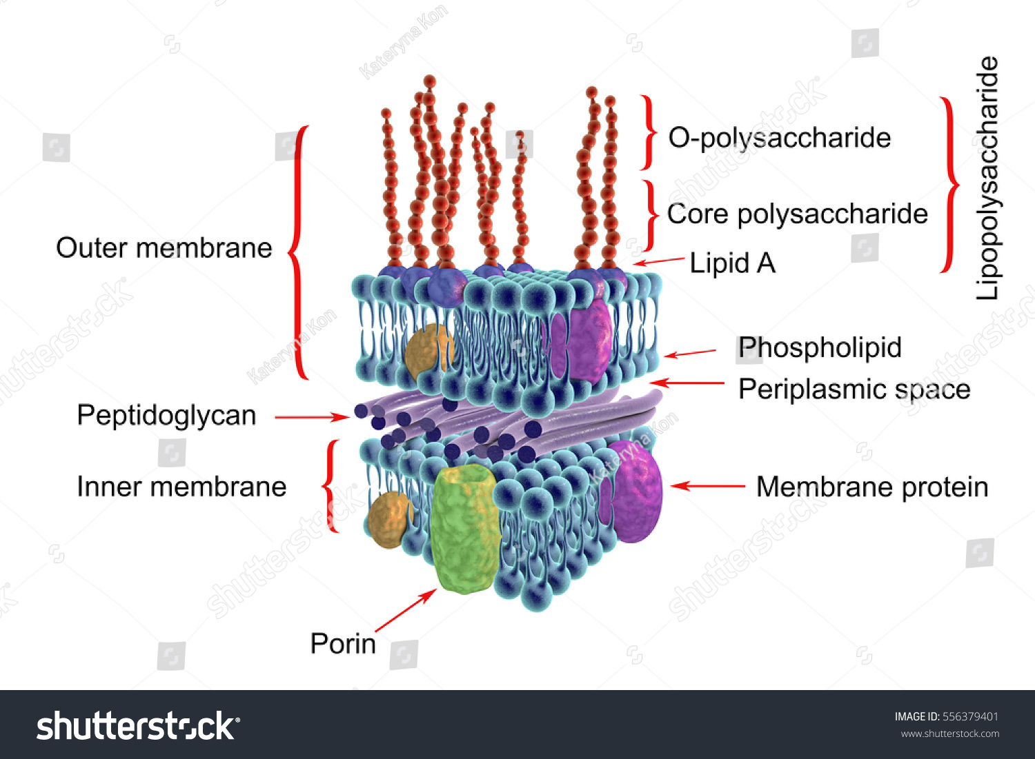
Structure Of Cell Wall Of Gram Negative Bacteria Royalty Free Stock Photo 556379401 Avopix Com

Gram Positive Vs Gram Negative Technology Networks

Structure Cell Wall Image Photo Free Trial Bigstock

Cell Wall Structures Of Gram Positive And Gram Negative Bacteria And Download Scientific Diagram
Science And Education Biology Biology Diagrams Microbiology Education

Pin Em Medimoon Com

Slime Layer An Overview Sciencedirect Topics
:max_bytes(150000):strip_icc()/gram_positive_bacteria-5b7f3032c9e77c0050f88457.jpg)
Gram Positive Vs Gram Negative Bacteria
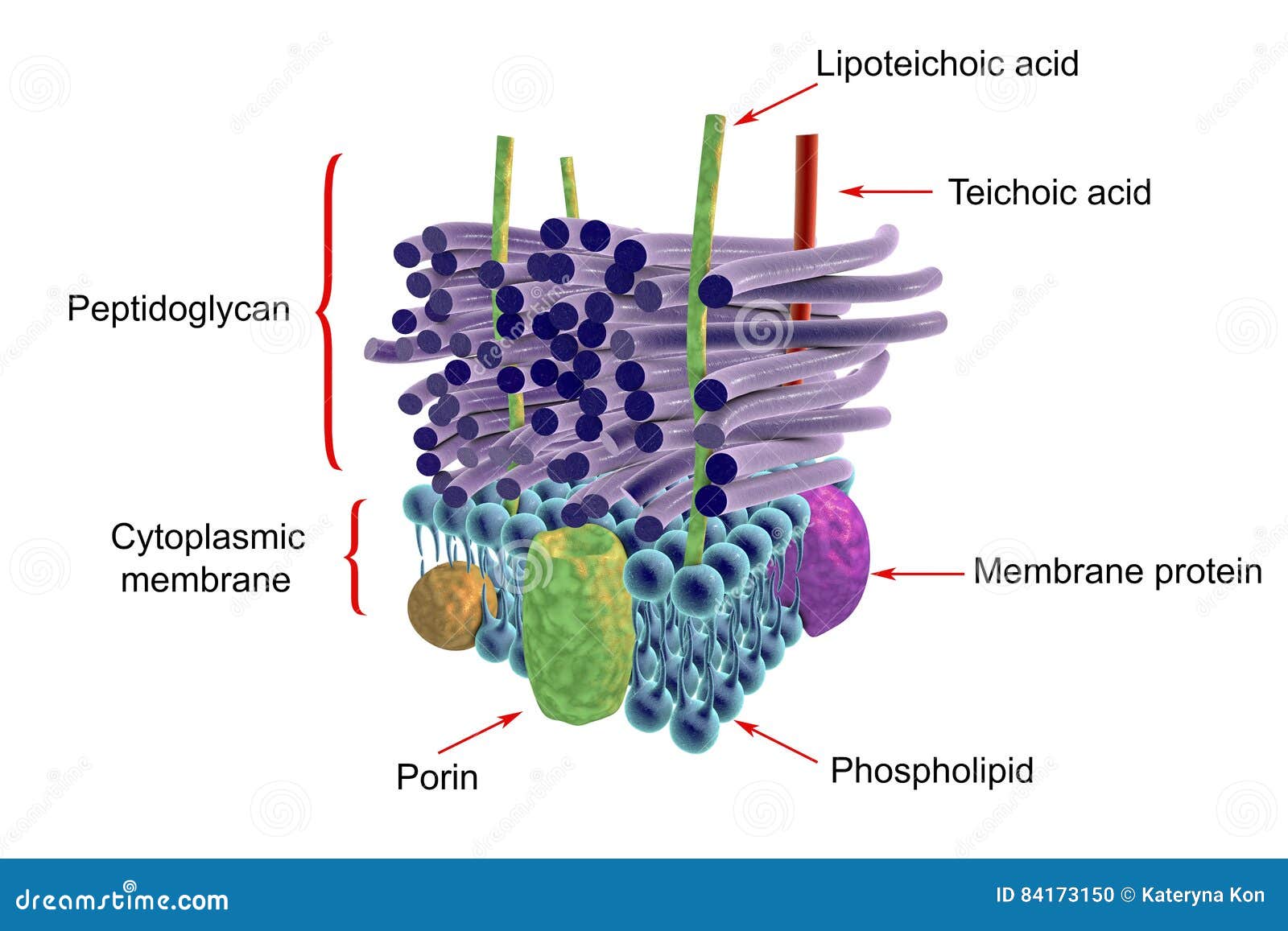
Structure Of Gram Positive Bacteria Cell Wall Stock Illustration Illustration Of Corynebacterium Microbiology 84173150
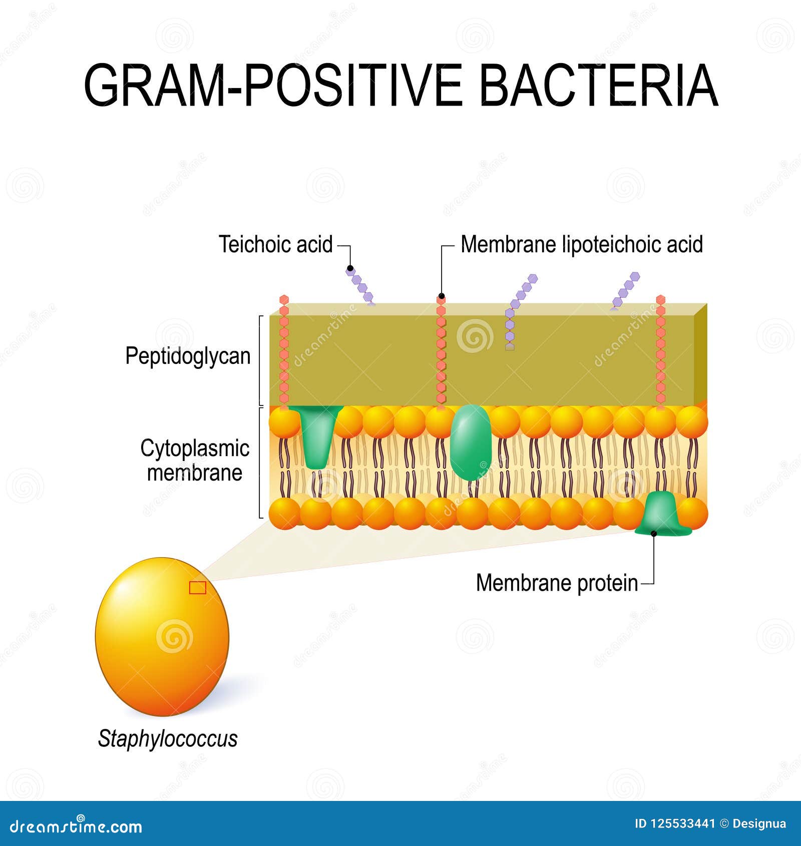
Cell Wall Structure Of Gram Positive Bacteria For Example Staphylococcus Stock Vector Illustration Of Crystal Bacterial 125533441